



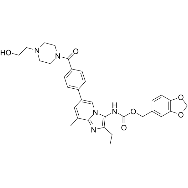
ATX inhibitor 11

Based on 1 Customer Validation
ATX inhibitor 11 (compound 13c) is a potent ATX (autotaxin) inhibitor, with an IC50 of 2.7 nM. ATX inhibitor 11 can typically alleviate the severity of fibrosis tissues and effectively reduce the deposition of fibrotic biomarker α-SMA in mice fibrosis model. ATX inhibitor 11 can be used for lung fibrosis research.
For research use only. We do not sell to patients.
- Purity : 99.44%
- CAS No.: 2485779-27-7
- Formula: C32H35N5O6
- Molecular Weight:585.65
-
Storage:Powder -20°C, 3 years , 4°C, 2 years ; In solvent -80°C, 6 months , -20°C, 1 month

To place orders, for customer services and technical support, please contact: MedChemExpress USA
Tel: 609-228-6898 E-mail: [email protected] [email protected]


-
 Biological Activity
-
Chemical Information
-
Solvent & Solubility
- Protocols
- Purity & Documentation
- References
-
 Help & FAQs




Biological Activity
Description
IC50 & Target
|
Autotaxin 2.7 ± 1 nM (IC50) |


Chemical Information
-
CAS No. 2485779-27-7
-
Appearance Solid
-
Molecular Weight 585.65
-
Formula C32H35N5O6
-
Color White to off-white
-
SMILES
CCC1=C(N2C=C(C=C(C2=N1)C)C3=CC=C(C=C3)C(N4CCN(CC4)CCO)=O)NC(OCC5=CC6=C(OCO6)C=C5)=O
-
Shipping
Room temperature in continental US; may vary elsewhere.
-
Storage
Powder -20°C 3 years 4°C 2 years In solvent -80°C 6 months -20°C 1 month


Solvent & Solubility
In Vitro:
DMSO : 100 mg/mL (170.75 mM; Need ultrasonic; Hygroscopic DMSO has a significant impact on the solubility of product, please use newly opened DMSO)
Please refer to the solubility information to select the appropriate solvent. Once prepared, please aliquot and store the solution to prevent product inactivation from repeated freeze-thaw cycles.
Storage method and period of stock solution: -80°C, 6 months; -20°C, 1 month. When stored at -80°C, please use it within 6 months. When stored at -20°C, please use it within 1 month.
Please refer to the solubility information to select the appropriate solvent. Once prepared, please aliquot and store the solution to prevent product inactivation from repeated freeze-thaw cycles.
Storage method and period of stock solution: -80°C, 6 months; -20°C, 1 month. When stored at -80°C, please use it within 6 months. When stored at -20°C, please use it within 1 month.
Concentration (start) × Volume (start) = Concentration (final) × Volume (final)

Protocols
-
Fibrosis/Collagen Morphometry
Fibrosis and collagen morphometry is based on the quantitative visualization of fibrillar collagen deposition in tissue sections using histochemical stains such as Sirius Red (Picrosirius Red) or Masson's trichrome, followed by image-based or polarization-enhanced analysis to estimate collagen proportional area as a surrogate of extracellular matrix accumulation during fibrotic remodeling. Sirius Red combined with polarized light microscopy enhances detection of collagen fibers due to birefringence properties, enabling more specific visualization of collagen type I and III fibrils compared to conventional bright-field histology, while whole-section or region-restricted digital morphometry reduces field-selection bias in fibrosis assessment. Alternative quantitative approaches include second harmonic generation (SHG) and two-photon excited fluorescence microscopy, which enable label-free detection of fibrillar collagen and have been validated against histological staining and biochemica
-
Collagen: Sirius Red Staining
Sirius Red or picrosirius red staining is a histochemical method for visualizing collagen-rich extracellular matrix in tissue sections, and collagen fibers are detected as red-stained structures under bright-field microscopy with enhanced birefringence under polarized light. Picrosirius red is useful for assessing total collagen organization, distribution, and fibrosis burden, but polarized color should not be interpreted as a definitive collagen type I versus type III readout because color is affected by fiber orientation, thickness, and packing.
-
Cell Cytotoxicity Assay
Cytotoxicity assays are usually based on the assessment of cell membrane damage, which can also be indirectly detected by measuring cell viability. Detection methods include MTT assay, CKK-8 assay, LDH assay and ATP assay, etc.
-
Connective Tissue: Masson's Trichrome/Collagen Trichrome Staining
Masson’s Trichrome (collagen/trichrome staining) is a histological technique that differentially stains tissue compartments using sequential acidic dyes to distinguish collagen from muscle and cytoplasmic components based on dye affinity and tissue permeability differences, enabling visualization of fibrosis and connective tissue architecture in histological sections. The classical formulation typically uses Weigert's iron hematoxylin for nuclear staining, Biebrich scarlet-acid fuchsin for cytoplasm and muscle, and aniline blue (or light green variants) for collagen, producing a characteristic blue/green collagen signal contrasted against red cytoplasm and dark nuclei. The staining principle relies on selective displacement of smaller dye molecules by larger anionic dyes in collagen-rich regions under controlled acidified conditions, which enhances collagen-specific dye retention. This property makes the method widely used for fibrosis assessment in organs such as heart, liver, lung, a


Purity & Documentation
-
Data Sheet (270 KB)
-
SDS (251 KB)
- English - EN (251 KB)
- Français - FR (251 KB)
- Deutsch - DE (251 KB)
- Norwegian - NO (251 KB)
- Español - ES (251 KB)
- Swedish - SV (251 KB)
- Italian - IT (251 KB)
- Korean - KR (251 KB)
- Portuguese - PT (251 KB)
-
Handling Instructions (2659 KB)
References


Complete Stock Solution Preparation Table
Please refer to the solubility information to select the appropriate solvent. Once prepared, please aliquot and store the solution to prevent product inactivation from repeated freeze-thaw cycles.
Storage method and period of stock solution: -80°C, 6 months; -20°C, 1 month. When stored at -80°C, please use it within 6 months. When stored at -20°C, please use it within 1 month.
| Optional Solvent | Concentration Solvent Mass | 1 mg | 5 mg | 10 mg | 25 mg |
|---|---|---|---|---|---|
| DMSO | 1 mM | 1.7075 mL | 8.5375 mL | 17.0750 mL | 42.6876 mL |
| 5 mM | 0.3415 mL | 1.7075 mL | 3.4150 mL | 8.5375 mL | |
| 10 mM | 0.1708 mL | 0.8538 mL | 1.7075 mL | 4.2688 mL | |
| 15 mM | 0.1138 mL | 0.5692 mL | 1.1383 mL | 2.8458 mL | |
| 20 mM | 0.0854 mL | 0.4269 mL | 0.8538 mL | 2.1344 mL | |
| 25 mM | 0.0683 mL | 0.3415 mL | 0.6830 mL | 1.7075 mL | |
| 30 mM | 0.0569 mL | 0.2846 mL | 0.5692 mL | 1.4229 mL | |
| 40 mM | 0.0427 mL | 0.2134 mL | 0.4269 mL | 1.0672 mL | |
| 50 mM | 0.0342 mL | 0.1708 mL | 0.3415 mL | 0.8538 mL | |
| 60 mM | 0.0285 mL | 0.1423 mL | 0.2846 mL | 0.7115 mL | |
| 80 mM | 0.0213 mL | 0.1067 mL | 0.2134 mL | 0.5336 mL | |
| 100 mM | 0.0171 mL | 0.0854 mL | 0.1708 mL | 0.4269 mL |


ATX inhibitor 11 Related Classifications
HY-143237 Related Classifications
 Powered by Bioz