



Aurein 1.2

Based on 1 Customer Validation
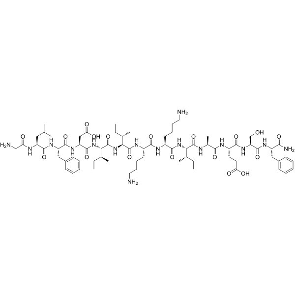
Aurein 1.2 is a C-terminally amidated antimicrobial peptide found in Litoria raniformis. Aurein 1.2 exhibits moderate, broad-spectrum antimicrobial activity against a variety of bacteria, with a MIC of 12-100 μg/mL. Aurein 1.2 forms an amphipathic α-helix with distinct hydrophilic and hydrophobic faces, and may exert antimicrobial and cytotoxic effects on tumor cells by disrupting cell membranes. Aurein 1.2 can be used in studies related to antimicrobial peptides, membrane-active peptides, and tumor cytotoxicity.
商品は「研究用試薬」です。人や動物の医療用・臨床診断用・食品用の製品ではありません。
研究用途以外に使用した場合、当社は一切の責任を負いかねます。
- 純度 : 99.21%
- CAS 番号: 302343-01-7
- 分子式: C71H114N16O18
- 分子量:1479.76
-
保管条件:
Sealed storage, away from moisture.
Powder -80°C, 2 years , -20°C, 1 year* In solvent : -80°C, 6 months; -20°C, 1 month (sealed storage, away from moisture)

To place orders, for customer services and technical support, please contact: MedChemExpress USA
Tel: 609-228-6898 E-mail: [email protected] [email protected]


-
 生物活性
生物活性
-
化学情報
-
溶剤 & 溶解度
- プロトコル
- 純度とドキュメンテーション
- 参考文献
-
 Help & FAQs


Antibiotic アイソフォーム固有の製品をすべて表示
More

生物活性
製品説明
体外実験
Aurein 1.2 (9.7 mM; 70% trifluoroethanol/H2O; pH 2.04; 25°C) forms a regular linear amphipathic α-helix with clearly separated hydrophilic and hydrophobic surfaces in NMR structural analysis[1].
MedChemExpress (MCE) has not independently confirmed the accuracy of these methods. They are for reference only.


化学情報
-
CAS 番号 302343-01-7
-
性状 Solid
-
分子量 1479.76
-
分子式 C71H114N16O18
-
Color White to off-white
-
配列
Gly-Leu-Phe-Asp-Ile-Ile-Lys-Lys-Ile-Ala-Glu-Ser-Phe-NH2
-
シーケンスの短縮
GLFDIIKKIAESF-NH2
-
輸送条件
Room temperature in continental US; may vary elsewhere.
-
保管条件
Sealed storage, away from moisture
Powder -80°C 2 years -20°C 1 year * In solvent : -80°C, 6 months; -20°C, 1 month (sealed storage, away from moisture)


溶剤 & 溶解度
体外:
DMSO : 100 mg/mL (67.58 mM; Need ultrasonic; Hygroscopic DMSO has a significant impact on the solubility of product, please use newly opened DMSO)
Please refer to the solubility information to select the appropriate solvent. Once prepared, please aliquot and store the solution to prevent product inactivation from repeated freeze-thaw cycles.
Storage method and period of stock solution: -80°C, 6 months; -20°C, 1 month (sealed storage, away from moisture). When stored at -80°C, please use it within 6 months. When stored at -20°C, please use it within 1 month.
Please refer to the solubility information to select the appropriate solvent. Once prepared, please aliquot and store the solution to prevent product inactivation from repeated freeze-thaw cycles.
Storage method and period of stock solution: -80°C, 6 months; -20°C, 1 month (sealed storage, away from moisture). When stored at -80°C, please use it within 6 months. When stored at -20°C, please use it within 1 month.
濃度 (開始) × 体積 (開始) = 濃度 (終了) × 体積 (終了)


純度とドキュメンテーション
-
データシート (272 KB)
-
SDS (252 KB)
- English - EN (252 KB)
- Français - FR (252 KB)
- Deutsch - DE (252 KB)
- Norwegian - NO (252 KB)
- Español - ES (252 KB)
- Swedish - SV (252 KB)
- Italian - IT (252 KB)
- Korean - KR (252 KB)
- Portuguese - PT (252 KB)
-
取扱説明書 (2659 KB)
参考文献


Complete Stock Solution Preparation Table
Please refer to the solubility information to select the appropriate solvent. Once prepared, please aliquot and store the solution to prevent product inactivation from repeated freeze-thaw cycles.
Storage method and period of stock solution: -80°C, 6 months; -20°C, 1 month (sealed storage, away from moisture). When stored at -80°C, please use it within 6 months. When stored at -20°C, please use it within 1 month.
| Optional Solvent | Concentration Solvent Mass | 1 mg | 5 mg | 10 mg | 25 mg |
|---|---|---|---|---|---|
| DMSO | 1 mM | 0.6758 mL | 3.3789 mL | 6.7579 mL | 16.8946 mL |
| 5 mM | 0.1352 mL | 0.6758 mL | 1.3516 mL | 3.3789 mL | |
| 10 mM | 0.0676 mL | 0.3379 mL | 0.6758 mL | 1.6895 mL | |
| 15 mM | 0.0451 mL | 0.2253 mL | 0.4505 mL | 1.1263 mL | |
| 20 mM | 0.0338 mL | 0.1689 mL | 0.3379 mL | 0.8447 mL | |
| 25 mM | 0.0270 mL | 0.1352 mL | 0.2703 mL | 0.6758 mL | |
| 30 mM | 0.0225 mL | 0.1126 mL | 0.2253 mL | 0.5632 mL | |
| 40 mM | 0.0169 mL | 0.0845 mL | 0.1689 mL | 0.4224 mL | |
| 50 mM | 0.0135 mL | 0.0676 mL | 0.1352 mL | 0.3379 mL | |
| 60 mM | 0.0113 mL | 0.0563 mL | 0.1126 mL | 0.2816 mL |


Aurein 1.2 関連分類
HY-P5726 関連分類
 Powered by Bioz