



Beauveriolide I

Based on 1 Customer Validation
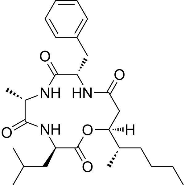
Beauveriolide I is an ACAT/SOAT inhibitor. Beauveriolide I selectively inhibits macrophage cholesteryl ester synthesis, blocks cholesteryl ester recycling, reduces cholesterol concentration, lipid droplet accumulation, and post-lysosomal cholesterol metabolism, and promotes the degradation of cholesteryl esters preferentially over triglycerides. Beauveriolide I reduces amyloid β peptide secretion. Beauveriolide I exhibits insecticidal and weak antibacterial activities. Beauveriolide I can be used in research on atherosclerosis, Alzheimer's disease, and cancer.
For research use only. We do not sell to patients.
- Purity : 99.6%
- CAS No.: 154491-55-1
- Formula: C27H41N3O5
- Molecular Weight:487.63
-
Storage:Powder -20°C, 3 years ; In solvent -80°C, 6 months , -20°C, 1 month

To place orders, for customer services and technical support, please contact: MedChemExpress USA
Tel: 609-228-6898 E-mail: [email protected] [email protected]


-
 Biological Activity
-
Chemical Information
- Protocols
- Purity & Documentation
- References
-
 Help & FAQs


-
HY-114295A
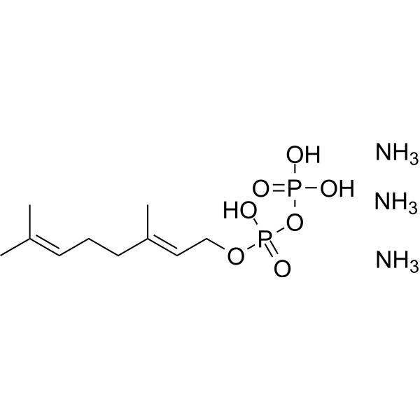
Geranyl diphosphate triammoniumIsoprenoid -

HY-114649
19-Hydroxy cholesterolEster Product -
HY-18366A
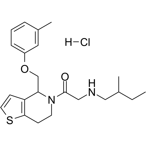
RU-SKI 43 hydrochlorideSelective Hhat Inhibitor -
HY-118486
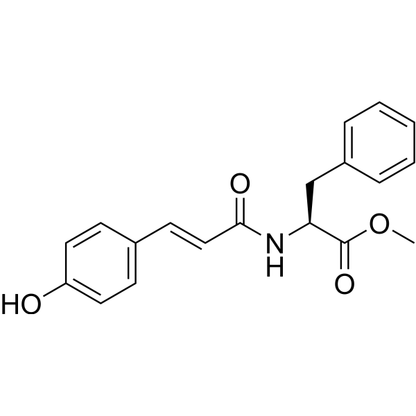
LDL-IN-1Antioxidant -
HY-P2792
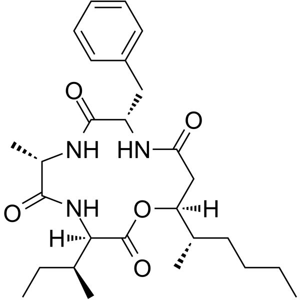
Beauveriolide IIILipid Droplet Formation Inhibitor -

HY-124064
TMP-153ACAT Inhibitor -

HY-120984
Oleic Acid-2,6-diisopropylanilide -
HY-126455
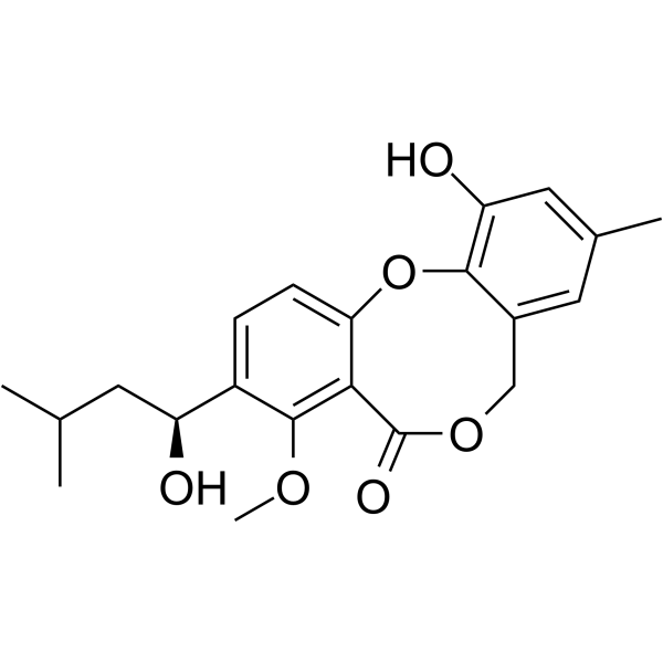
PenicillideACAT Inhibitor


Biological Activity
Description
|
ACAT |
SOAT |
Cellular Effect
|
Cell Line

|
Type | Value | Description | References |
|---|---|---|---|---|
| KB 3-1 | IC50 |
13.3 μM
|
Cytotoxicity against human endocervical adenocarcinoma KB3.1 cells assessed as IC50 by serial dilution assay in 96-well microtiter plates.
Cytotoxicity against human endocervical adenocarcinoma KB3.1 cells assessed as IC50 by serial dilution assay in 96-well microtiter plates.
|
41424705 |
| A549 | IC50 |
0.9 μM
|
Cytotoxicity against human lung carcinoma A549 cells assessed as IC50 by serial dilution assay in 96-well microtiter plates.
Cytotoxicity against human lung carcinoma A549 cells assessed as IC50 by serial dilution assay in 96-well microtiter plates.
|
41424705 |
| A-431 | IC50 |
1.2 μM
|
Cytotoxicity against human epidermoid carcinoma A431 cells assessed as IC50 by serial dilution assay in 96-well microtiter plates.
Cytotoxicity against human epidermoid carcinoma A431 cells assessed as IC50 by serial dilution assay in 96-well microtiter plates.
|
41424705 |
| PC-3 | IC50 |
3.5 μM
|
Cytotoxicity against human prostate carcinoma PC-3 cells assessed as IC50 by serial dilution assay in 96-well microtiter plates.
Cytotoxicity against human prostate carcinoma PC-3 cells assessed as IC50 by serial dilution assay in 96-well microtiter plates.
|
41424705 |
In Vitro
Beauveriolide I inhibits cytosolic lipid droplet accumulation in mouse peritoneal macrophages, with no cytotoxicity at concentrations up to 20 µM[1].
Beauveriolide I selectively inhibits [14C]CE synthesis in mouse peritoneal macrophages with an IC50 of 0.78 µM, while not inhibiting [14C]TG synthesis[1].
Beauveriolide I inhibits the metabolism of lysosomal [14C]cholesterol to [14C]CE in mouse peritoneal macrophages, with an IC50 of 0.37 µM[1].
Beauveriolide I inhibits ACAT activity in mouse macrophage microsomes (IC50 6.0 µM), mouse liver microsomes (IC50 1.5 µM), and human Caco-2 microsomes (IC50 2.5 µM)[1].
Beauveriolide I selectively enhances [14C]CE degradation in CHO-K1 cells with an EC50 of 0.02 µM; its EC50 for [14C]TG degradation is >2.0 µM[2].
Beauveriolide I inhibits cell-based [14C]CE synthesis in CHO-K1 cells with an IC50 of 0.01 µM; its IC50 for [14C]TG synthesis is >2.0 µM[2].
Beauveriolide I inhibits SOAT activity in CHO-K1 microsomes with an IC50 of 0.05 µM[2].
Beauveriolide I (compound 1) (0.08 μM; 96 h) reduces cholesteryl ester levels in 7WD10 CHO cells with an IC50 of 0.08 μM and decreases lipid droplet accumulation[5].
Beauveriolide I (1 μM; 4 days) reduces Aβ40 and Aβ42 secretion in 7WD10 CHO cells, producing approximately 25% and approximately 10% reductions, respectively[5].
Beauveriolide I showed weak antibacterial activity against Staphylococcus aureus and Bacillus subtilis, with an MIC of 66.6 µg/mL[4].
Beauveriolide I exhibits cytotoxicity against KB3.1, A549, A431, and PC-3 cancer cell lines, while showing no significant toxicity to mouse fibroblasts[4].
MedChemExpress (MCE) has not independently confirmed the accuracy of these methods. They are for reference only. Further protocols information, click here.
-
Cell Line:stable transgenic CHO cell line 7WD10 expressing human APP751
-
Concentration:1 μM (up to 4 days incubation); 1 and 5 μM (96 h incubation)
-
Incubation Time:up to 4 days (Aβ40/Aβ42 secretion); 96 h (concentration-effect study)
-
Result:Decreased Aβ40 and Aβ42 secretion at 1 μM for up to four days; after four days, reduced Aβ40 by ~25% and Aβ42 by ~10% relative to vehicle control cells.
Reached maximum effect after one to two days.
Reduced Aβ_total (Aβ40 + Aβ42) after 96 h at 1 and 5 μM.
In Vivo
Beauveriolide I (10 μg/body; injection) produces 40% insecticidal activity against female Callosobruchus chinensis and no damage to males[3].
MedChemExpress (MCE) has not independently confirmed the accuracy of these methods. They are for reference only.
-
Animal Model:larva[3]
-
Dosage:10 μg/body (DMSO solution, 0.4 μl); 1 μg/body (DMSO solution, 0.4 μl)
-
Administration:injection
-
Result:Resulted in 12 survival, 0 agony, and 3 death among 15 larvae at 10 μg/body.
Resulted in 14 survival, 0 agony, and 1 death among 15 larvae at 1 μg/body.
Exhibited ca. 20% insecticidal activity against Spodoptera litura larvae at 10 μg/body.
-
Animal Model:adult male and female[3]
-
Dosage:10 μg/body (MeOH-CHCl3 1/1 solution, 0.5 μl)
-
Administration:injection
-
Result:Resulted in 9 survival, 1 agony, and 0 death among 10 male adults at 10 μg/body.
Resulted in 5 survival, 1 agony, and 4 death among 10 female adults at 10 μg/body.
Showed 40% insecticidal activity against females and no damage to males at 10 μg/body.


Chemical Information
-
CAS No. 154491-55-1
-
Appearance Solid
-
Molecular Weight 487.63
-
Formula C27H41N3O5
-
Color White to light yellow
-
SMILES
O=C(N[C@H](C(N[C@@H]1CC(C)C)=O)C)[C@@H](NC(C[C@@](OC1=O)([H])[C@@H](C)CCCC)=O)CC2=CC=CC=C2
-
Structure Classification
-
Initial Source
Beauveria sp.
-
Shipping
Room temperature in continental US; may vary elsewhere.
-
Storage
Powder -20°C 3 years In solvent -80°C 6 months -20°C 1 month

Protocols
-
Research Protocol for Infectious Diseases
Infectious-disease experiments test how pathogens interact with host barriers, innate immune receptors, inflammatory signaling, pathogen replication, and tissue injury; pattern-recognition receptors such as TLRs, RIG-I-like receptors, NOD-like receptors, and inflammasomes detect microbial molecules and activate NF-κB, interferon, and cytokine responses. The central hypothesis is that infection severity reflects the balance between pathogen burden and host response: protective inflammation restricts pathogen growth, whereas excessive or mislocalized inflammation contributes to tissue damage and disease phenotype. Unresolved questions include which host pathways are protective versus pathogenic, why some infection models fail to translate to human disease, and which combined readouts best predict clinically relevant infection outcomes.
-
Research Protocol for Cardiovascular Diseases
Cardiovascular disease can be modeled as maladaptive cardiac remodeling, where ischemic injury or pressure overload activates inflammatory signaling, fibroblast activation, extracellular-matrix deposition, cardiomyocyte hypertrophy, vascular remodeling, and progressive ventricular dysfunction. The TGF-β/SMAD axis is a central profibrotic pathway after myocardial injury and pressure overload, while innate immune and cytokine pathways regulate leukocyte recruitment, scar formation, and adverse remodeling. Key unresolved questions include which inflammatory signals are reparative versus harmful, when fibrosis is protective versus maladaptive, and whether pathway inhibition improves function without weakening necessary infarct healing or compensatory remodeling.
-
Lysosome and acidic-vesicle live-cell staining
Lysosome and acidic-vesicle live-cell staining detects acidic intracellular compartments by using membrane-permeant acidotropic probes that accumulate in low-pH vesicles, including lysosomes, late endosomes, autolysosomes, and acidic phagosomes. LysoTracker staining is commonly used as an intensity-based readout of acidic lysosomal compartment abundance or enlargement, while acridine orange produces green fluorescence in less concentrated compartments and red fluorescence after concentration-dependent accumulation in acidic vesicular organelles. Loss or reduction of acridine-orange red signal can be used as a readout of lysosomal membrane permeabilization or reduced acidic-vesicle integrity. This protocol is designed for live cultured cells and can be adapted for fluorescence microscopy, high-content imaging, plate-reader readout, or flow cytometry when the selected literature supports the readout. Because these dyes report acidotropic accumulation rather than lysosome identity alone,
-
Amyloid: Congo Red Amyloid Staining
Congo red amyloid staining is a histochemical method used to detect extracellular amyloid deposits in tissue sections based on the affinity of Congo red dye for β-pleated sheet-rich protein aggregates. When bound to amyloid, Congo red produces characteristic apple-green birefringence under polarized light microscopy, which is widely regarded as a diagnostic feature of amyloid deposition in histopathology. The diagnostic principle relies on the combination of dye binding (congophilia) and optical anisotropy under polarized illumination, which distinguishes amyloid from most non-amyloid eosinophilic extracellular deposits in routine histological evaluation. Amyloid identification by Congo red staining remains a cornerstone in diagnostic pathology despite the availability of adjunct methods such as immunohistochemistry and mass spectrometry, particularly because of its ability to localize deposits directly within tissue architecture. The specificity of Congo red-positive deposits is incre
-
Alzheimer’s Disease Modeling
Alzheimer’s Disease (AD) is a neurodegenerative disorder characterized by a progressive decline in cognitive functions and loss of specific types of neurons and synapses. Alzheimer's symptoms can be simulated in mice by injecting drugs (such as Aβ) or genetically modified.


Purity & Documentation
-
Data Sheet (300 KB)
-
SDS (252 KB)
- English - EN (252 KB)
- Français - FR (252 KB)
- Deutsch - DE (252 KB)
- Norwegian - NO (252 KB)
- Español - ES (252 KB)
- Swedish - SV (252 KB)
- Italian - IT (252 KB)
- Korean - KR (252 KB)
- Portuguese - PT (252 KB)
-
Handling Instructions (2659 KB)
References

Calculators
Concentration (start) × Volume (start) = Concentration (final) × Volume (final)


Beauveriolide I Related Classifications
HY-N8355 Related Classifications
 Powered by Bioz