



Vps34-IN-1
Based on 18 publication(s) in Google Scholar
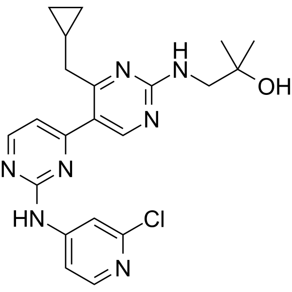
Vps34-IN-1 is a potent and selective inhibitor of class III Vps34 PI3K. Vps34-IN-1 inhibits phosphorylation of PtdIns by recombinant insect cell expressed Vps34-Vps15 complex with an IC50 of ~25 nM. Vps34-IN-1 can suppress SGK3 activation by reducing PtdIns(3)P levels via lowering phosphorylation of T-loop and hydrophobic motifs. Vps34-IN-1 modulates autophagy.
Nos produits utilisent uniquement pour la recherche. Nous ne vendons pas aux patients.
- Pureté : 99.79%
- CAS No.: 1383716-33-3
- Formule: C21H24ClN7O
- Masse moléculaire:425.91
-
Stockage:Powder -20°C, 3 years , 4°C, 2 years ; In solvent -80°C, 1 year , -20°C, 6 months

To place orders, for customer services and technical support, please contact: MedChemExpress USA
Tel: 609-228-6898 E-mail: [email protected] [email protected]


-
 Activité biologique
-
Chemical Information
-
Solvant et solubilité
- Protocole
- Pureté et documentation
- Références
-
 Help & FAQs


-
HY-12481
SAR405Vps34 Inhibitor -
HY-16966
SBI-0206965ULK1/AMPK Inhibitor -
HY-19312
3-MethyladenineAutophagy/PI3K Inhibitor -
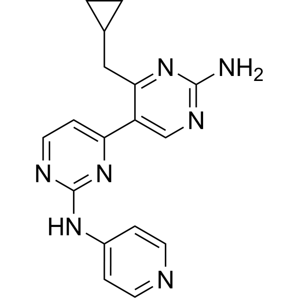
HY-12794
Vps34-PIK-IIIVps35 Inhibitor -
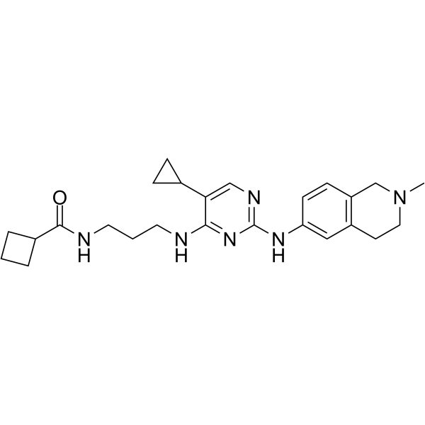
HY-100006
MRT68921NUAK1/ULK1 Inhibitor -
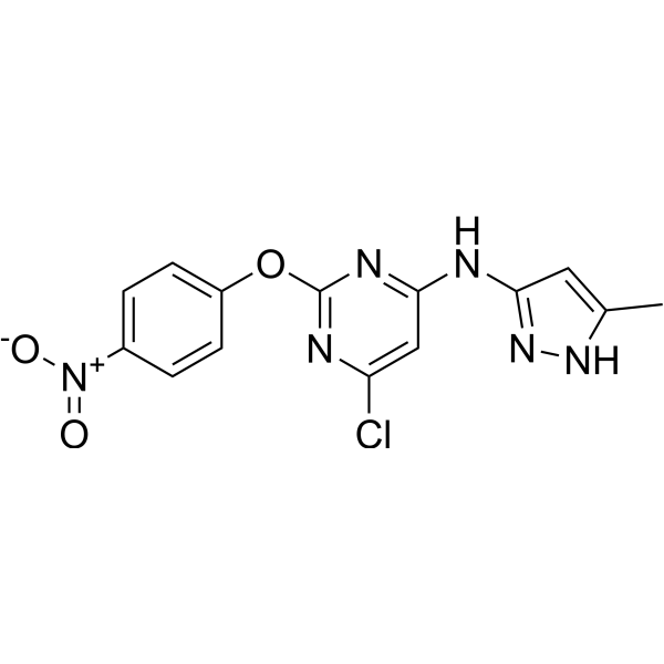
HY-101920
AutophinibVPS34 Inhibitor -
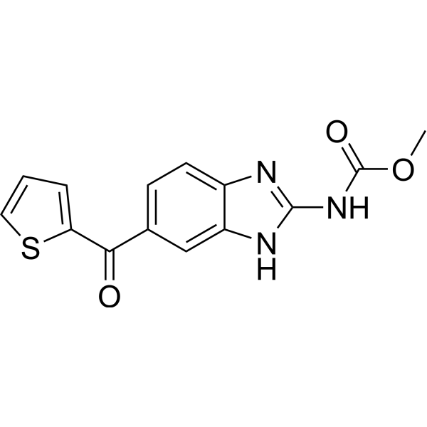
HY-13520
NocodazoleMicrotubule Inhibitor -
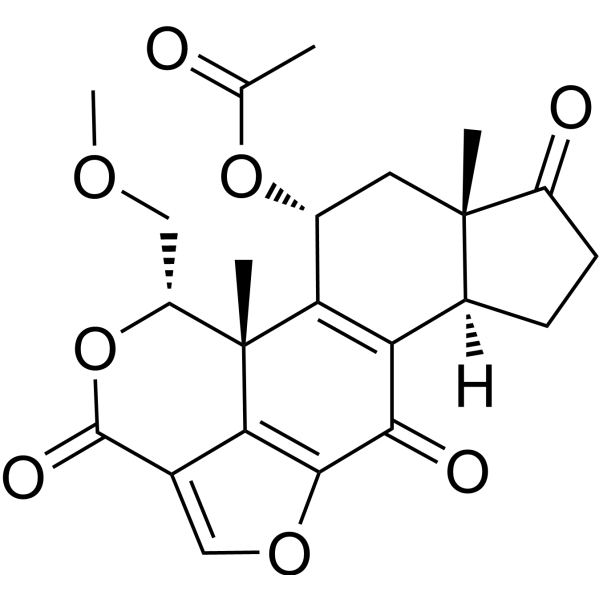
HY-10197
WortmanninPI3K Inhibitor -

HY-100558
Bafilomycin A1V-ATPase Inhibitor -
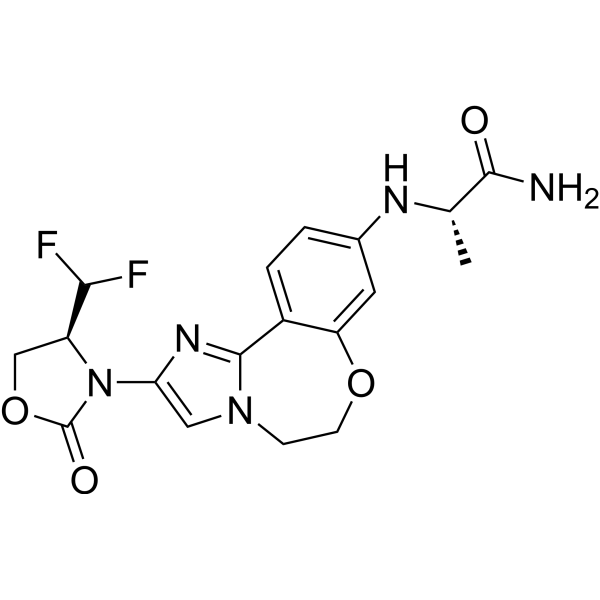
HY-101562
InavolisibPI3Kα Inhibitor -
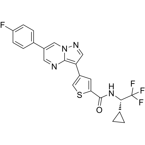
HY-114490
ULK-101ULK1 Inhibitor -
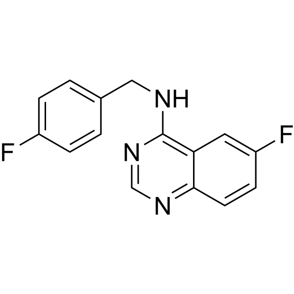
HY-12990
Spautin-1Autophagy Inhibitor
Publications Citing Use of MedChemExpress (MCE) Vps34-IN-1
More- Autophagy. 2026 May 7:1-17. [Abstract]
- Autophagy. 2025 Aug 28:1-15. [Abstract]
- Nat Chem Biol. 2025 May 26. [Abstract]
- Acta Pharm Sin B. 2025 Dec 18.
- J Exp Clin Cancer Res. 2018 Jun 25;37(1):122. [Abstract]
- Int J Biol Sci. 2025 Mar 19;21(6):2462-2475. [Abstract]
- Cell Rep. 2026 Feb 24;45(2):116914. [Abstract]
- Biochem Pharmacol. 2023 Aug:214:115634. [Abstract]
- Plant Cell Rep. 2022 Feb;41(2):463-471. [Abstract]
- PLoS Pathog. 2024 Aug 22;20(8):e1012444. [Abstract]
- Sports Med Health Sci. 2024 Mar 8;7(1):37-47. [Abstract]
- Thorac Cancer. 2026 Mar;17(5):e70255. [Abstract]
- Nephrology (Carlton). 2026 Jul;31(7):e70246.
- bioRxiv. 2026 Jan 24.
- Res Sq. 2025 Feb 11:rs.3.rs-4138879. [Abstract]
- bioRxiv. 2024 October 31.
- bioRxiv. 2024 Mar 7:2024.03.04.583390. [Abstract]
- Oncotarget. 2016 Aug 16;7(33):53515-53525. [Abstract]
 Customer Validation & Images

-
Cell Proliferation/Viability Assay
-
IF
-
RT-PCR
-
Cell Proliferation/Viability Assay
-
WB


Activité biologique
Description
IC50 & Target
[1]|
Vps34 25 nM (IC50) |
In Vitro
Vps34-IN-1 does not significantly inhibit any of the lipid kinases including class I (p110α, p110β p110γ and p110δ) and all three members of the class II PI3Ks (PI3KC2α, PI3KC2β and PI3KC2γ) enzymes[1].
Vps34-IN-1 (0.01, 0.1, 1 μM; for 1 h) inhibits PtdIns(3)P levels at endosomes in a dose-dependent manner[1].
Vps34-IN-1 (0.01, 0.1, 1 μM; for 1 h) induces a dose-dependent reduction in SGK3 activity that is maximally lowered by ~60% at 1 μM and ~40% at 0.1 μM. The suppression of SGK3 activity is accompanied by a commensurate decrease in T-loop and hydrophobic motif phosphorylation[1].
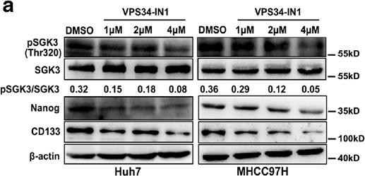
Vps34-IN-1 (1, 2, 4 μM; 24 h) induces a dose-dependent inhibition of SGK3 phosphorylation[2].
MedChemExpress (MCE) has not independently confirmed the accuracy of these methods. They are for reference only. Further protocols information, click here.
-
Cell Line:MHH97H and Huh7 cells
-
Concentration:1, 2, 4 μM
-
Incubation Time:24 h
-
Result:Induced a dose-dependent inhibition of SGK3 phosphorylation in MHH97H and Huh7 cells.
Reduced the expression of CSC-related markers CD133 and Nanog.


Chemical Information
-
CAS No. 1383716-33-3
-
Appearance Solid
-
Masse moléculaire 425.91
-
Formule C21H24ClN7O
-
Color White to off-white
-
SMILES
CC(C)(O)CNC1=NC=C(C2=CC=NC(NC3=CC=NC(Cl)=C3)=N2)C(CC4CC4)=N1
-
Livraison
Room temperature in continental US; may vary elsewhere.
-
Stockage
Powder -20°C 3 years 4°C 2 years In solvent -80°C 1 year -20°C 6 months


Publications (18)
-
Journal Impact Factor
-
Most Recent
-
Autophagy
2026 May 7:1-17. PMID: 42080372 -
Autophagy
2025 Aug 28:1-15. PMID: 40851277 -
Nat Chem Biol
2025 May 26. PMID: 40419770 -
Vps34-IN-1 purchased from MedChemExpress. Usage Cited in: Acta Pharm Sin B. 2025 Dec 18.
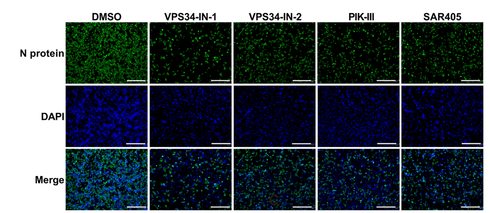
VPS34-IN-1, VPS34-IN-2, PIK-III, and SAR405 (all 1 μM, 48 h) exhibited similar inhibition of 0.02 MOI WT SARS-CoV-2 infection in Vero E6 cells.
Vps34-IN-1 purchased from MedChemExpress. Usage Cited in: Acta Pharm Sin B. 2025 Dec 18.
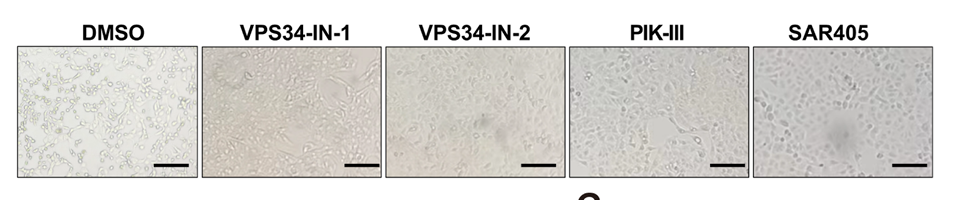
VPS34-IN-1, VPS34-IN-2, PIK-III, and SAR405 (all 1 μM, 48 h) protected cells from SARS-CoV-2-induced CPE, highlighting the critical role of VPS34 in SARS-CoV-2 infection.
Vps34-IN-1 purchased from MedChemExpress. Usage Cited in: Acta Pharm Sin B. 2025 Dec 18.
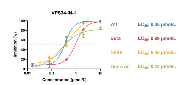
Pharmacological profiling of VPS34 inhibitors against SARS-CoV-2 variants demonstrated that the EC50 of VPS34-IN-1 was 0.36, 0.99, 0.46, and 0.24 μM against WT, Beta, Delta, and Omicron variants, respectively.
Vps34-IN-1 purchased from MedChemExpress. Usage Cited in: Acta Pharm Sin B. 2025 Dec 18.
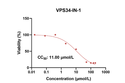
Cytotoxicity assessments at 48 h in Vero E6 cells revealed that the CC50 values of VPS34-IN-1 was 11.00 μM.
-
J Exp Clin Cancer Res
Prolonged inhibition of class I PI3K promotes liver cancer stem cell expansion by augmenting SGK3/GSK-3β/β-catenin signalling. [Abstract]2018 Jun 25;37(1):122. PMID: 29940988
Vps34-IN-1 purchased from MedChemExpress. Usage Cited in: J Exp Clin Cancer Res. 2018 Jun 25;37(1):122. [Abstract]
Huh7 and MHCC97H cells are treated with VPS34-IN1 for 24 h, and cell lysates are subjected to western blot analysis with the indicated antibodies.
-
Int J Biol Sci
The SGK3/GSK3β/β-catenin signaling promotes breast cancer stemness and confers resistance to alpelisib therapy. [Abstract]2025 Mar 19;21(6):2462-2475. PMID: 40303291 -
Cell Rep
HRD1 negatively regulates autolysosome formation by inhibiting liquid-liquid phase separation of SNAP29. [Abstract]2026 Feb 24;45(2):116914. PMID: 41615796 -
Biochem Pharmacol
VPS34-IN1 induces apoptosis of ER + breast cancer cells via activating PERK/ATF4/CHOP pathway. [Abstract]2023 Aug:214:115634. PMID: 37290596 -
Plant Cell Rep
Autophagy inducers lead to transient accumulation of autophagosomes in Arabidopsis roots. [Abstract]2022 Feb;41(2):463-471. PMID: 34977975 -
PLoS Pathog
Genome-wide CRISPR/Cas9 screen identifies SLC39A9 and PIK3C3 as crucial entry factors for Ebola virus infection. [Abstract]2024 Aug 22;20(8):e1012444. PMID: 39173055 -
Sports Med Health Sci
Exercise improves systemic metabolism in a monocrotaline model of pulmonary hypertension. [Abstract]2024 Mar 8;7(1):37-47. PMID: 39649790 -
Thorac Cancer
Effects of Autophagy Inhibition by SAR405, a Selective VPS34 Inhibitor, on Pleural Mesothelioma Cells. [Abstract]2026 Mar;17(5):e70255. PMID: 41771171 -
-
-
Res Sq
2025 Feb 11:rs.3.rs-4138879. PMID: 39989975 -
-
bioRxiv
2024 Mar 7:2024.03.04.583390. PMID: 38496520 -
Oncotarget
Simultaneous inhibition of Vps34 kinase would enhance PI3Kδ inhibitor cytotoxicity in the B-cell malignancies. [Abstract]2016 Aug 16;7(33):53515-53525. PMID: 27447747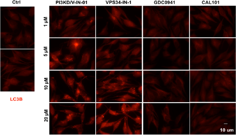
Vps34-IN-1 purchased from MedChemExpress. Usage Cited in: Oncotarget. 2016 Aug 16;7(33):53515-53525. [Abstract]
PI3KD/V-IN-01 affects autophagy HeLa cells are treated with different concentrations of PI3KD/V-IN-01, VPS34-IN-1, GDC-0941 or CAL-101 for 16 hours before they are fixed and stained for the autophagy marker LC3B.


Solvant et solubilité
In Vitro:
DMSO : ≥ 31 mg/mL (72.79 mM; Hygroscopic DMSO has a significant impact on the solubility of product, please use newly opened DMSO)
* "≥" means soluble, but saturation unknown.
Please refer to the solubility information to select the appropriate solvent. Once prepared, please aliquot and store the solution to prevent product inactivation from repeated freeze-thaw cycles.
Storage method and period of stock solution: -80°C, 1 year; -20°C, 6 months. When stored at -80°C, please use it within 1 year. When stored at -20°C, please use it within 6 months.
Please refer to the solubility information to select the appropriate solvent. Once prepared, please aliquot and store the solution to prevent product inactivation from repeated freeze-thaw cycles.
Storage method and period of stock solution: -80°C, 1 year; -20°C, 6 months. When stored at -80°C, please use it within 1 year. When stored at -20°C, please use it within 6 months.
Concentration (start) × Volume (start) = Concentration (final) × Volume (final)
In Vivo:
Select the appropriate dissolution method based on your experimental animal and administration route.
- For the following dissolution methods, please ensure to first prepare a clear stock solution using an In Vitro approach and then sequentially add co-solvents:
- To ensure reliable experimental results, the clarified stock solution can be appropriately stored based on storage conditions. As for the working solution for In Vivo experiments, it is recommended to prepare freshly and use it on the same day.
- The percentages shown for the solvents indicate their volumetric ratio in the final prepared solution. If precipitation or phase separation occurs during preparation, heat and/or sonication can be used to aid dissolution.
Add each solvent one by one: 10% DMSO 40% PEG300 5% Tween-80 45% Saline
Solubility: ≥ 2.5 mg/mL (5.87 mM); Clear solution
This protocol yields a clear solution of ≥ 2.5 mg/mL (saturation unknown).
Taking 1 mL working solution as an example, add 100 μL DMSO stock solution (25.0 mg/mL) to 400 μL PEG300, and mix evenly; then add 50 μL Tween-80 and mix evenly; then add 450 μL Saline to adjust the volume to 1 mL.
Preparation of Saline: Dissolve 0.9 g sodium chloride in ddH₂O and dilute to 100 mL to obtain a clear Saline solution.
Add each solvent one by one: 10% DMSO 90% (20% SBE-β-CD in Saline)
Solubility: ≥ 2.5 mg/mL (5.87 mM); Clear solution
This protocol yields a clear solution of ≥ 2.5 mg/mL (saturation unknown).
Taking 1 mL working solution as an example, add 100 μL DMSO stock solution (25.0 mg/mL) to 900 μL 20% SBE-β-CD in Saline, and mix evenly.
Preparation of 20% SBE-β-CD in Saline (4°C, storage for one week): 2 g SBE-β-CD powder is dissolved in 10 mL Saline, completely dissolve until clear.
In Vivo Dissolution Calculator
Please enter the basic information of animal experiments:
-
-
-
-
Recommended: Prepare an additional quantity of animals to account for potential losses during experiments.
Please enter your animal formula composition:
-
%DMSO +
Recommended: Keep the proportion of DMSO in working solution below 2% if your animal is weak.
-
%+
-
+%Tween-80 + +
-
%Saline +
The co-solvents required include: DMSO, . All of co-solvents are available by MedChemExpress (MCE). , Tween 80. All of co-solvents are available by MedChemExpress (MCE).
Working solution concentration: 0.22 mg/mL
Method for preparing stock solution: mg drug dissolved in μL DMSO. Stock solution concentration: mg/mL.
1. Take μL DMSO stock solution;
2. Add μL .
μL , mix evenly;
3. Then add μL Tween 80, mix evenly;
4. Then add μL
Please ensure that the stock solution in the first step is dissolved to a clear state, and add co-solvents in sequence. You can use ultrasonic heating (ultrasonic cleaner, recommended frequency 20-40 kHz), vortexing, etc. to assist dissolution.

Protocole
-
Kinase activity and phosphorylation assays
Kinase activity assays measure the ability of kinases to transfer phosphate groups from ATP to specific substrates, while phosphorylation assays detect the presence and levels of phosphorylated proteins. Common methods include radiolabeled ATP incorporation (e. g. ,), ADP release detection via bioluminescence (e. g. ,[3]), enzyme-linked immunosorbent assays (ELISA) for phospho-specific epitopes (e. g. ,[6]), and microtiter-based formats for high-throughput screening (e. g. ,[8]). The ADP-Glo assay quantifies kinase activity by measuring ADP produced during phosphorylation using a luciferase-based system. Radiometric assays involve autoradiography or scintillation counting after incorporation of 32P-labeled ATP into substrate proteins. ELISA-based approaches rely on phospho-specific antibodies to detect activated kinases in cell lysates or purified samples.
-
Western Blot
Western blotting (WB) is a commonly used experimental method in molecular biology, biochemistry, and immunogenetics for identifying and quantifying target proteins. It combines gel electrophoresis with immunoassay, enabling researchers to analyze protein expression, post-translational modifications, and molecular weight.
-
Cell Cytotoxicity Assay
Cytotoxicity assays are usually based on the assessment of cell membrane damage, which can also be indirectly detected by measuring cell viability. Detection methods include MTT assay, CKK-8 assay, LDH assay and ATP assay, etc.
-
Autophagy
Autophagy is a process in which eukaryotic cells use lysosomes to degrade their own cytoplasmic proteins and damaged organelles under the regulation of autophagy related gene (Atg). Microtubule-associated proteins light chain 3 (LC3) is recognized as autophagy marker, which transfers from cytoplasmic LC3 (LC3-I) to membrane type (LC3-II). LC3-II/I ratio could be detected by Western Blot and fluorescence microscopy.
-
Protocol for Kinase activity and phosphorylation assays
Kinase activity assays measure transfer of phosphate from ATP to a protein or peptide substrate, generating phosphorylated substrate, ADP, or incorporated radiolabeled phosphate as the readout; phosphorylation assays measure site-specific phosphorylation in cells or tissues as a proxy for kinase-pathway activation, inhibition, or substrate regulation. Phosphorylation can be detected by phospho-specific Western blot, immunoprecipitation kinase assay, phospho-immunofluorescence, phospho-flow cytometry, luminescent ADP detection, radiolabeled ATP incorporation, or reporter-based pathway assays, and these readouts can be applied to cancer cells, primary neurons, mouse tumors, organoids, inflammatory macrophages, ferroptosis studies, and mitophagy studies when the kinase target is biologically relevant.
-
Lysosome and acidic-vesicle live-cell staining
Lysosome and acidic-vesicle live-cell staining detects acidic intracellular compartments by using membrane-permeant acidotropic probes that accumulate in low-pH vesicles, including lysosomes, late endosomes, autolysosomes, and acidic phagosomes. LysoTracker staining is commonly used as an intensity-based readout of acidic lysosomal compartment abundance or enlargement, while acridine orange produces green fluorescence in less concentrated compartments and red fluorescence after concentration-dependent accumulation in acidic vesicular organelles. Loss or reduction of acridine-orange red signal can be used as a readout of lysosomal membrane permeabilization or reduced acidic-vesicle integrity. This protocol is designed for live cultured cells and can be adapted for fluorescence microscopy, high-content imaging, plate-reader readout, or flow cytometry when the selected literature supports the readout. Because these dyes report acidotropic accumulation rather than lysosome identity alone,
-
Macroautophagy Solutions
Macroautophagy is a conserved lysosome-dependent degradation pathway in which cytoplasmic material is sequestered into double-membrane autophagosomes and delivered to lysosomes for degradation and recycling. The pathway supports cellular homeostasis during nutrient limitation, organelle stress, protein-aggregate accumulation, infection, differentiation, and tissue remodeling by coupling cargo sequestration, autophagosome maturation, lysosomal fusion, and degradation of cargo-derived macromolecules. The core molecular sequence includes initiation by nutrient- and stress-regulated autophagy machinery, autophagosome nucleation, LC3/ATG8-family conjugation to autophagosomal membranes, cargo selection through receptors such as SQSTM1/p62, autophagosome-lysosome fusion, and lysosomal degradation. LC3 was identified as a mammalian homolog of yeast Atg8 that localizes to autophagosomal membranes after processing, and p62/SQSTM1 was shown to connect ubiquitinated cargo with autophagic degradati


Pureté et documentation
-
Fiche technique (280 KB)
-
SDS (393 KB)
- English - EN (393 KB)
- Français - FR (393 KB)
- Deutsch - DE (393 KB)
- Norwegian - NO (393 KB)
- Español - ES (393 KB)
- Swedish - SV (393 KB)
- Italian - IT (393 KB)
- Korean - KR (393 KB)
- Portuguese - PT (393 KB)
-
Instruction de manipulation (2659 KB)
Références
[1]. Ruzica Bago, et al. Characterization of VPS34-IN1, a selective inhibitor of Vps34, reveals that the phosphatidylinositol 3-phosphate-binding SGK3 protein kinase is a downstream target of class III phosphoinositide 3-kinase. Biochem J. 2014 Nov 1;463(3):413-27. [Content Brief]
[2]. Fengchao Liu, et al. Prolonged inhibition of class I PI3K promotes liver cancer stem cell expansion by augmenting SGK3/GSK-3β/β-catenin signaling. J Exp Clin Cancer Res. 2018 Jun 25;37(1):122. [Content Brief]
[3]. Limpert AS, et al. Autophagy in Cancer: Regulation by Small Molecules. Trends Pharmacol Sci. 2018 Dec;39(12):1021-1032 [Content Brief]


Complete Stock Solution Preparation Table
Please refer to the solubility information to select the appropriate solvent. Once prepared, please aliquot and store the solution to prevent product inactivation from repeated freeze-thaw cycles.
Storage method and period of stock solution: -80°C, 1 year; -20°C, 6 months. When stored at -80°C, please use it within 1 year. When stored at -20°C, please use it within 6 months.
| Optional Solvent | Concentration Solvent Mass | 1 mg | 5 mg | 10 mg | 25 mg |
|---|---|---|---|---|---|
| DMSO | 1 mM | 2.3479 mL | 11.7396 mL | 23.4791 mL | 58.6978 mL |
| 5 mM | 0.4696 mL | 2.3479 mL | 4.6958 mL | 11.7396 mL | |
| 10 mM | 0.2348 mL | 1.1740 mL | 2.3479 mL | 5.8698 mL | |
| 15 mM | 0.1565 mL | 0.7826 mL | 1.5653 mL | 3.9132 mL | |
| 20 mM | 0.1174 mL | 0.5870 mL | 1.1740 mL | 2.9349 mL | |
| 25 mM | 0.0939 mL | 0.4696 mL | 0.9392 mL | 2.3479 mL | |
| 30 mM | 0.0783 mL | 0.3913 mL | 0.7826 mL | 1.9566 mL | |
| 40 mM | 0.0587 mL | 0.2935 mL | 0.5870 mL | 1.4674 mL | |
| 50 mM | 0.0470 mL | 0.2348 mL | 0.4696 mL | 1.1740 mL | |
| 60 mM | 0.0391 mL | 0.1957 mL | 0.3913 mL | 0.9783 mL |


Vps34-IN-1 Related Classifications
HY-12795 Related Classifications
 Powered by Bioz