



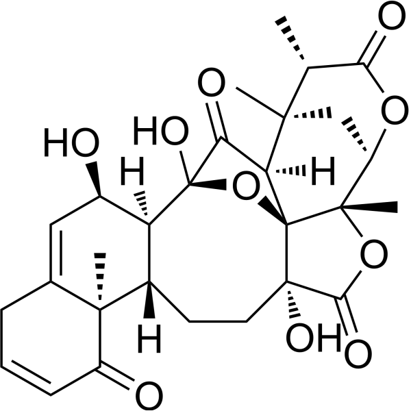
Physalin O

Based on 1 Customer Validation
Physalin O is a physalin that can be isolated from Physalis angulata. Physalin O shows cytotoxicity to Hep G2 and MCF-7 cancer cells with IC50 values of 31.1 and 11.4 µM, respectively. Physalin O inhibits the production of nitric oxide (NO) and shows anti-inflammatory activities.
Nos produits utilisent uniquement pour la recherche. Nous ne vendons pas aux patients.
- Pureté : 99.35%
- CAS No.: 120849-18-5
- Formule: C28H32O10
- Masse moléculaire:528.55
-
Stockage:
-20°C, protect from light
* In solvent : -80°C, 6 months; -20°C, 1 month (protect from light)

To place orders, for customer services and technical support, please contact: MedChemExpress USA
Tel: 609-228-6898 E-mail: [email protected] [email protected]


-
 Activité biologique
-
Chemical Information
-
Solvant et solubilité
- Protocole
- Pureté et documentation
- Références
-
 Help & FAQs




Activité biologique
Description
Cellular Effect
|
Cell Line

|
Type | Value | Description | References |
|---|---|---|---|---|
| HeLa | IC50 |
>50 μM
Compound: PC-2
|
Inhibition of TNFalpha-induced IkappaBalpha (unknown origin) phosphorylation expressed in human HeLa cells pretreated 30 mins before TNFalpha addition measured up to 120 mins by ELISA in presence of proteasome inhibitor MG-132
Inhibition of TNFalpha-induced IkappaBalpha (unknown origin) phosphorylation expressed in human HeLa cells pretreated 30 mins before TNFalpha addition measured up to 120 mins by ELISA in presence of proteasome inhibitor MG-132
|
[PMID: 24900739] |
| HeLa | IC50 |
>50 μM
Compound: PC-2
|
Inhibition of TNFalpha-induced NF-kappaB activation (unknown origin) expressed in human HeLa cells pretreated 30 mins before TNFalpha addition after 7 hrs by luciferase reporter gene assay
Inhibition of TNFalpha-induced NF-kappaB activation (unknown origin) expressed in human HeLa cells pretreated 30 mins before TNFalpha addition after 7 hrs by luciferase reporter gene assay
|
[PMID: 24900739] |
| RAW264.7 | IC50 |
17.11 μM
Compound: 12
|
Inhibition of LPS-induced nitric oxide production in mouse RAW264.7 cells assessed as nitrite formation
Inhibition of LPS-induced nitric oxide production in mouse RAW264.7 cells assessed as nitrite formation
|
[PMID: 18348534] |


Chemical Information
-
CAS No. 120849-18-5
-
Appearance Powder
-
Masse moléculaire 528.55
-
Formule C28H32O10
-
Color white
-
SMILES
C[C@@]12[C@]34[C@](C([C@](O3)([C@@]5([H])[C@]([C@@]6(C(CC=CC6=O)=C[C@H]5O)C)([H])CC[C@@]4(C(O2)=O)O)O)=O)([H])[C@@]7(C[C@H]1OC([C@H]7C)=O)C
-
Structure Classification
-
Initial Source
-
Livraison
Room temperature in continental US; may vary elsewhere.
-
Stockage
-20°C, protect from light
* In solvent : -80°C, 6 months; -20°C, 1 month (protect from light)


Solvant et solubilité
In Vitro:
DMSO : 25 mg/mL (47.30 mM; Need ultrasonic; Hygroscopic DMSO has a significant impact on the solubility of product, please use newly opened DMSO)
Please refer to the solubility information to select the appropriate solvent. Once prepared, please aliquot and store the solution to prevent product inactivation from repeated freeze-thaw cycles.
Storage method and period of stock solution: -80°C, 6 months; -20°C, 1 month (protect from light). When stored at -80°C, please use it within 6 months. When stored at -20°C, please use it within 1 month.
Please refer to the solubility information to select the appropriate solvent. Once prepared, please aliquot and store the solution to prevent product inactivation from repeated freeze-thaw cycles.
Storage method and period of stock solution: -80°C, 6 months; -20°C, 1 month (protect from light). When stored at -80°C, please use it within 6 months. When stored at -20°C, please use it within 1 month.
Concentration (start) × Volume (start) = Concentration (final) × Volume (final)


Pureté et documentation
-
Fiche technique (288 KB)
-
SDS (251 KB)
- English - EN (251 KB)
- Français - FR (251 KB)
- Deutsch - DE (251 KB)
- Norwegian - NO (251 KB)
- Español - ES (251 KB)
- Swedish - SV (251 KB)
- Italian - IT (251 KB)
- Korean - KR (251 KB)
- Portuguese - PT (251 KB)
-
Instruction de manipulation (2659 KB)
Références
[1]. Kuo PC, et al. Physanolide A, a novel skeleton steroid, and other cytotoxic principles from Physalis angulata. Org Lett. 2006 Jul 6;8(14):2953-6. [Content Brief]
[2]. Ji L, et al. Physalins with anti-inflammatory activity are present in Physalis alkekengi var. franchetii and can function as Michael reaction acceptors. Steroids. 2012 Apr;77(5):441-7. [Content Brief]


Complete Stock Solution Preparation Table
Please refer to the solubility information to select the appropriate solvent. Once prepared, please aliquot and store the solution to prevent product inactivation from repeated freeze-thaw cycles.
Storage method and period of stock solution: -80°C, 6 months; -20°C, 1 month (protect from light). When stored at -80°C, please use it within 6 months. When stored at -20°C, please use it within 1 month.
| Optional Solvent | Concentration Solvent Mass | 1 mg | 5 mg | 10 mg | 25 mg |
|---|---|---|---|---|---|
| DMSO | 1 mM | 1.8920 mL | 9.4598 mL | 18.9197 mL | 47.2992 mL |
| 5 mM | 0.3784 mL | 1.8920 mL | 3.7839 mL | 9.4598 mL | |
| 10 mM | 0.1892 mL | 0.9460 mL | 1.8920 mL | 4.7299 mL | |
| 15 mM | 0.1261 mL | 0.6307 mL | 1.2613 mL | 3.1533 mL | |
| 20 mM | 0.0946 mL | 0.4730 mL | 0.9460 mL | 2.3650 mL | |
| 25 mM | 0.0757 mL | 0.3784 mL | 0.7568 mL | 1.8920 mL | |
| 30 mM | 0.0631 mL | 0.3153 mL | 0.6307 mL | 1.5766 mL | |
| 40 mM | 0.0473 mL | 0.2365 mL | 0.4730 mL | 1.1825 mL |


Physalin O Related Classifications
HY-N10023 Related Classifications
 Powered by Bioz