



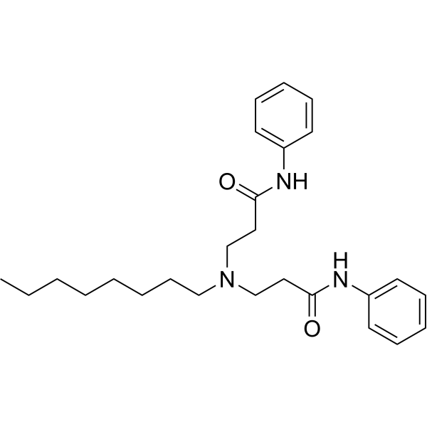
UCM-1336

Based on 1 Customer Validation
UCM-1336 is a potent ICMT inhibitor, with an IC50 of 2 μM. UCM-1336 induces mislocalization of endogenous Ras, decreases Ras activation and induces cell death by autophagy and apoptosis.
Nos produits utilisent uniquement pour la recherche. Nous ne vendons pas aux patients.
- Pureté : 99.86%
- CAS No.: 1621535-90-7
- Formule: C26H37N3O2
- Masse moléculaire:423.59
-
Stockage:Powder -20°C, 3 years ; In solvent -80°C, 6 months , -20°C, 1 month

To place orders, for customer services and technical support, please contact: MedChemExpress USA
Tel: 609-228-6898 E-mail: [email protected] [email protected]


-
 Activité biologique
-
Chemical Information
-
Solvant et solubilité
- Protocole
- Pureté et documentation
- Références
-
 Help & FAQs




Activité biologique
Description
IC50 & Target
IC50: 2 μM (2 μM)[1].
Cellular Effect
|
Cell Line

|
Type | Value | Description | References |
|---|---|---|---|---|
| HL-60 | IC50 |
2 μM
Compound: 3; UCM-1336
|
Cytotoxicity against human HL60 cells harboring N-Ras Q61K mutant assessed as reduction in cell viability after 48 hrs by MTT assay
Cytotoxicity against human HL60 cells harboring N-Ras Q61K mutant assessed as reduction in cell viability after 48 hrs by MTT assay
|
[PMID: 31181882] |
| MDA-MB-231 | IC50 |
5 μM
Compound: 3; UCM-1336
|
Cytotoxicity against human MDA-MB-231 cells assessed as reduction in cell viability after 72 hrs by XTT assay
Cytotoxicity against human MDA-MB-231 cells assessed as reduction in cell viability after 72 hrs by XTT assay
|
[PMID: 31181882] |
| MIA PaCa-2 | IC50 |
2 μM
Compound: 3; UCM-1336
|
Cytotoxicity against human MIAPaCa2 cells harboring K-Ras G12C mutant assessed as reduction in cell viability after 72 hrs by XTT assay
Cytotoxicity against human MIAPaCa2 cells harboring K-Ras G12C mutant assessed as reduction in cell viability after 72 hrs by XTT assay
|
[PMID: 31181882] |
| NIH3T3 | IC50 |
>50 μM
Compound: 3; UCM-1336
|
Cytotoxicity against mouse NIH/3T3 cells harboring N-Ras Q61K mutant assessed as reduction in cell viability after 48 hrs by MTT assay
Cytotoxicity against mouse NIH/3T3 cells harboring N-Ras Q61K mutant assessed as reduction in cell viability after 48 hrs by MTT assay
|
[PMID: 31181882] |
| PANC-1 | IC50 |
7 μM
Compound: 3; UCM-1336
|
Cytotoxicity against human PANC1 cells harboring K-Ras G12D mutant assessed as reduction in cell viability after 72 hrs by XTT assay
Cytotoxicity against human PANC1 cells harboring K-Ras G12D mutant assessed as reduction in cell viability after 72 hrs by XTT assay
|
[PMID: 31181882] |
| Sf9 | IC50 |
2 μM
Compound: 3; UCM-1336
|
Inhibition of recombinant human ICMT expressed in Sf9 insect cell membranes using biotin-farnesyl-L-cysteine as substrate preincubated for 15 mins followed by substrate addition and measured after 30 mins by scintillation counting analysis
Inhibition of recombinant human ICMT expressed in Sf9 insect cell membranes using biotin-farnesyl-L-cysteine as substrate preincubated for 15 mins followed by substrate addition and measured after 30 mins by scintillation counting analysis
|
[PMID: 31181882] |
| SW-620 | IC50 |
3 μM
Compound: 3; UCM-1336
|
Cytotoxicity against human SW620 cells harboring K-Ras G12V mutant assessed as reduction in cell viability after 72 hrs by XTT assay
Cytotoxicity against human SW620 cells harboring K-Ras G12V mutant assessed as reduction in cell viability after 72 hrs by XTT assay
|
[PMID: 31181882] |


Chemical Information
-
CAS No. 1621535-90-7
-
Appearance Solid
-
Masse moléculaire 423.59
-
Formule C26H37N3O2
-
Color Off-white to light yellow
-
SMILES
O=C(CCN(CCCCCCCC)CCC(NC1=CC=CC=C1)=O)NC2=CC=CC=C2
-
Livraison
Room temperature in continental US; may vary elsewhere.
-
Stockage
Powder -20°C 3 years In solvent -80°C 6 months -20°C 1 month


Solvant et solubilité
In Vitro:
DMSO : 100 mg/mL (236.08 mM; Need ultrasonic; Hygroscopic DMSO has a significant impact on the solubility of product, please use newly opened DMSO)
Please refer to the solubility information to select the appropriate solvent. Once prepared, please aliquot and store the solution to prevent product inactivation from repeated freeze-thaw cycles.
Storage method and period of stock solution: -80°C, 6 months; -20°C, 1 month. When stored at -80°C, please use it within 6 months. When stored at -20°C, please use it within 1 month.
Please refer to the solubility information to select the appropriate solvent. Once prepared, please aliquot and store the solution to prevent product inactivation from repeated freeze-thaw cycles.
Storage method and period of stock solution: -80°C, 6 months; -20°C, 1 month. When stored at -80°C, please use it within 6 months. When stored at -20°C, please use it within 1 month.
Concentration (start) × Volume (start) = Concentration (final) × Volume (final)


Pureté et documentation
-
Fiche technique (269 KB)
-
SDS (251 KB)
- English - EN (251 KB)
- Français - FR (251 KB)
- Deutsch - DE (251 KB)
- Norwegian - NO (251 KB)
- Español - ES (251 KB)
- Swedish - SV (251 KB)
- Italian - IT (251 KB)
- Korean - KR (251 KB)
- Portuguese - PT (251 KB)
-
Instruction de manipulation (2659 KB)
Références


Complete Stock Solution Preparation Table
Please refer to the solubility information to select the appropriate solvent. Once prepared, please aliquot and store the solution to prevent product inactivation from repeated freeze-thaw cycles.
Storage method and period of stock solution: -80°C, 6 months; -20°C, 1 month. When stored at -80°C, please use it within 6 months. When stored at -20°C, please use it within 1 month.
| Optional Solvent | Concentration Solvent Mass | 1 mg | 5 mg | 10 mg | 25 mg |
|---|---|---|---|---|---|
| DMSO | 1 mM | 2.3608 mL | 11.8039 mL | 23.6077 mL | 59.0193 mL |
| 5 mM | 0.4722 mL | 2.3608 mL | 4.7215 mL | 11.8039 mL | |
| 10 mM | 0.2361 mL | 1.1804 mL | 2.3608 mL | 5.9019 mL | |
| 15 mM | 0.1574 mL | 0.7869 mL | 1.5738 mL | 3.9346 mL | |
| 20 mM | 0.1180 mL | 0.5902 mL | 1.1804 mL | 2.9510 mL | |
| 25 mM | 0.0944 mL | 0.4722 mL | 0.9443 mL | 2.3608 mL | |
| 30 mM | 0.0787 mL | 0.3935 mL | 0.7869 mL | 1.9673 mL | |
| 40 mM | 0.0590 mL | 0.2951 mL | 0.5902 mL | 1.4755 mL | |
| 50 mM | 0.0472 mL | 0.2361 mL | 0.4722 mL | 1.1804 mL | |
| 60 mM | 0.0393 mL | 0.1967 mL | 0.3935 mL | 0.9837 mL | |
| 80 mM | 0.0295 mL | 0.1475 mL | 0.2951 mL | 0.7377 mL | |
| 100 mM | 0.0236 mL | 0.1180 mL | 0.2361 mL | 0.5902 mL |


UCM-1336 Related Classifications
HY-155954 Related Classifications
 Powered by Bioz