



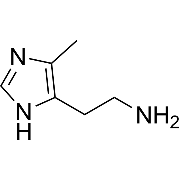
4-Methylhistamine
Based on 1 publication(s) in Google Scholar
4-Methylhistamine is a potent and selective agonist of histamine 4 receptor (H4R) with a Ki of 50 nM. 4-Methylhistamine has a >100-fold selectivity for the hH4R over the other histamine receptor subtypes. 4-Methylhistamine can potently activate the hH4R (pEC50 = 7.4). 4-Methylhistamine can be used for the researches of cancer, inflammation and immunology, such as lung cancer and skin inflammation.
Nos produits utilisent uniquement pour la recherche. Nous ne vendons pas aux patients.
- CAS No.: 36507-31-0
- Formule: C6H11N3
- Masse moléculaire:125.17
-
Stockage:Powder -20°C, 3 years ; In solvent -80°C, 6 months , -20°C, 1 month

To place orders, for customer services and technical support, please contact: MedChemExpress USA
Tel: 609-228-6898 E-mail: [email protected] [email protected]


-
 Activité biologique
-
Chemical Information
- Protocole
- Pureté et documentation
- Références
-
 Help & FAQs


Publications Citing Use of MedChemExpress (MCE) 4-Methylhistamine
MoreVoir tous les produits spécifiques à Isoform Histamine Receptor
More

Activité biologique
Description
IC50 & Target
[1]|
H4 receptor 50 nM (Ki) |
Cellular Effect
|
Cell Line

|
Type | Value | Description | References |
|---|---|---|---|---|
| Sf9 | EC50 |
12400 nM
Compound: 58
|
Agonist activity at human histamine H3 receptor expressed in sf9 cell membrane co-expressing mammalian Galphai2 and Gbeta1gamma2 incubated for 90 mins by [35S]GTPgammaS binding assay
Agonist activity at human histamine H3 receptor expressed in sf9 cell membrane co-expressing mammalian Galphai2 and Gbeta1gamma2 incubated for 90 mins by [35S]GTPgammaS binding assay
|
[PMID: 27007611] |
In Vitro
4-Methylhistamine (1-10 μM, 12 h) inhibits cell proliferation in A549, H157, H460 and H322 cells[4].
4-Methylhistamine (1 μM, 12 h) decreases the TGF-β1 mRNA expression in A549 cells[4].
MedChemExpress (MCE) has not independently confirmed the accuracy of these methods. They are for reference only.
-
Cell Line:A549, H157, H460 and H322 cells
-
Concentration:1 μM
-
Incubation Time:12 h
-
Result:Increased E-cadherin levels and decreased Vimentin levels.
In Vivo
4-Methylhistamine (20-40 mg/kg, i.p., once a day for 10 days) attenuates Imiquimod (HY-B0180)-induced psoriasis-like skin inflammation in mice[3].
4-Methylhistamine (100 μmol/kg, i.v., every other day for 33 days) decreases the average tumour volume and prolongs survival in A549 tumor mice models[4].
MedChemExpress (MCE) has not independently confirmed the accuracy of these methods. They are for reference only.
-
Animal Model:Balb/c mice with chronic restraint[2]
-
Dosage:30 mg/kg
-
Administration:Intraperitoneally injection, twice a day before 2 days prior to stress
-
Result:Increased in the CD4+ T cells as well as in IFN-γ production.
Decreased IL-4 expression.
Upregulated expression of IL-1β, IFN-γ and TNF-α mRNA.
-
Animal Model:Imiquimod (HY-B0180)-induced psoriasis-like dermatitis mice[3]
-
Dosage:20 and 40 mg/kg
-
Administration:Intraperitoneally injection, once a day for 10 days
-
Result:Ameliorated the total clinical severity scores.
Attenuated the psoriatic phenotypes, including epidermal hyperplasis, hyperkeratosis and lymphocytes infiltration.
Led to reductions in the levels of Th1 cytokines (TNF-a, IFN-a, and IL-27).
Increased CD4+CD25+FoxP3+ regulatory T (Treg) cells.
-
Animal Model:A549 tumor mice models[4]
-
Dosage:100 μmol/kg
-
Administration:Intravenously injection, every other day for 33 days
-
Result:Decreased tumor volume and the proliferation of tumour cells.
Prolonged survival of mice.
Increased the mRNA expression of E-cadherin and decreased Vimentin levels.


Chemical Information
-
CAS No. 36507-31-0
-
Appearance Solid
-
Masse moléculaire 125.17
-
Formule C6H11N3
-
Color White to off-white
-
SMILES
NCCC1=C(C)N=CN1
-
Livraison
Room temperature in continental US; may vary elsewhere.
-
Stockage
Powder -20°C 3 years In solvent -80°C 6 months -20°C 1 month


Publications (1)
-
Journal Impact Factor
-
Most Recent


Pureté et documentation
-
Fiche technique (275 KB)
-
SDS (392 KB)
- English - EN (392 KB)
- Français - FR (392 KB)
- Deutsch - DE (392 KB)
- Norwegian - NO (392 KB)
- Español - ES (392 KB)
- Swedish - SV (392 KB)
- Italian - IT (392 KB)
- Korean - KR (392 KB)
- Portuguese - PT (392 KB)
-
Instruction de manipulation (2659 KB)
Références
[1]. Lim HD, et al. Evaluation of histamine H1-, H2-, and H3-receptor ligands at the human histamine H4 receptor: identification of 4-methylhistamine as the first potent and selective H4 receptor agonist. J Pharmacol Exp Ther. 2005 Sep;314(3):1310-21. [Content Brief]
[2]. Ahmad SF, et al. Stimulation of the histamine 4 receptor with 4-methylhistamine modulates the effects of chronic stress on the Th1/Th2 cytokine balance. Immunobiology. 2015 Mar;220(3):341-9. [Content Brief]
[3]. Kim CH, et al. Inhibitory Effect of Imiquimod-Induced Psoriasis-Like Skin Inflammation in Mice by Histamine H4 Receptor Agonist 4-Methylhistamine. Scand J Immunol. 2016 Jun;83(6):409-17. [Content Brief]
[4]. Cai WK, et al. Activation of histamine H4 receptors decreases epithelial-to-mesenchymal transition progress by inhibiting transforming growth factor-β1 signalling pathway in non-small cell lung cancer. Eur J Cancer. 2014 Apr;50(6):1195-206. [Content Brief]

Calculators
Concentration (start) × Volume (start) = Concentration (final) × Volume (final)


4-Methylhistamine Related Classifications
HY-W580721 Related Classifications
 Powered by Bioz