



ACBI3

Based on 1 Customer Validation
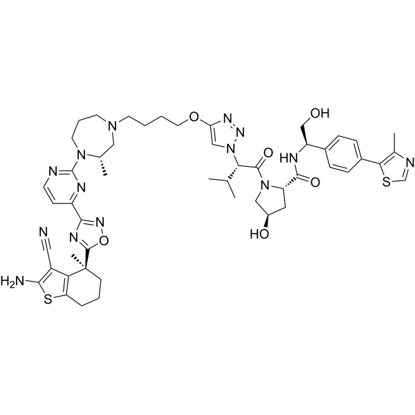
ACBI3 (compound 7), a chemical probe, is a PROTAC targeting KRAS. ACBI3 achieves in vivo degradation of oncogenic KRAS, resulting in durable pathway modulation and tumor regressions in KRAS mutant xenograft mouse models.
(Pink: K-Ras WT ligand (HY-162960); Blue: VHL ligand (HY-401613); Black: linker (HY-169992)).
Nos produits utilisent uniquement pour la recherche. Nous ne vendons pas aux patients.
The ACBI3 was designed by Boehringer Ingelheim and could be obtained free of charge through the Boehringer Ingelheim open innovation portal opnMe.com, associated with its negative control.
- Pureté : 99.47%
- CAS No.: 2938169-76-5
- Formule: C50H62N14O6S2
- Masse moléculaire:1019.25
-
Stockage:Powder -20°C, 3 years , 4°C, 2 years ; In solvent -80°C, 6 months , -20°C, 1 month

To place orders, for customer services and technical support, please contact: MedChemExpress USA
Tel: 609-228-6898 E-mail: [email protected] [email protected]


-
 Activité biologique
-
Chemical Information
-
Solvant et solubilité
- Protocole
- Pureté et documentation
- Références
-
 Help & FAQs






Voir tous les produits spécifiques à Isoform PROTACs
More

Activité biologique
Description
In Vitro
ACBI3 (0.01-10000 nM, 5 days) exhibits antiproliferative activity in KRAS mutant versus KRASWT cell lines (geometric mean IC50 =478 nM versus 8.3 μM, respectively). ACBI3 exhibits high efflux in the Caco-2 assay in LS513 cells[1][2].
MedChemExpress (MCE) has not independently confirmed the accuracy of these methods. They are for reference only.
In Vivo
MedChemExpress (MCE) has not independently confirmed the accuracy of these methods. They are for reference only.
-
Animal Model:KRAS mutant xenograft mouse models[1]
-
Dosage:2 mg/kg i.v. or 30 mg/kg s.c. or 30 mg/kg i.p.
-
Administration:once a day for 3 days
-
Result:Resulted in pronounced tumor regressions with a tumor growth inhibition of 127% in KRAS mutant xenograft mouse models.


Chemical Information
-
CAS No. 2938169-76-5
-
Appearance Solid
-
Masse moléculaire 1019.25
-
Formule C50H62N14O6S2
-
Color White to yellow
-
SMILES
NC(SC1=C2[C@](C3=NC(C4=NC(N5CCCN(CCCCOC(N=N6)=CN6[C@@H](C(C)C)C(N7[C@H](C(N[C@@H](CO)C8=CC=C(C=C8)C9=C(N=CS9)C)=O)C[C@@H](O)C7)=O)C[C@@H]5C)=NC=C4)=NO3)(C)CCC1)=C2C#N
-
Livraison
Room temperature in continental US; may vary elsewhere.
-
Stockage
Powder -20°C 3 years 4°C 2 years In solvent -80°C 6 months -20°C 1 month


Solvant et solubilité
In Vitro:
DMSO : 140 mg/mL (137.36 mM; Need ultrasonic; Hygroscopic DMSO has a significant impact on the solubility of product, please use newly opened DMSO)
Please refer to the solubility information to select the appropriate solvent. Once prepared, please aliquot and store the solution to prevent product inactivation from repeated freeze-thaw cycles.
Storage method and period of stock solution: -80°C, 6 months; -20°C, 1 month. When stored at -80°C, please use it within 6 months. When stored at -20°C, please use it within 1 month.
Please refer to the solubility information to select the appropriate solvent. Once prepared, please aliquot and store the solution to prevent product inactivation from repeated freeze-thaw cycles.
Storage method and period of stock solution: -80°C, 6 months; -20°C, 1 month. When stored at -80°C, please use it within 6 months. When stored at -20°C, please use it within 1 month.
Concentration (start) × Volume (start) = Concentration (final) × Volume (final)


Pureté et documentation
-
Fiche technique (274 KB)
-
SDS (251 KB)
- English - EN (251 KB)
- Français - FR (251 KB)
- Deutsch - DE (251 KB)
- Norwegian - NO (251 KB)
- Español - ES (251 KB)
- Swedish - SV (251 KB)
- Italian - IT (251 KB)
- Korean - KR (251 KB)
- Portuguese - PT (251 KB)
-
Instruction de manipulation (2659 KB)
Références


Complete Stock Solution Preparation Table
Please refer to the solubility information to select the appropriate solvent. Once prepared, please aliquot and store the solution to prevent product inactivation from repeated freeze-thaw cycles.
Storage method and period of stock solution: -80°C, 6 months; -20°C, 1 month. When stored at -80°C, please use it within 6 months. When stored at -20°C, please use it within 1 month.
| Optional Solvent | Concentration Solvent Mass | 1 mg | 5 mg | 10 mg | 25 mg |
|---|---|---|---|---|---|
| DMSO | 1 mM | 0.9811 mL | 4.9056 mL | 9.8111 mL | 24.5278 mL |
| 5 mM | 0.1962 mL | 0.9811 mL | 1.9622 mL | 4.9056 mL | |
| 10 mM | 0.0981 mL | 0.4906 mL | 0.9811 mL | 2.4528 mL | |
| 15 mM | 0.0654 mL | 0.3270 mL | 0.6541 mL | 1.6352 mL | |
| 20 mM | 0.0491 mL | 0.2453 mL | 0.4906 mL | 1.2264 mL | |
| 25 mM | 0.0392 mL | 0.1962 mL | 0.3924 mL | 0.9811 mL | |
| 30 mM | 0.0327 mL | 0.1635 mL | 0.3270 mL | 0.8176 mL | |
| 40 mM | 0.0245 mL | 0.1226 mL | 0.2453 mL | 0.6132 mL | |
| 50 mM | 0.0196 mL | 0.0981 mL | 0.1962 mL | 0.4906 mL | |
| 60 mM | 0.0164 mL | 0.0818 mL | 0.1635 mL | 0.4088 mL | |
| 80 mM | 0.0123 mL | 0.0613 mL | 0.1226 mL | 0.3066 mL | |
| 100 mM | 0.0098 mL | 0.0491 mL | 0.0981 mL | 0.2453 mL |


ACBI3 Related Classifications
HY-157228 Related Classifications
 Powered by Bioz