



DeepRed Nucleus Dye

Based on 1 Customer Validation
DeepRed Nucleus Dye is a novel cell permeant and far red-fluorescing DNA probe. DeepRed Nucleus Dye excites at a wavelength of 647 nm, close to the Ex, and produces a fluorescence spectrum extending from 665 nm out to beyond 780 nm wavelengths. DeepRed Nucleus Dye fluorescence reflects cellular DNA content. DeepRed Nucleus Dye can be used in combination with FITC and RPE-labelled antibodies, without the need for fluorescence compensation.
Nos produits utilisent uniquement pour la recherche. Nous ne vendons pas aux patients.
- Masse moléculaire:412.50
-
Stockage:
Solution, -20°C, protect from light, 2 years

To place orders, for customer services and technical support, please contact: MedChemExpress USA
Tel: 609-228-6898 E-mail: [email protected] [email protected]


-
 Activité biologique
-
Chemical Information
- Protocole
- Pureté et documentation
- Références
-
 Help & FAQs




Activité biologique
Description
In Vitro
Guidelines (Following is our recommended protocol. This protocol only provides a guideline, and should be modified according to your specific needs).
Mammalian cell in full culture medium staining methods[2]:
(1) Cell planking: Digestive separation of cells and resuspend in complete medium to a concentration of 2-4 × 10⁵ cells/mL.
Note: Attached cell cultures (e.g., coverslip cultures or chambered wells) can be stained in a 1-2-mL staining volume overlayering a 4-cm2 surface area.
(2) Prepare staining solution: Add 4 μL of 5 mM DeepRed Nucleus Dye acidified stock per ml culture medium (20 μM final).
Note: Nuclear discrimination is achievable at 2.5 to 5 μM, and it is unlikely that concentrations >30 μM would be required.
(3) Fluorescence staining: Incubate 5 to 15 min at 37°C.
Note: Overstaining cannot occur.
(4) Wash (optional): Centrifuge cells 3 to 5 min at 800 × g, 37°C. Discard supernatant and resuspend in complete medium with 10 mM HEPES (HY-D0857) at 4 × 10⁵ cells/mL .
(5) For flow cytometry: Use conventional pulse analysis for doublet discrimination and analyze parameters using appropriate software.
(6) For laser scanning microscopy: Collect fluorescence images using a 695 nm long-pass filter.
Fixed cells staining methods[2]:
(1) Fixed cells: Use 4% paraformaldehyde in PBS for 30 min with resuspension in an aqueous buffer (e.g., PBS).
(2) Fluorescence staining: similar concentrations of dye and similar incubation conditions can be used as for live cells.
MedChemExpress (MCE) has not independently confirmed the accuracy of these methods. They are for reference only.


Chemical Information
-
Appearance Liquid
-
Masse moléculaire 412.50
-
Color Blue to dark blue
-
SMILES
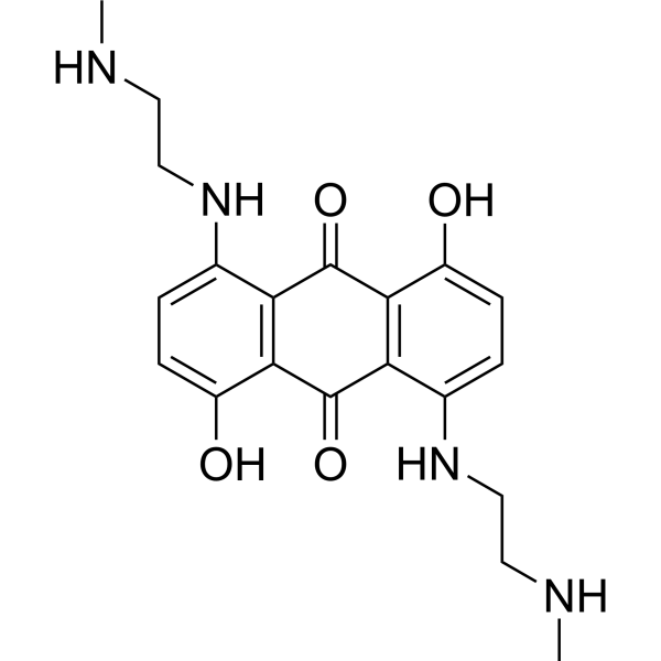
[DeepRed Nucleus Dye]
-
Livraison
Room temperature in continental US; may vary elsewhere.
-
Stockage
Solution, -20°C, protect from light, 2 years


Pureté et documentation
-
Fiche technique (270 KB)
-
SDS (252 KB)
- English - EN (252 KB)
- Français - FR (252 KB)
- Deutsch - DE (252 KB)
- Norwegian - NO (252 KB)
- Español - ES (252 KB)
- Swedish - SV (252 KB)
- Italian - IT (252 KB)
- Korean - KR (252 KB)
- Portuguese - PT (252 KB)
-
Instruction de manipulation (2659 KB)
Références
[1]. Smith PJ, et al. A novel cell permeant and far red-fluorescing DNA probe, DRAQ5, for blood cell discrimination by flow cytometry. J Immunol Methods. 1999 Oct 29;229(1-2):131-9. [Content Brief]
[2]. Smith PJ, et al. DRAQ5 labeling of nuclear DNA in live and fixed cells. Curr Protoc Cytom. 2004 May;Chapter 7:Unit 7.25. [Content Brief]

Calculators
Concentration (start) × Volume (start) = Concentration (final) × Volume (final)


DeepRed Nucleus Dye Related Classifications
HY-D1742 Related Classifications
 Powered by Bioz