



Chloranil

Based on 1 Customer Validation
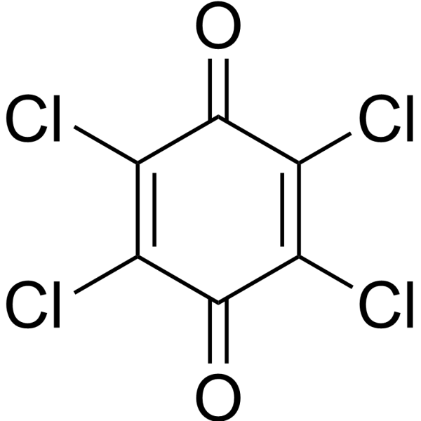
Chloranil (Tetrachloro-p-benzoquinone), an orally active metabolite of pentachlorophenol and hexachlorobenzene, is a widely used fungicide. Chloranil can induce ROS production. Chloranil induces neutrophil extracellular traps through the ROS-JNK-NOX2 pathway. Chloranil induces ferroptosis and neuroinflammation. Chloranil induces apoptosis of mouse embryonic stem cells .
연구목적의 판매만을 진행합니다. 환자를 대상으로 한 판매는 하지 않습니다.
- Purity : 98%
- CAS No.: 118-75-2
- 화학식: C6Cl4O2
- 분자량:245.88
-
보관:Powder -20°C, 3 years , 4°C, 2 years ; In solvent -80°C, 6 months , -20°C, 1 month

To place orders, for customer services and technical support, please contact: MedChemExpress USA
Tel: 609-228-6898 E-mail: [email protected] [email protected]


-
 Biological Activity
-
Chemical Information
-
용액&용해도
- Protocol
- 순도&문서
- References
-
 Help & FAQs


-

HY-Y0278R
Chloranil (Standard)ROS Inducer -
HY-N0165
Methyl-HesperidinVasodilating Agent -
HY-Y0786
1,3-Dibromo-5,5-dimethylhydantoin -

HY-Y0532
N-Chlorosuccinimide生化试剂 -

HY-W145053
ChloraMine-T hydrate -

HY-Y0532R
N-Chlorosuccinimide (Standard)Reference Standard -

HY-W145076
2-Azaadamantane-N-oxyl -

HY-N0165R
Methyl-Hesperidin (Standard)Vasodilating Agent


Biological Activity
제품 설명
IC50 & Target
[1]|
TLR4 |
Cellular Effect
|
Cell Line

|
Type | Value | Description | References |
|---|---|---|---|---|
| MRC5 | IC50 |
>100 μM
Compound: 16
|
Cytotoxicity against human MRC5 cells assessed as cell growth inhibition after 48 hrs by alamar blue assay
Cytotoxicity against human MRC5 cells assessed as cell growth inhibition after 48 hrs by alamar blue assay
|
[PMID: 26638044] |
In Vitro
Chloranil (20 μM; 12-24 h) induces ferroptosis in PC12 cells[1].
Chloranil (0-50 μM; 24 h) induces apoptosis in mouse embryonic stem cells in a dose-dependent manner[2].
Chloranil (10 μM; 1.5-3 h) induces neutrophil extracellular traps by ROS-JNK-NOX2 signaling pathway in mouse neutrophils[3].
Chloranil (25 μM; 6 h) decreases the viability of PC12 cells, increases the expression and interaction of TLR4 and MyD88, and up-regulates the expression and interaction of CD14 and MD2[4].
Chloranil (25 μM; 6 h) stimulates the expression of inflammatory factors and activates MAPK signaling pathway in PC12 cells[4].
MedChemExpress (MCE) has not independently confirmed the accuracy of these methods. They are for reference only.
-
Cell Line:Mouse neutrophil
-
Concentration:10 μM
-
Incubation Time:1.5 h
-
Result:Significantly increased the phosphorylation level of JNK.
Increased the level of NOX2.
-
Cell Line:PC12 cells
-
Concentration:25 μM
-
Incubation Time:6 h
-
Result:Enhanced the expression levels of TLR4 and MyD88.
Enhanced the expression levels of CD14 and MD2.
Enhanced the expression levels of TNF-a, IL-1β and IL-6.
Enhanced the expression levels of c-fos, c-jun and AP-1.
Increased the phosphorylation levels of p38, JNK and ERK.
In Vivo
Chloranil (1 mg/kg; Intraperitoneal injection; 3 days) has a strong hepatotoxic effect in mice, but can be alleviated by chlorogenic acid (HY-N0055)[5].
MedChemExpress (MCE) has not independently confirmed the accuracy of these methods. They are for reference only.
-
Animal Model:Male C57BL/6 TLR4 wild-type mice (5-8 weeks old)[4]
-
Dosage:10 mg/kg
-
Administration:Oral administration (p.o.); 2 weeks
-
Result:Caused severe coagulation necrosis, nuclear pyknosis, anachromasis and shrunken neuronal bodies in both cortical and the hippocampus region.
Caused cortical damage.
Decreased NeuN-positive neurons and increased highly activated microglia in cortex.
-
Animal Model:Male Kunming mice (22 ± 2 g)[5]
-
Dosage:1 mg/kg
-
Administration:Intraperitoneal injection (i.p.); 3 days
-
Result:Caused marked liver cell necrosis and inflammation but not apoptosis, and this damage was alleviated by Chlorogenic acid (HY-N0055).
Enhanced serum ALT, AST activities, TBIL content, hepatic oxidative stress and lipid peroxidation, decreased GSH content and inhibited the activities of antioxidant enzymes.
Up-regulated HO-1 and NQO1 expression.


Chemical Information
-
CAS No. 118-75-2
-
Appearance Solid
-
분자량 245.88
-
화학식 C6Cl4O2
-
Color Light yellow to green yellow
-
SMILES
O=C1C(Cl)=C(Cl)C(C(Cl)=C1Cl)=O
-
Synonyms
Tetrachloro-p-benzoquinone; TCBQ
-
선적
Room temperature in continental US; may vary elsewhere.
-
보관
Powder -20°C 3 years 4°C 2 years In solvent -80°C 6 months -20°C 1 month


용액&용해도
In Vitro:
DMSO : 5 mg/mL (20.34 mM; ultrasonic and warming and heat to 60°C; Hygroscopic DMSO has a significant impact on the solubility of product, please use newly opened DMSO)
Please refer to the solubility information to select the appropriate solvent. Once prepared, please aliquot and store the solution to prevent product inactivation from repeated freeze-thaw cycles.
Storage method and period of stock solution: -80°C, 6 months; -20°C, 1 month. When stored at -80°C, please use it within 6 months. When stored at -20°C, please use it within 1 month.
Please refer to the solubility information to select the appropriate solvent. Once prepared, please aliquot and store the solution to prevent product inactivation from repeated freeze-thaw cycles.
Storage method and period of stock solution: -80°C, 6 months; -20°C, 1 month. When stored at -80°C, please use it within 6 months. When stored at -20°C, please use it within 1 month.
Concentration (start) × Volume (start) = Concentration (final) × Volume (final)


순도&문서
-
Data Sheet (283 KB)
-
SDS (758 KB)
- English - EN (758 KB)
- Français - FR (758 KB)
- Deutsch - DE (758 KB)
- Norwegian - NO (758 KB)
- Español - ES (758 KB)
- Swedish - SV (758 KB)
- Italian - IT (758 KB)
- Korean - KR (758 KB)
- Portuguese - PT (758 KB)
-
Handling Instructions (2659 KB)
References
[1]. Liu Z, et al. Tetrachlorobenzoquinone exposure triggers ferroptosis contributing to its neurotoxicity. Chemosphere. 2021 Feb;264(Pt 1):128413. [Content Brief]
[2]. Zuehlke A, et al. Elevated 5-hydroxymethycytosine and cell apoptosis induced by tetrachloro-1,4-benzoquinone in mouse embryonic stem cells. J Environ Sci (China). 2017 Jan;51:1-4. [Content Brief]
[3]. Lv X, et al. Tetrachlorobenzoquinone exhibits immunotoxicity by inducing neutrophil extracellular traps through a mechanism involving ROS-JNK-NOX2 positive feedback loop. Environ Pollut. 2021 Jan 1;268(Pt B):115921. [Content Brief]
[4]. Fu J, et al.The acute exposure of tetrachloro-p-benzoquinone (a.k.a. chloranil) triggers inflammation and neurological dysfunction via Toll-like receptor 4 signaling: The protective role of melatonin preconditioning. Toxicology. 2017 Apr 15;381:39-50. [Content Brief]
[5]. Xu D, et al. Tetrachlorobenzoquinone induces acute liver injury, up-regulates HO-1 and NQO1 expression in mice model: the protective role of chlorogenic acid. Environ Toxicol Pharmacol. 2014 May;37(3):1212-20. [Content Brief]


Complete Stock Solution Preparation Table
Please refer to the solubility information to select the appropriate solvent. Once prepared, please aliquot and store the solution to prevent product inactivation from repeated freeze-thaw cycles.
Storage method and period of stock solution: -80°C, 6 months; -20°C, 1 month. When stored at -80°C, please use it within 6 months. When stored at -20°C, please use it within 1 month.
| Optional Solvent | Concentration Solvent Mass | 1 mg | 5 mg | 10 mg | 25 mg |
|---|---|---|---|---|---|
| DMSO | 1 mM | 4.0670 mL | 20.3351 mL | 40.6702 mL | 101.6756 mL |
| 5 mM | 0.8134 mL | 4.0670 mL | 8.1340 mL | 20.3351 mL | |
| 10 mM | 0.4067 mL | 2.0335 mL | 4.0670 mL | 10.1676 mL | |
| 15 mM | 0.2711 mL | 1.3557 mL | 2.7113 mL | 6.7784 mL | |
| 20 mM | 0.2034 mL | 1.0168 mL | 2.0335 mL | 5.0838 mL |


Chloranil Related Classifications
HY-Y0278 Related Classifications
 Powered by Bioz