



Dapivirine-d11

Based on 1 Customer Validation
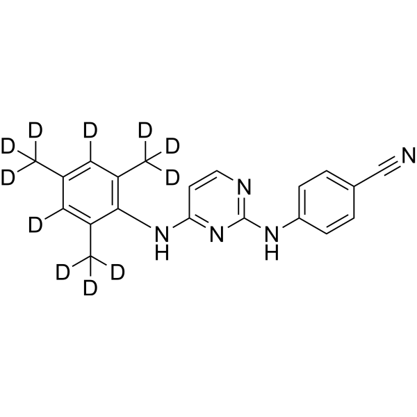
Dapivirine-d11 is the deuterium labeled Dapivirine. Dapivirine (TMC120), the prototype of diarylpyrimidines (DAPY), is an orally active and nonnucleoside reverse transcriptase inhibitor (NRTI). Dapivirine (TMC120) binds directly to HIV-1 reverse transcriptase. Dapivirine (TMC120) regulates autophagy and induced Akt, Bad and SAPK/JNK activations.
For research use only. We do not sell to patients.
- CAS No.: 1329613-10-6
- Formula: C20H8D11N5
- Molecular Weight:340.47
-
Storage:Powder -20°C, 3 years , 4°C, 2 years ; In solvent -80°C, 6 months , -20°C, 1 month

To place orders, for customer services and technical support, please contact: MedChemExpress USA
Tel: 609-228-6898 E-mail: [email protected] [email protected]


-
 Biological Activity
-
Chemical Information
- Protocols
- Purity & Documentation
- References
-
 Help & FAQs




Biological Activity
Description
In Vitro
Stable heavy isotopes of hydrogen, carbon, and other elements have been incorporated into drug molecules, largely as tracers for quantitation during the drug development process. Deuteration has gained attention because of its potential to affect the pharmacokinetic and metabolic profiles of drugs[1].
MedChemExpress (MCE) has not independently confirmed the accuracy of these methods. They are for reference only.
Application
1. This compound can be used as a tracer
2. This compound can be used as an internal standard for quantitative analysis by NMR, GC-MS, or LC-MS.


Chemical Information
-
CAS No. 1329613-10-6
-
Unlabeled CAS 244767-67-7
-
Appearance Solid
-
Molecular Weight 340.47
-
Formula C20H8D11N5
-
Color White to off-white
-
SMILES
N#CC1=CC=C(NC2=NC=CC(NC3=C(C([2H])([2H])[2H])C([2H])=C(C([2H])([2H])[2H])C([2H])=C3C([2H])([2H])[2H])=N2)C=C1
-
Synonyms
TMC120-d11; R147681-d11
-
Shipping
Room temperature in continental US; may vary elsewhere.
-
Storage
Powder -20°C 3 years 4°C 2 years In solvent -80°C 6 months -20°C 1 month


Purity & Documentation
-
Data Sheet (269 KB)
-
SDS (252 KB)
- Français - FR (252 KB)
- Deutsch - DE (252 KB)
- Norwegian - NO (252 KB)
- Español - ES (252 KB)
- Swedish - SV (252 KB)
- Italian - IT (252 KB)
- Korean - KR (252 KB)
- Portuguese - PT (252 KB)
-
Handling Instructions (2659 KB)
References
[1]. Russak EM, et al. Impact of Deuterium Substitution on the Pharmacokinetics of Pharmaceuticals. Ann Pharmacother. 2019;53(2):211-216. [Content Brief]
[2]. Weiwen Liu, et al. Antitumor Activity and Mechanism of a Reverse Transcriptase Inhibitor, Dapivirine, in Glioblastoma. J Cancer. 2018 Jan 1;9(1):117-128. [Content Brief]
[3]. Bríd Devlin, et al. Development of dapivirine vaginal ring for HIV prevention. Antiviral Res. 2013 Dec;100 Suppl:S3-8. [Content Brief]
[4]. Yven Van Herrewege, et al. In vitro evaluation of nonnucleoside reverse transcriptase inhibitors UC-781 and TMC120-R147681 as human immunodeficiency virus microbicides. Antimicrob Agents Chemother. 2004 Jan;48(1):337-9. [Content Brief]
[5]. Michael E Halwes, et al. Pharmacokinetic modeling of a gel-delivered dapivirine microbicide in humans. Eur J Pharm Sci. 2016 Oct 10;93:410-8. [Content Brief]

Calculators
Concentration (start) × Volume (start) = Concentration (final) × Volume (final)


Dapivirine-d11 Related Classifications
HY-14266S Related Classifications
 Powered by Bioz