



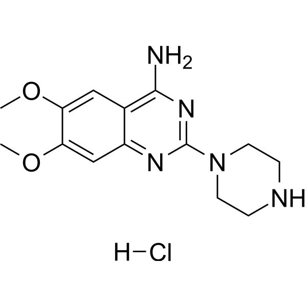
DPQ hydrochloride
Based on 4 publication(s) in Google Scholar
DPQ hydrochloride is a blood-brain barrier permeable and selective PARP-1 inhibitor that blocks PARP-1-mediated DNA damage repair and NAD+/ATP consumption, thereby inhibiting excessive inflammatory responses. DPQ hydrochloride inhibits NF-κB pathway activation, reduces the expression of pro-inflammatory factors (such as TNF-α, IL-6) and oxidative stress. DPQ hydrochloride can be used in inflammation-related studies of acute lung injury, myocardial infarction, and neurodegenerative diseases.
Nur für Forschungszwecke. Wir verkaufen nicht an Patienten.
- Reinheit : 98.97%
- CAS. Nr.: 84050-22-6
- Formel: C14H20ClN5O2
- Molecular Weight:325.79
-
Speicherung:
4°C, sealed storage, away from moisture and light
* In solvent : -80°C, 6 months; -20°C, 1 month (sealed storage, away from moisture and light)

To place orders, for customer services and technical support, please contact: MedChemExpress USA
Tel: 609-228-6898 E-mail: [email protected] [email protected]


-
 Biologische Aktivität
-
Chemical Information
-
Lösungsmittel & Löslichkeit
- Protokoll
- Reinheit & Dokumentation
- Verweise
-
 Help & FAQs


-
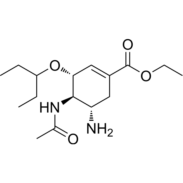
HY-13317
OseltamivirInfluenza Virus Inhibitor -
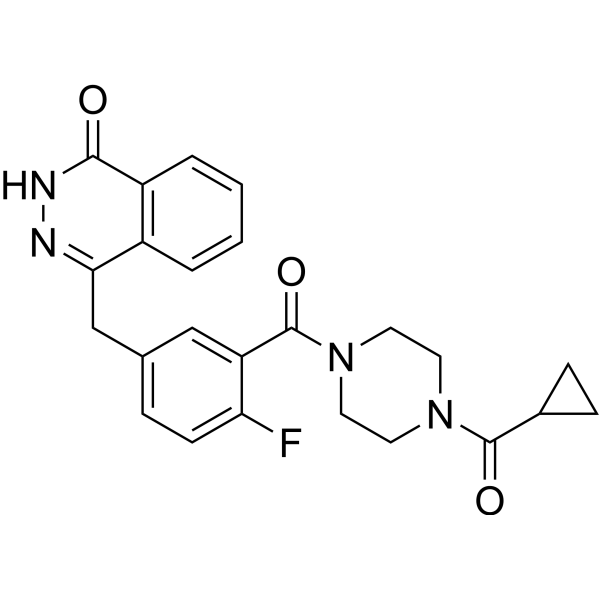
HY-10162
OlaparibPARP Inhibitor -
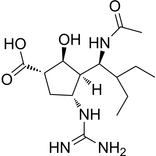
HY-17015A
PeramivirCyclopentane neuraminidase Inhibitor -

HY-10162R
Olaparib (Standard)PARP Inhibitor -
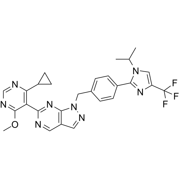
HY-145471
KSQ-4279USP1 Inhibitor -
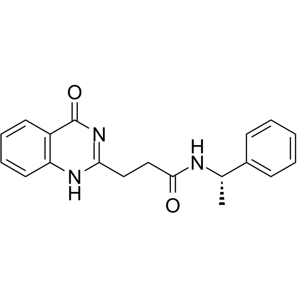
HY-100225
ME0328ARTD3/PARP3 Inhibitor -
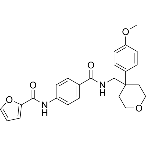
HY-13968
JW 55PARP Inhibitor -
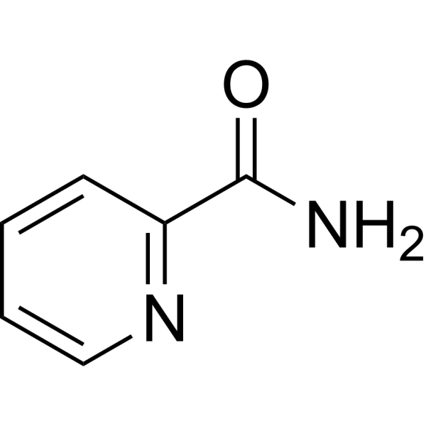
HY-101020
PicolinamidePARS Inhibitor -

HY-13317R
Oseltamivir (Standard)Influenza Virus Inhibitor -
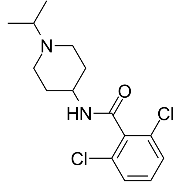
HY-131059
CBS1117H1N1 Inhibitor -
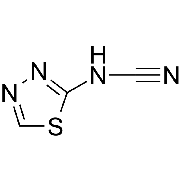
HY-106817
AmitivirAntiinfluenza Agent -
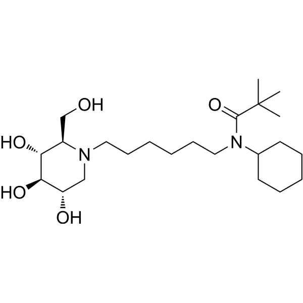
HY-139663
IHVR-17028Anti-Viral Agent
Publications Citing Use of MedChemExpress (MCE) DPQ hydrochloride
More

Biologische Aktivität
Beschreibung
IC50 & Target
|
PARP-1 |
In Vitro
DPQ hydrochloride (10 μM; pretreatment 30 min, treatment 2-8 h) significantly inhibited the mRNA expression of TNF-α, IL-1β, IL-6, CXCL-1, MIP-2 and iNOS in mouse peritoneal macrophages stimulated by LPS (100 ng/mL) [2].
DPQ hydrochloride (10 μM; pretreatment 30 min, treatment 2-8 h) inhibited the degradation of IkB-α and phosphorylation of NF-κB p65 in macrophages induced by LPS (100 ng/mL), blocking the inflammatory signaling pathway[2].
MedChemExpress (MCE) has not independently confirmed the accuracy of these methods. They are for reference only. Further protocols information, click here.
-
Cell Line:Mixed murine cortical neurons
-
Concentration:10 μM
-
Incubation Time:10 min pretreatment + 20 min NMDA exposure
-
Result:Reduced NMDA-induced neuronal apoptosis by 84% at 6 h and 50% at 24 h, restored ATP levels from 4% to 72% of control, and suppressed PARP activation.
-
Cell Line:Murine peritoneal macrophages
-
Concentration:10 μM
-
Incubation Time:30 min pretreatment + 2-8 h LPS stimulation
-
Result:Significantly decreased LPS-induced mRNA expression of TNF-α (50% reduction), IL-1β (40%), IL-6 (45%), CXCL-1 (35%), MIP-2 (40%), and iNOS (50%).
-
Cell Line:Murine peritoneal macrophages
-
Concentration:10 μM
-
Incubation Time:30 min pretreatment + 15-60 min LPS stimulation
-
Result:Blocked LPS-induced IkB-α degradation (50% inhibition at 15 min) and NF-κB p65 phosphorylation (40% reduction at 30 min).
-
Cell Line:Murine peritoneal macrophages
-
Concentration:10 μM
-
Incubation Time:30 min pretreatment + 1 h LPS stimulation
-
Result:Reduced LPS-induced PARP activation (40% decrease in PAR fluorescence intensity) and nitrotyrosine formation (35% reduction).
In Vivo
DPQ hydrochloride (10 mg/kg; intraperitoneal injection; single dose; 4 weeks) improves cardiac function and reduced apoptosis and oxidative stress in myocardial infarction model of Wistar rats[3].
MedChemExpress (MCE) has not independently confirmed the accuracy of these methods. They are for reference only.
-
Animal Model:LPS-Induced Acute Lung Injury Model in C57BL/6 mice (male, 8-10 weeks old)[2]
-
Dosage:10 μg/kg (dissolved in 0.01% DMSO (PBS)
-
Administration:Intraperitoneal injection, 30 min after LPS chanllenge (7.5 mg/kg; ip; single dose)
-
Result:Reduced neutrophil infiltration (50% decrease), MPO activity (40% decrease), and pro-inflammatory cytokines (TNF-α, IL-1β, IL-6) in lungs. Restored vascular permeability (Evans blue extravasation reduced by 35%), and inhibited apoptotic cell death (TUNEL-positive cells decreased by 45%).
Suppressed NF-κB activation with reduced IkB-α degradation and p65 phosphorylation.
-
Animal Model:Wistar rats (male, 4 months old) + MI via coronary artery ligation[3]
-
Dosage:10 mg/kg (dissolved in DMSO)
-
Administration:Intraperitoneal injection, single dose immediately after MI induction
-
Result:Improved cardiac function (FS increased by 25%, EDD/ESD reduced by 15%), decreased apoptotic cardiomyocytes (TUNEL-positive cells reduced by 40%), and suppressed cleaved caspase-3 and PARP1 expression. Oxidative stress markers (O2-, nitrotyrosine) were reduced by 30-40% in infarcted myocardium.


Chemical Information
-
CAS. Nr. 84050-22-6
-
Appearance Solid
-
Molecular Weight 325.79
-
Formel C14H20ClN5O2
-
Color White to off-white
-
SMILES
NC1=C2C=C(OC)C(OC)=CC2=NC(N3CCNCC3)=N1.[H]Cl
-
Synonyms
6,7-Dimethoxy-2-(1-piperazinyl)-4-quinazolinamine hydrochloride
-
Versand
Room temperature in continental US; may vary elsewhere.
-
Speicherung
4°C, sealed storage, away from moisture and light
* In solvent : -80°C, 6 months; -20°C, 1 month (sealed storage, away from moisture and light)


Publications (4)
-
Journal Impact Factor
-
Most Recent
-
J Adv Res
Targeting myofibroblast copper vulnerability reverses pulmonary fibrosis via METTL3-directed STAT6 m6A driving cuproptosis. [Abstract]2026 May 21:S2090-1232(26)00436-4. PMID: 42173361 -
Sci Immunol
Schlafen 11 triggers innate immune responses through its ribonuclease activity upon detection of single-stranded DNA. [Abstract]2024 Jun 14;9(96):eadj5465. PMID: 38875319 -
Adv Sci (Weinh)
Cuproptosis and Disulfidptosis Converge to Empower PD-L1 Checkpoint Therapy via Cadict-Induced PD-L1 Translation. [Abstract]2026 May;13(25):e15367. PMID: 41722054 -
Stem Cell Res Ther
MSC-derived exosomes attenuate cell death through suppressing AIF nucleus translocation and enhance cutaneous wound healing. [Abstract]2020 May 11;11(1):174. PMID: 32393338


Lösungsmittel & Löslichkeit
In Vitro:
DMSO : 50 mg/mL (153.47 mM; Need ultrasonic; Hygroscopic DMSO has a significant impact on the solubility of product, please use newly opened DMSO)
Please refer to the solubility information to select the appropriate solvent. Once prepared, please aliquot and store the solution to prevent product inactivation from repeated freeze-thaw cycles.
Storage method and period of stock solution: -80°C, 6 months; -20°C, 1 month (sealed storage, away from moisture and light). When stored at -80°C, please use it within 6 months. When stored at -20°C, please use it within 1 month.
Please refer to the solubility information to select the appropriate solvent. Once prepared, please aliquot and store the solution to prevent product inactivation from repeated freeze-thaw cycles.
Storage method and period of stock solution: -80°C, 6 months; -20°C, 1 month (sealed storage, away from moisture and light). When stored at -80°C, please use it within 6 months. When stored at -20°C, please use it within 1 month.
Konzentration (Stammlösung) × Volumen (Stammlösung) = Konzentration (Ziellösung) × Volumen (Ziellösung)
In Vivo:
Select the appropriate dissolution method based on your experimental animal and administration route.
- For the following dissolution methods, please ensure to first prepare a clear stock solution using an In Vitro approach and then sequentially add co-solvents:
- To ensure reliable experimental results, the clarified stock solution can be appropriately stored based on storage conditions. As for the working solution for In Vivo experiments, it is recommended to prepare freshly and use it on the same day.
- The percentages shown for the solvents indicate their volumetric ratio in the final prepared solution. If precipitation or phase separation occurs during preparation, heat and/or sonication can be used to aid dissolution.
Add each solvent one by one: 10% DMSO 40% PEG300 5% Tween-80 45% Saline
Solubility: ≥ 1.25 mg/mL (3.84 mM); Clear solution
This protocol yields a clear solution of ≥ 1.25 mg/mL (saturation unknown).
Taking 1 mL working solution as an example, add 100 μL DMSO stock solution (12.5 mg/mL) to 400 μL PEG300, and mix evenly; then add 50 μL Tween-80 and mix evenly; then add 450 μL Saline to adjust the volume to 1 mL.
Preparation of Saline: Dissolve 0.9 g sodium chloride in ddH₂O and dilute to 100 mL to obtain a clear Saline solution.
Add each solvent one by one: 10% DMSO 90% (20% SBE-β-CD in Saline)
Solubility: ≥ 1.25 mg/mL (3.84 mM); Clear solution
This protocol yields a clear solution of ≥ 1.25 mg/mL (saturation unknown).
Taking 1 mL working solution as an example, add 100 μL DMSO stock solution (12.5 mg/mL) to 900 μL 20% SBE-β-CD in Saline, and mix evenly.
Preparation of 20% SBE-β-CD in Saline (4°C, storage for one week): 2 g SBE-β-CD powder is dissolved in 10 mL Saline, completely dissolve until clear.
In Vivo Dissolution Calculator
Please enter the basic information of animal experiments:
-
-
-
-
Recommended: Prepare an additional quantity of animals to account for potential losses during experiments.
Please enter your animal formula composition:
-
%DMSO +
Recommended: Keep the proportion of DMSO in working solution below 2% if your animal is weak.
-
%+
-
+%Tween-80 + +
-
%Saline +
The co-solvents required include: DMSO, . All of co-solvents are available by MedChemExpress (MCE). , Tween 80. All of co-solvents are available by MedChemExpress (MCE).
Working solution concentration: 0.22 mg/mL
Method for preparing stock solution: mg drug dissolved in μL DMSO. Stock solution concentration: mg/mL. * In solvent : -80°C, 6 months; -20°C, 1 month (sealed storage, away from moisture and light)
1. Take μL DMSO stock solution;
2. Add μL .
μL , mix evenly;
3. Then add μL Tween 80, mix evenly;
4. Then add μL
Please ensure that the stock solution in the first step is dissolved to a clear state, and add co-solvents in sequence. You can use ultrasonic heating (ultrasonic cleaner, recommended frequency 20-40 kHz), vortexing, etc. to assist dissolution.

Protokoll
-
RNA extraction experimental
By lysing cells, releasing RNA, and removing impurities such as proteins and DNA, high-purity RNA products are finally obtained. The commonly used traditional method is the guanidine isothiocyanate/phenol/chloroform method (Trizol), which is suitable for a variety of animal materials including animal tissues, microorganisms, cultured cells, etc., and most plant materials.
-
ROS/oxidative-stress fluorescent staining
ROS/oxidative-stress fluorescent staining uses cell-permeant fluorogenic probes that become fluorescent after oxidation inside cells or tissues; commonly used examples include DCFH-DA/DCFDA for broad cellular oxidant detection, DHE for superoxide-related signal detection, MitoSOX for mitochondrial superoxide-related signal detection, and CellROX probes for oxidative-stress-associated fluorescence readouts. The assay detects probe oxidation rather than a single ROS species unless the probe and analysis method have been chemically validated for that species. DCFH-DA enters cells, is deacetylated by intracellular esterases to DCFH, and produces fluorescent DCF after oxidation, so the readout is used as an operational measure of total cellular oxidative stress rather than a species-specific ROS measurement. DHE and MitoSOX can report superoxide-related oxidation, but red fluorescence alone can include non-specific ethidium-like oxidation products; HPLC or optimized spectral approaches are
-
Research Protocol for Cardiovascular Diseases
Cardiovascular disease can be modeled as maladaptive cardiac remodeling, where ischemic injury or pressure overload activates inflammatory signaling, fibroblast activation, extracellular-matrix deposition, cardiomyocyte hypertrophy, vascular remodeling, and progressive ventricular dysfunction. The TGF-β/SMAD axis is a central profibrotic pathway after myocardial injury and pressure overload, while innate immune and cytokine pathways regulate leukocyte recruitment, scar formation, and adverse remodeling. Key unresolved questions include which inflammatory signals are reparative versus harmful, when fibrosis is protective versus maladaptive, and whether pathway inhibition improves function without weakening necessary infarct healing or compensatory remodeling.
-
Research Protocol for Inflammation-related Diseases
The NLRP3 inflammasome is a cytosolic innate immune signaling platform that integrates priming signals and danger-signal activation to promote caspase-1 activation, maturation of IL-1β and IL-18, and gasdermin D-mediated pyroptotic cell death. The core experimental logic is to determine whether inflammatory disease phenotypes are driven by increased NLRP3 expression, ASC-containing inflammasome assembly, caspase-1 cleavage, GSDMD cleavage, and extracellular release of IL-1β/IL-18 rather than by nonspecific cell injury alone. The pathway is strongly linked to inflammation-related disease phenotypes because monosodium urate crystals activate NALP3/NLRP3 inflammasome signaling in gout-like crystal inflammation, cholesterol crystals activate NLRP3 inflammasomes in atherogenesis models, and DSS-induced intestinal inflammation has been reported to involve NLRP3 inflammasome activity. However, experimental colitis studies also show context-dependent protective effects of NLRP3 inflammasome co
-
Inhalation Toxicity Study
Inhalation toxicity studies expose rodents to a controlled aerosol, vapor, gas, or smoke atmosphere and assess respiratory and systemic toxicity using exposure-atmosphere characterization, clinical observations, body and organ weights, bronchoalveolar lavage fluid, histopathology, blood chemistry, hematology, and, when included, molecular endpoints such as transcriptomics, proteomics, lipidomics, or tissue burden analysis. The primary biological readouts are airway irritation, pulmonary inflammation, cytotoxicity, altered surfactant or lipid homeostasis, impaired particle clearance, and tissue remodeling, reflected by BALF cell differentials, BALF protein, LDH, phosphatase activities, cytokines, lung weight, microscopic respiratory-tract lesions, and retained lung burden.
-
Genotoxicity/Mutagenicity Study
The bacterial reverse mutation assay detects point mutations that restore amino-acid prototrophy in auxotrophic Salmonella typhimurium or Escherichia coli tester strains; after exposure to a test article, mutagenic activity is read out as an increased number of revertant colonies on minimal agar compared with the vehicle control. The assay uses tester strains with different mutation targets so that base-substitution and frameshift mutagens can be detected, and testing is performed with and without exogenous mammalian metabolic activation because some chemicals require biotransformation to become mutagenic.


Reinheit & Dokumentation
-
Data Sheet (283 KB)
-
SDS (252 KB)
- English - EN (252 KB)
- Français - FR (252 KB)
- Deutsch - DE (252 KB)
- Norwegian - NO (252 KB)
- Español - ES (252 KB)
- Swedish - SV (252 KB)
- Italian - IT (252 KB)
- Korean - KR (252 KB)
- Portuguese - PT (252 KB)
-
Handling Instructions (2659 KB)
Verweise
[1]. Meli E, et al. Differential role of poly (ADP-ribose) polymerase-1in apoptotic and necrotic neuronal death induced by mild or intense NMDA exposure in vitro. Mol Cell Neurosci. 2004;25 (1) :172-180. [Content Brief]
[2]. Wang G, et al. PARP-1 inhibitor, DPQ, attenuates LPS-induced acute lung injury through inhibiting NF-κB-mediated inflammatory response. PLoS One. 2013 Nov 21;8 (11) :e79757. [Content Brief]
[3]. Wang J, et al. Inhibition of poly (ADP-ribose) polymerase and inducible nitric oxide synthase protects against ischemic myocardial damage by reduction of apoptosis. Mol Med Rep. 2015 Mar;11 (3) :1768-76. [Content Brief]


Complete Stock Solution Preparation Table
Please refer to the solubility information to select the appropriate solvent. Once prepared, please aliquot and store the solution to prevent product inactivation from repeated freeze-thaw cycles.
Storage method and period of stock solution: -80°C, 6 months; -20°C, 1 month (sealed storage, away from moisture and light). When stored at -80°C, please use it within 6 months. When stored at -20°C, please use it within 1 month.
| Optional Solvent | Concentration Solvent Mass | 1 mg | 5 mg | 10 mg | 25 mg |
|---|---|---|---|---|---|
| DMSO | 1 mM | 3.0695 mL | 15.3473 mL | 30.6946 mL | 76.7365 mL |
| 5 mM | 0.6139 mL | 3.0695 mL | 6.1389 mL | 15.3473 mL | |
| 10 mM | 0.3069 mL | 1.5347 mL | 3.0695 mL | 7.6737 mL | |
| 15 mM | 0.2046 mL | 1.0232 mL | 2.0463 mL | 5.1158 mL | |
| 20 mM | 0.1535 mL | 0.7674 mL | 1.5347 mL | 3.8368 mL | |
| 25 mM | 0.1228 mL | 0.6139 mL | 1.2278 mL | 3.0695 mL | |
| 30 mM | 0.1023 mL | 0.5116 mL | 1.0232 mL | 2.5579 mL | |
| 40 mM | 0.0767 mL | 0.3837 mL | 0.7674 mL | 1.9184 mL | |
| 50 mM | 0.0614 mL | 0.3069 mL | 0.6139 mL | 1.5347 mL | |
| 60 mM | 0.0512 mL | 0.2558 mL | 0.5116 mL | 1.2789 mL | |
| 80 mM | 0.0384 mL | 0.1918 mL | 0.3837 mL | 0.9592 mL | |
| 100 mM | 0.0307 mL | 0.1535 mL | 0.3069 mL | 0.7674 mL |


DPQ hydrochloride Related Classifications
HY-W424851 Related Classifications
 Powered by Bioz