



dGTP
Based on 1 publication(s) in Google Scholar
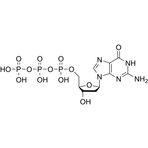
dGTP (2'-Deoxyguanosine-5'-triphosphate) is one of the precursors for DNA synthesis, and serves as a direct substrate for DNA replication and repair. dGTP is prone to oxidative damage; under the action of reactive oxygen species and other factors, dGTP is oxidized to form 8-oxo-dGTP.
For research use only. We do not sell to patients.
- CAS No.: 2564-35-4
- Formula: C10H16N5O13P3
- Molecular Weight:507.18
-
Storage:
4°C, protect from light, stored under nitrogen
* In solvent : -80°C, 6 months; -20°C, 1 month (protect from light, stored under nitrogen)

To place orders, for customer services and technical support, please contact: MedChemExpress USA
Tel: 609-228-6898 E-mail: [email protected] [email protected]


-
 Biological Activity
-
Chemical Information
- Protocols
- Purity & Documentation
- References
-
 Help & FAQs


Publications Citing Use of MedChemExpress (MCE) dGTP
MoreAll DNA/RNA Synthesis Isoforms
More

Biological Activity
Description
In Vitro
dGTP can be hydrolyzed to deoxyguanosine and triphosphate by dGTP triphosphohydrolase in E. coli, which acts as a key factor in regulating the size of the dGTP pool[1].
dGTP is oxidized to form 8-oxo-dGTP under the action of reactive oxygen species and other factors. During DNA replication, 8-oxo-dGTP can pair with C on the template strand, and also mispair with A. If it is incorporated into DNA without timely clearance, it will cause transversion mutations from A to C (or T to G), which is one of the main pathways of mutations induced by oxidative stress[2].
MedChemExpress (MCE) has not independently confirmed the accuracy of these methods. They are for reference only. Further protocols information, click here.


Chemical Information
-
CAS No. 2564-35-4
-
Appearance Solid
-
Molecular Weight 507.18
-
Formula C10H16N5O13P3
-
Color White to off-white
-
SMILES
O[C@H]1C[C@H](N2C(N=C(N)NC3=O)=C3N=C2)O[C@@H]1COP(OP(OP(O)(O)=O)(O)=O)(O)=O
-
Synonyms
2'-Deoxyguanosine-5'-triphosphate
-
Structure Classification
-
Initial Source
-
Shipping
Room temperature in continental US; may vary elsewhere.
-
Storage
4°C, protect from light, stored under nitrogen
* In solvent : -80°C, 6 months; -20°C, 1 month (protect from light, stored under nitrogen)


Publications (1)
-
Journal Impact Factor
-
Most Recent

Protocols
-
EdU Incorporation Assay (Click Chemistry-Based DNA Synthesis Measurement)
The EdU incorporation assay measures DNA synthesis by adding the thymidine analog 5-ethynyl-2′-deoxyuridine to cells or tissues, where it is incorporated into newly synthesized DNA during S phase. Incorporated EdU is detected by copper-catalyzed azide-alkyne cycloaddition, in which a fluorescent azide covalently reacts with the ethynyl group on EdU, allowing S-phase cells to be detected by fluorescence microscopy, flow cytometry, or high-content imaging. EdU detection does not require DNA denaturation or anti-BrdU antibody access, which preserves sample structure and improves compatibility with immunostaining and multiparameter cytometry compared with BrdU-based detection. EdU can be cytotoxic in a cell-type- and exposure-dependent manner, so pulse duration, concentration, and continuous-labeling designs should be validated for each cell type.
-
PCNA Immunodetection Proliferation Assay
PCNA immunodetection measures proliferative activity by detecting proliferating cell nuclear antigen, a nuclear protein associated with DNA polymerase δ function and DNA replication. The assay readout is the proportion of PCNA-positive nuclei among total counted cells, but PCNA labeling is not identical to BrdU labeling because PCNA can mark late G1/early S-associated replication competence and may persist beyond active DNA synthesis depending on fixation and extraction conditions.
-
BrdU Incorporation Assay
Bromodeoxyuridine (BrdU) incorporation assay is based on the principle that BrdU, a thymidine analog, is incorporated into newly synthesized DNA during the S phase of the cell cycle, thereby serving as a marker of DNA replication and cellular proliferation. Incorporated BrdU can be detected using anti-BrdU antibodies following DNA denaturation, enabling visualization or quantification of proliferating cells through immunochemical detection methods such as immunofluorescence or immunohistochemistry.
-
ROS/oxidative-stress fluorescent staining
ROS/oxidative-stress fluorescent staining uses cell-permeant fluorogenic probes that become fluorescent after oxidation inside cells or tissues; commonly used examples include DCFH-DA/DCFDA for broad cellular oxidant detection, DHE for superoxide-related signal detection, MitoSOX for mitochondrial superoxide-related signal detection, and CellROX probes for oxidative-stress-associated fluorescence readouts. The assay detects probe oxidation rather than a single ROS species unless the probe and analysis method have been chemically validated for that species. DCFH-DA enters cells, is deacetylated by intracellular esterases to DCFH, and produces fluorescent DCF after oxidation, so the readout is used as an operational measure of total cellular oxidative stress rather than a species-specific ROS measurement. DHE and MitoSOX can report superoxide-related oxidation, but red fluorescence alone can include non-specific ethidium-like oxidation products; HPLC or optimized spectral approaches are


Purity & Documentation
-
Data Sheet (279 KB)
-
SDS (252 KB)
- English - EN (252 KB)
- Français - FR (252 KB)
- Deutsch - DE (252 KB)
- Norwegian - NO (252 KB)
- Español - ES (252 KB)
- Swedish - SV (252 KB)
- Italian - IT (252 KB)
- Korean - KR (252 KB)
- Portuguese - PT (252 KB)
-
Handling Instructions (2659 KB)
References
[1]. Kapoor I, et al. Nucleoside Diphosphate Kinase Escalates A-to-C Mutations in MutT-Deficient Strains of Escherichia coli. J Bacteriol. 2019;202(1):e00567-19. Published 2019 Dec 6. [Content Brief]
[2]. Itsko M, et al. dGTP starvation in Escherichia coli provides new insights into the thymineless-death phenomenon. PLoS Genet. 2014;10(5):e1004310. Published 2014 May 8. [Content Brief]

Calculators
Concentration (start) × Volume (start) = Concentration (final) × Volume (final)


dGTP Related Classifications
HY-138616 Related Classifications
 Powered by Bioz