



(DHQD)2PHAL

Based on 1 Customer Validation
(DHQD)2PHAL (C-61) is a SYK tyrosine kinase inhibitor. (DHQD)2PHAL blocks SYK-dependent STAT3 signaling and prevents oxidative stress-induced STAT3 tyrosine phosphorylation and DNA-binding activity. (DHQD)2PHAL promotes oxidative stress-induced apoptosis. (DHQD)2PHAL can be used in research on B-cell precursor leukemia and B-lineage acute lymphoblastic leukemia.
For research use only. We do not sell to patients.
- Purity : 98.0%
- CAS No.: 140853-10-7
- Formula: C48H54N6O4
- Molecular Weight:778.98
-
Storage:
4°C, protect from light, stored under nitrogen
* In solvent : -80°C, 6 months; -20°C, 1 month (protect from light, stored under nitrogen)


To place orders, for customer services and technical support, please contact: MedChemExpress USA
Tel: 609-228-6898 E-mail: [email protected] [email protected]


-
 Biological Activity
-
Chemical Information
-
Solvent & Solubility
- Protocols
- Purity & Documentation
- References
-
 Help & FAQs


-

HY-W002502
(DHQ)2PHALBiochemical Assay Reagent -
HY-B0997
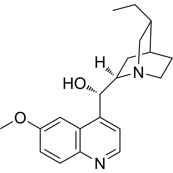
HydroquinidineAntiarrhythmic Agent -
HY-W040233
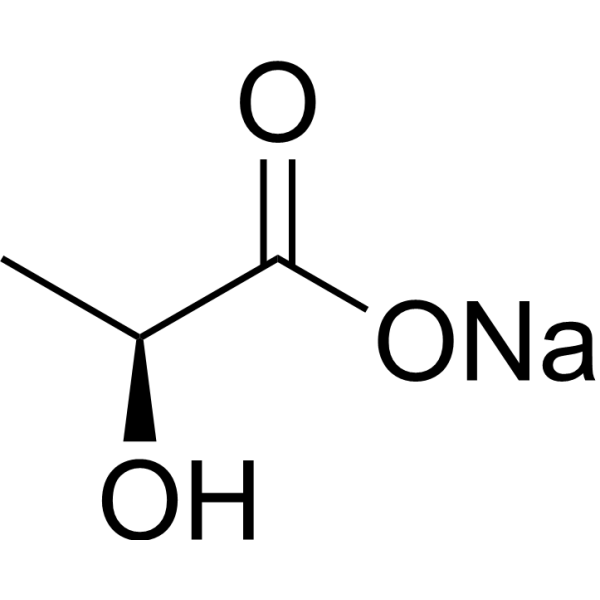
L-Lactic acid sodiumBuildiing Block -

HY-W007880
(R)-D2PMPyrrolidine Analog -
HY-I0718
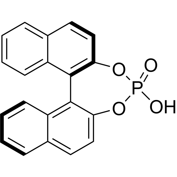
(R)-1,1'-Binaphthyl-2,2'-diyl hydrogenphosphateBiochemical Reagent -

HY-W040233R
L-Lactic acid sodium (Standard)PPLA Precursor -
HY-W035709
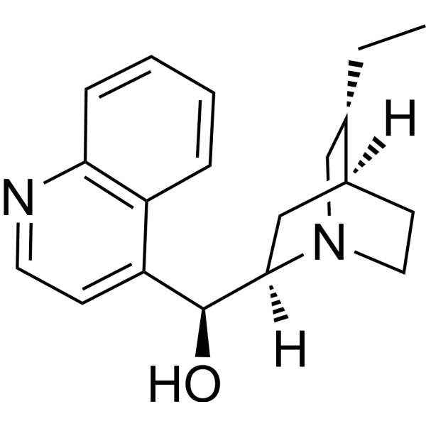
HydrocinchonineMDR Reversal Agent -
HY-N1467
(-)-α-TerpineolMnoterpene Compound -

HY-B0997R
Hydroquinidine (Standard)Antiarrhythmic Agent -
![(R)-[1,1'-Binaphthalene]-2,2'-diamine](https://file.medchemexpress.com/product_pic/hy-w007978.gif)
HY-W007978
(R)-[1,1'-Binaphthalene]-2,2'-diamineIsomer -
HY-W017757
(R)-BINAPBiochemical Reagent -

HY-I0606B
(R)-(+)-2,2-Dimethyl-1,3-dioxolane-4-carboxaldehydeStarting Material


Biological Activity
Description
|
Syk |
STAT3 |
In Vitro
(DHQD) 2PHAL (C-61) induces apoptosis in SYK+ primary leukemia cells[1].
(DHQD) 2PHAL (SYKINH-61) (1-100 nM; 30 min RAMOS; 1-10 min BCL-1) prevents oxidative stress-induced SYK tyrosine phosphorylation in RAMOS and BCL-1 B-lineage lymphocytes[2].
(DHQD) 2PHAL (100 nM; 1-10 min) inhibits H2O2-induced STAT3 Y705 phosphorylation in RAMOS Burkitt leukemia/lymphoma cells[2].
(DHQD) 2PHAL eliminates PV-induced STAT3 DNA-binding activity in human B-cell lines[2].
(DHQD) 2PHAL inhibits constitutively active native SYK in NALM-6 B-lineage ALL cells without altering SYK protein levels[2].
MedChemExpress (MCE) has not independently confirmed the accuracy of these methods. They are for reference only. Further protocols information, click here.
-
Cell Line:RAMOS Burkitt’s leukemia/lymphoma cells and BCL-1 cells
-
Concentration:1, 10, 50, 100 nM
-
Incubation Time:30 min (RAMOS); 1, 5, 10 min (BCL-1)
-
Result:Prevented PV-induced and H2O2-induced Y-phosphorylation of SYK in RAMOS and BCL-1 cells, respectively.
-
Cell Line:RAMOS Burkitt’s leukemia/lymphoma cells
-
Concentration:100 nM
-
Incubation Time:1 min; 5 min; 10 min
-
Result:Inhibited H2O2-induced Y-phosphorylation of STAT3 in RAMOS cells.
In Vivo
(DHQD) 2PHAL (10 mg/kg; intravenous bolus; two consecutive days; administration starting 1 day after inoculation) prevents overt leukemia and prolonged survival in SCID mice bearing RS4;11 or LC1;19 human BPL xenografts[1].
(DHQD) 2PHAL (10 mg/kg; intravenous bolus; two consecutive days; dosing initiated 1 day after leukemia cell inoculation) produces durable event-free survival in SCID mouse xenograft models of multiple chemotherapy-resistant human BPL[1].
MedChemExpress (MCE) has not independently confirmed the accuracy of these methods. They are for reference only.
-
Animal Model:CB.17 SCID (female, 6-8 weeks of age)[1]
-
Dosage:10 mg/kg per dose
-
Administration:i.v. bolus injection (0.2 mL/mouse); two consecutive days; starting 1 day after NALM-6 inoculation
-
Result:In mice inoculated with 1×10⁶ NALM-6 cells, C-61 produced a median EFS >120 d; cumulative proportion surviving event-free was 100% at 30 d, 100% at 60 d, 80% at 90 d, and 80% at 120 d.
Only 2 of 10 mice given C-61 developed leukemia with delayed-onset paraplegia at 75 and 80 d, whereas the remaining 8 became long-term survivors with no overt leukemia (paraplegia or hepatosplenomegaly) at elective killing at 121 d; median survival >121 d.
-
Animal Model:CB.17 SCID (female, 6-8 weeks of age)[1]
-
Dosage:10 mg/kg per dose
-
Administration:i.v. bolus injection (0.2 mL/mouse); two consecutive days; starting 1 day after inoculation
-
Result:In the combined RS4;11 and LC1;19 groups given C-61, all 20 mice remained healthy and had no signs of overt leukemia (paraplegia or hepatosplenomegaly) when electively killed at 121 d; median survival >121 d.
-
Animal Model:CB.17 SCID (female, 6-8 weeks of age)[1]
-
Dosage:10 mg/kg per dose
-
Administration:i.v. bolus injection (0.2 mL/mouse); two consecutive days; starting 1 day after leukemia cell inoculation
-
Result:In the composite C-61 group, median survival was >121 d and the 4-month EFS rate was 93%; cumulative proportion surviving event-free was 100% at 30 d, 100% at 60 d, 93% at 90 d, and 93% at 120 d.
Destroyed >99.9% of leukemia-initiating BPL cells in vivo across the three xenograft models.


Chemical Information
-
CAS No. 140853-10-7
-
Appearance Solid
-
Molecular Weight 778.98
-
Formula C48H54N6O4
-
Color White to off-white
-
SMILES
CC[C@H]1C[N@]2[C@@](C[C@]1([H])CC2)([H])[C@H](C3=CC=NC(C=C4)=C3C=C4OC)OC(C5=C6C=CC=C5)=NN=C6O[C@@H](C7=CC=NC(C=C8)=C7C=C8OC)[C@](C[C@]9([H])CC%10)([H])N%10C[C@@H]9CC
-
Synonyms
C-61; SYKINH-61
-
Shipping
Room temperature in continental US; may vary elsewhere.
-
Storage
4°C, protect from light, stored under nitrogen
* In solvent : -80°C, 6 months; -20°C, 1 month (protect from light, stored under nitrogen)


Solvent & Solubility
In Vitro:
DMSO : 5 mg/mL (6.42 mM; ultrasonic and warming and heat to 60°C; Hygroscopic DMSO has a significant impact on the solubility of product, please use newly opened DMSO)
Please refer to the solubility information to select the appropriate solvent. Once prepared, please aliquot and store the solution to prevent product inactivation from repeated freeze-thaw cycles.
Storage method and period of stock solution: -80°C, 6 months; -20°C, 1 month (protect from light, stored under nitrogen). When stored at -80°C, please use it within 6 months. When stored at -20°C, please use it within 1 month.
Please refer to the solubility information to select the appropriate solvent. Once prepared, please aliquot and store the solution to prevent product inactivation from repeated freeze-thaw cycles.
Storage method and period of stock solution: -80°C, 6 months; -20°C, 1 month (protect from light, stored under nitrogen). When stored at -80°C, please use it within 6 months. When stored at -20°C, please use it within 1 month.
Concentration (start) × Volume (start) = Concentration (final) × Volume (final)

Protocols
-
Kinase activity and phosphorylation assays
Kinase activity assays measure the ability of kinases to transfer phosphate groups from ATP to specific substrates, while phosphorylation assays detect the presence and levels of phosphorylated proteins. Common methods include radiolabeled ATP incorporation (e. g. ,), ADP release detection via bioluminescence (e. g. ,[3]), enzyme-linked immunosorbent assays (ELISA) for phospho-specific epitopes (e. g. ,[6]), and microtiter-based formats for high-throughput screening (e. g. ,[8]). The ADP-Glo assay quantifies kinase activity by measuring ADP produced during phosphorylation using a luciferase-based system. Radiometric assays involve autoradiography or scintillation counting after incorporation of 32P-labeled ATP into substrate proteins. ELISA-based approaches rely on phospho-specific antibodies to detect activated kinases in cell lysates or purified samples.
-
Western Blot
Western blotting (WB) is a commonly used experimental method in molecular biology, biochemistry, and immunogenetics for identifying and quantifying target proteins. It combines gel electrophoresis with immunoassay, enabling researchers to analyze protein expression, post-translational modifications, and molecular weight.
-
Apoptosis
Apoptosis, also called programmed cell death, is generally characterized by distinct morphological characteristics.
-
TUNEL staining for apoptotic DNA fragmentation
TUNEL staining detects DNA strand breaks by using terminal deoxynucleotidyl transferase to add labeled nucleotides to exposed 3′-OH DNA termini, generating either microscopic staining in fixed cells or tissue sections, or fluorescence/cytometric signal in cell suspensions. TUNEL positivity reflects DNA fragmentation but should not be interpreted alone as definitive apoptosis, because TUNEL can also label necrotic, autolytic, mechanically damaged, or DNA-repair-associated DNA breaks.
-
Annexin V plus membrane-impermeant dye apoptosis staining
Annexin V-based apoptosis assays rely on the detection of phosphatidylserine (PS) externalization from the inner leaflet of the plasma membrane to the outer leaflet, an early biochemical hallmark of apoptosis. Fluorescently labeled Annexin V binds PS in a calcium-dependent manner, enabling identification of early apoptotic cells by flow cytometry or fluorescence microscopy. When combined with a membrane-impermeant DNA-binding dye (e. g. , propidium iodide), this approach allows discrimination between viable (Annexin V−/dye−), early apoptotic (Annexin V+/dye−), and late apoptotic or necrotic (Annexin V+/dye+) cell populations by assessing membrane integrity and PS exposure.
-
ROS/oxidative-stress fluorescent staining
ROS/oxidative-stress fluorescent staining uses cell-permeant fluorogenic probes that become fluorescent after oxidation inside cells or tissues; commonly used examples include DCFH-DA/DCFDA for broad cellular oxidant detection, DHE for superoxide-related signal detection, MitoSOX for mitochondrial superoxide-related signal detection, and CellROX probes for oxidative-stress-associated fluorescence readouts. The assay detects probe oxidation rather than a single ROS species unless the probe and analysis method have been chemically validated for that species. DCFH-DA enters cells, is deacetylated by intracellular esterases to DCFH, and produces fluorescent DCF after oxidation, so the readout is used as an operational measure of total cellular oxidative stress rather than a species-specific ROS measurement. DHE and MitoSOX can report superoxide-related oxidation, but red fluorescence alone can include non-specific ethidium-like oxidation products; HPLC or optimized spectral approaches are
-
Apoptosis Solutions
Apoptosis is a regulated, generally non-lytic cell-death pathway that removes unwanted, damaged, infected, or abnormal cells through coordinated morphological changes, caspase activation, DNA fragmentation, and membrane remodeling. The intrinsic apoptosis pathway is controlled mainly by mitochondrial outer membrane permeabilization, BCL-2 family proteins, cytochrome c release, apoptosome formation, caspase-9 activation, and downstream executioner caspase-3/7 activation. The extrinsic apoptosis pathway is initiated by death receptors such as Fas, TNFR, and TRAIL receptors, which recruit adaptor proteins and activate caspase-8 before engaging executioner caspases or mitochondrial amplification through BID cleavage. Apoptosis is linked to many phenotypes, including cancer cell killing, tissue homeostasis, immune regulation, neurodegeneration, infection response, and treatment-induced cytotoxicity; unresolved questions include how apoptosis interacts with necroptosis, pyroptosis, ferroptos
-
Protocol for Kinase activity and phosphorylation assays
Kinase activity assays measure transfer of phosphate from ATP to a protein or peptide substrate, generating phosphorylated substrate, ADP, or incorporated radiolabeled phosphate as the readout; phosphorylation assays measure site-specific phosphorylation in cells or tissues as a proxy for kinase-pathway activation, inhibition, or substrate regulation. Phosphorylation can be detected by phospho-specific Western blot, immunoprecipitation kinase assay, phospho-immunofluorescence, phospho-flow cytometry, luminescent ADP detection, radiolabeled ATP incorporation, or reporter-based pathway assays, and these readouts can be applied to cancer cells, primary neurons, mouse tumors, organoids, inflammatory macrophages, ferroptosis studies, and mitophagy studies when the kinase target is biologically relevant.


Purity & Documentation
-
Data Sheet (303 KB)
-
SDS (396 KB)
- English - EN (396 KB)
- Français - FR (396 KB)
- Deutsch - DE (396 KB)
- Norwegian - NO (396 KB)
- Español - ES (396 KB)
- Swedish - SV (396 KB)
- Italian - IT (396 KB)
- Korean - KR (396 KB)
- Portuguese - PT (396 KB)
-
Handling Instructions (2659 KB)
References


Complete Stock Solution Preparation Table
Please refer to the solubility information to select the appropriate solvent. Once prepared, please aliquot and store the solution to prevent product inactivation from repeated freeze-thaw cycles.
Storage method and period of stock solution: -80°C, 6 months; -20°C, 1 month (protect from light, stored under nitrogen). When stored at -80°C, please use it within 6 months. When stored at -20°C, please use it within 1 month.
| Optional Solvent | Concentration Solvent Mass | 1 mg | 5 mg | 10 mg | 25 mg |
|---|---|---|---|---|---|
| DMSO | 1 mM | 1.2837 mL | 6.4187 mL | 12.8373 mL | 32.0933 mL |
| 5 mM | 0.2567 mL | 1.2837 mL | 2.5675 mL | 6.4187 mL |


(DHQD)2PHAL Related Classifications
HY-W008033 Related Classifications
 Powered by Bioz