



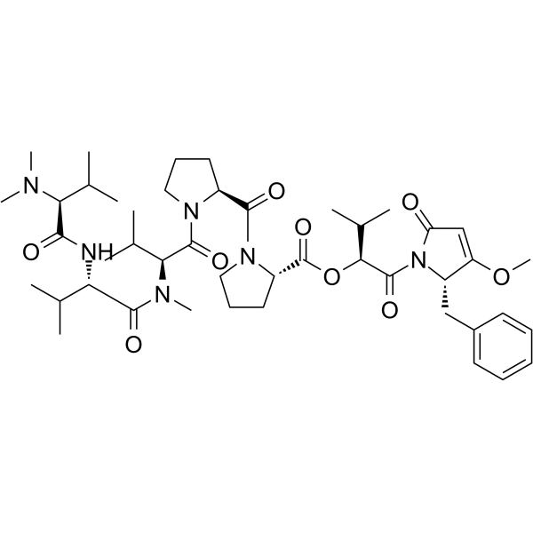
Dolastatin 15
Dolastatin 15 (DLS 15), a depsipeptide derived from Dolabella auricularia, is a potent antimitotic agent structurally related to the antitubulin agent Dolastatin 10. Dolastatin 15 induces cell cycle arrest and apoptosis in multiple myeloma cells. Dolastatin 15 can be used as an ADC cytotoxin.
商品は「研究用試薬」です。人や動物の医療用・臨床診断用・食品用の製品ではありません。
研究用途以外に使用した場合、当社は一切の責任を負いかねます。
- CAS 番号: 123884-00-4
- 分子式: C45H68N6O9
- 分子量:837.06
-
保管条件:
Please store the product under the recommended conditions in the Certificate of Analysis.

To place orders, for customer services and technical support, please contact: MedChemExpress USA
Tel: 609-228-6898 E-mail: [email protected] [email protected]


-
 生物活性
生物活性
-
化学情報
- プロトコル
- 純度とドキュメンテーション
- 参考文献
-
 Help & FAQs


ADC Payloads アイソフォーム固有の製品をすべて表示
More

生物活性
製品説明
IC50 & Target
|
Auristatin |
Cellular Effect
|
Cell Line

|
Type | Value | Description | References |
|---|---|---|---|---|
| CTLL | IC50 |
3 nM
Compound: 1 (4S,7S)(Dolastatin 15)
|
In vitro interleukin-2-dependent proliferation of the murine cytotoxlc T cell line (CTLL) was determined
In vitro interleukin-2-dependent proliferation of the murine cytotoxlc T cell line (CTLL) was determined
|
10.1016/S0960-894X(01)80540-0 |
| HT-29 | IC50 |
0.29 nM
Compound: dolastatin 15
|
Inhibitory activity against growth of HT-29 cell line
Inhibitory activity against growth of HT-29 cell line
|
[PMID: 9554885] |
| Jurkat | IC50 |
5 nM
Compound: 1 (4S,7S)(Dolastatin 15)
|
In vitro growth factor-independent proliferation against human leukemia T cell line jurkat was determined
In vitro growth factor-independent proliferation against human leukemia T cell line jurkat was determined
|
10.1016/S0960-894X(01)80540-0 |
| L1210 | IC50 |
0.13 nM
Compound: dolastatin 15
|
Inhibitory activity against growth of L1210 cell line
Inhibitory activity against growth of L1210 cell line
|
[PMID: 9554885] |
| L1210 | IC50 |
0.15 nM
Compound: 1 (4S,7S)(Dolastatin 15)
|
In vitro growth factor-independent proliferation against murine leukemia L1210 was determined
In vitro growth factor-independent proliferation against murine leukemia L1210 was determined
|
10.1016/S0960-894X(01)80540-0 |
| Lymphoma cell line | IC50 |
0.012 nM
Compound: 1 (4S,7S)(Dolastatin 15)
|
Antiproliferative effect against human lymphoma cells was determined; (0.012 to 0.12 nM)
Antiproliferative effect against human lymphoma cells was determined; (0.012 to 0.12 nM)
|
10.1016/S0960-894X(01)80540-0 |
| MCF7 | IC50 |
0.04 nM
Compound: dolastatin 15
|
Inhibitory activity against growth of MCF-7 cell line
Inhibitory activity against growth of MCF-7 cell line
|
[PMID: 9554885] |
| Osteoclast-like | IC50 |
17 nM
Compound: 1 (4S,7S)(Dolastatin 15)
|
In vitro growth factor-dependent proliferation against murine bone marrow cell was determined
In vitro growth factor-dependent proliferation against murine bone marrow cell was determined
|
10.1016/S0960-894X(01)80540-0 |
| P388 | ED50 |
2.4 ng/mL
Compound: Dolastatin15
|
Anticancer activity against lymphocytic leukemia P388 cells was determined
Anticancer activity against lymphocytic leukemia P388 cells was determined
|
[PMID: 9871690] |
| Panel leukemia (Carcinoma cell lines) | IC50 |
0.1 nM
Compound: 1 (4S,7S)(Dolastatin 15)
|
Antiproliferative effect against continuous human leukemia cell lines and peripheral blood cells from patients with acute myeloid leukemia was determined; (0.1 to 1 nM)
Antiproliferative effect against continuous human leukemia cell lines and peripheral blood cells from patients with acute myeloid leukemia was determined; (0.1 to 1 nM)
|
10.1016/S0960-894X(01)80540-0 |
体外実験
Dolastatin 15 (DLS 15) induces cell cycle arrest at the G2/M phase followed by apoptosis in various human myeloma cell lines (RPMI8226, U266, and IM9). Dolastatin 15 induces apoptosis of myeloma cells via activation of both mitochondrial- and Fas (CD95)/Fas-L (CD95-L)-mediated pathways[2].
Dolastatin 15 (DLS 15) displays growth inhibitory activity against all four SCLC cell lines (NCI-H69, NCI-H82, NCI-H345, NCI-H446) with IC50 values ranging from 0.039-28.8 nM, which were 2.7-9.2-fold higher than the values for dolastatin 10. All four SCLC cell lines underwent G2/M arrest within 24 hours of exposure to dolastatin 15[4].
MedChemExpress (MCE) has not independently confirmed the accuracy of these methods. They are for reference only.
体内実験
MedChemExpress (MCE) has not independently confirmed the accuracy of these methods. They are for reference only.


化学情報
-
CAS 番号 123884-00-4
-
分子量 837.06
-
分子式 C45H68N6O9
-
別名
DLS 15
-
輸送条件
Room temperature in continental US; may vary elsewhere.
-
保管条件
Please store the product under the recommended conditions in the Certificate of Analysis.


純度とドキュメンテーション
参考文献
[1]. Bai R, et al. Dolastatin 15, a potent antimitotic depsipeptide derived from Dolabella auricularia. Interaction with tubulin and effects of cellular microtubules. Biochem Pharmacol. 1992 Jun 23;43(12):2637-45. [Content Brief]
[2]. Sato M, et al. A natural peptide, dolastatin 15, induces G2/M cell cycle arrest and apoptosis of human multiple myeloma cells. Int J Oncol. 2007 Jun;30(6):1453-9. [Content Brief]
[3]. Gianolio DA, et al. Targeting HER2-positive cancer with dolastatin 15 derivatives conjugated to trastuzumab, novel antibody-drug conjugates. Cancer Chemother Pharmacol. 2012 Sep;70(3):439-49. [Content Brief]
[4]. Ali MA, et al. Dolastatin 15 induces apoptosis and BCL-2 phosphorylation in small cell lung cancer cell lines. Anticancer Res. 1998 Mar-Apr;18(2A):1021-6. [Content Brief]

Calculators
濃度 (開始) × 体積 (開始) = 濃度 (終了) × 体積 (終了)


Dolastatin 15 関連分類
HY-P1126 関連分類
 Powered by Bioz