



eCF506-d5
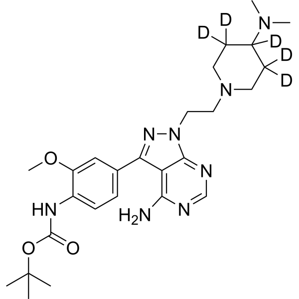
eCF506-d5 (NXP900-d5) is deuterated labeled eCF506 (HY-112096). eCF506 is a highly potent and orally active YES1/SRC kinase inhibitor with an IC50 of 0.47 nM. eCF506 locks its target into its native “closed” conformation, thereby inhibiting both kinase activity and complex formation with protein partners. eCF506 can be used for the study of esophageal squamous cancer and breast cancer.
Nos produits utilisent uniquement pour la recherche. Nous ne vendons pas aux patients.
- Formule: C26H33D5N8O3
- Masse moléculaire:515.66
-
Stockage:
Please store the product under the recommended conditions in the Certificate of Analysis.

To place orders, for customer services and technical support, please contact: MedChemExpress USA
Tel: 609-228-6898 E-mail: [email protected] [email protected]


-
 Activité biologique
-
Chemical Information
- Protocole
- Pureté et documentation
- Références
-
 Help & FAQs




Activité biologique
Description
In Vitro
Stable heavy isotopes of hydrogen, carbon, and other elements have been incorporated into drug molecules, largely as tracers for quantitation during the drug development process. Deuteration has gained attention because of its potential to affect the pharmacokinetic and metabolic profiles of drugs[1].
eCF506 induces a very potent antiproliferative effect in both MCF7 and MDA-MB-231 cells. eCF506 inhibits phosphorylation of SRC and FAK at low nanomolar levels, with complete inhibition observed at 100 nM. eCF506 significantly reduces cell motility at 10 nM as early as 6 h into the study, with equivalent efficacy to dasatinib. eCF506 exclusively inhibits SFK, with subnanomolar IC50 values against SRC and YES (IC50=0.5, 2.1 nM). It is important to highlight that eCF506 displays a vast difference in activity (>950-fold difference) between ABL and its primary target SRC[2].
MedChemExpress (MCE) has not independently confirmed the accuracy of these methods. They are for reference only.
In Vivo
MedChemExpress (MCE) has not independently confirmed the accuracy of these methods. They are for reference only.
Application
1. This compound can be used as a tracer
2. This compound can be used as an internal standard for quantitative analysis by NMR, GC-MS, or LC-MS.


Chemical Information
-
Unlabeled CAS 1914078-41-3
-
Masse moléculaire 515.66
-
Formule C26H33D5N8O3
-
SMILES
[2H]C1(CN(CC([2H])(C1(N(C)C)[2H])[2H])CCN2N=C(C(C2=NC=N3)=C3N)C4=CC=C(C(OC)=C4)NC(OC(C)(C)C)=O)[2H]
-
Synonyms
NXP900-d5
-
Livraison
Room temperature in continental US; may vary elsewhere.
-
Stockage
Please store the product under the recommended conditions in the Certificate of Analysis.


Pureté et documentation
Références
[1]. Russak EM, et al. Impact of Deuterium Substitution on the Pharmacokinetics of Pharmaceuticals. Ann Pharmacother. 2019 Feb;53(2):211-216. [Content Brief]
[2]. Fraser C, et al. Rapid Discovery and Structure-Activity Relationships of Pyrazolopyrimidines That Potently Suppress Breast Cancer Cell Growth via SRC Kinase Inhibition with Exceptional Selectivity over ABL Kinase. J Med Chem. 2016 May 26;59(10):4697-710. [Content Brief]

Calculators
Concentration (start) × Volume (start) = Concentration (final) × Volume (final)


eCF506-d5 Related Classifications
HY-112096S Related Classifications
 Powered by Bioz