



Fibronectin
Based on 11 publication(s) in Google Scholar
Fibronectin, a glycoprotein present in blood as well as in cells, is a biomarker of tissue injury. Fibronectin binds to membrane-spanning receptor proteins called integrins. Fibronectin also binds to other extracellular matrix proteins such as collagen, fibrin, and heparan sulfate proteoglycans.
This product is a human Fibronectin obtained by recombinant expression in a rice expression system.
For research use only. We do not sell to patients.
- Purity : 98.7%
- CAS No.: 86088-83-7
-
Storage:
Please store the product under the recommended conditions in the Certificate of Analysis.


To place orders, for customer services and technical support, please contact: MedChemExpress USA
Tel: 609-228-6898 E-mail: [email protected] [email protected]


-
 Biological Activity
-
Chemical Information
-
Solvent & Solubility
- Protocols
- Purity & Documentation
- References
-
 Help & FAQs


-

HY-P3160C
Fibronectin, bovine plasmaFibronectin Dderived From Bovine Plasma -

HY-D0842
BSA ProteinGlobular Protein -
HY-19424
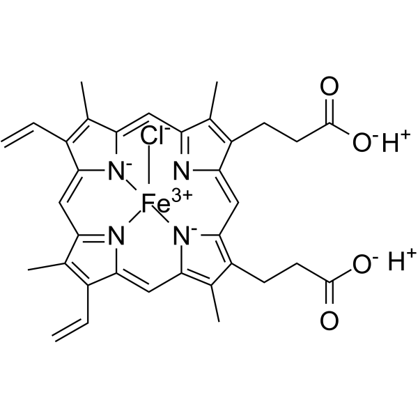
HeminHeme Oxygenase-1 Inducer -

HY-P2738
Collagen, Type I, from bovine achilles tendonCollagen Peptide -

HY-NP010
Type I Collagen, from rat tailCollagen -

HY-15371
ForskolinAdenylate Cyclase Activator -
HY-N0583
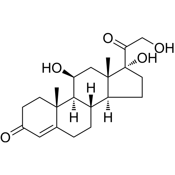
HydrocortisoneGlucocorticoid Receptor Agonist -

HY-10583
Y-27632 dihydrochlorideROCK Inhibitor -
HY-90006
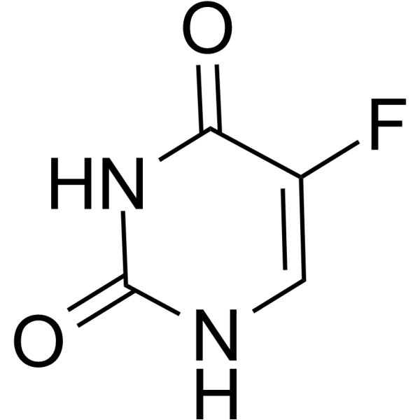
5-FluorouracilThymidylate Synthase Inhibitor -
HY-B0141
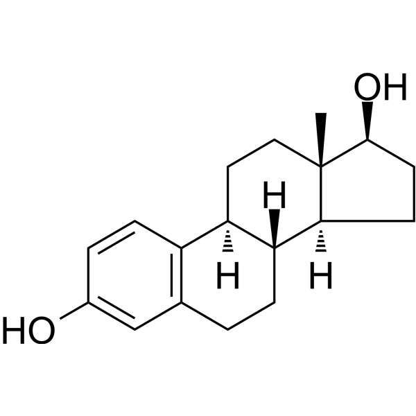
EstradiolHuman Endogenous Metabolite -

HY-15141
StaurosporinePKC/PKA Inhibitor
Publications Citing Use of MedChemExpress (MCE) Fibronectin
More- Nat Commun. 2025 Dec 15;16(1):11117. [Abstract]
- Nat Commun. 2024 Mar 22;15(1):2578. [Abstract]
- Acta Pharm Sin B. 2025 Jan;15(1):314-330. [Abstract]
- MedComm (2020). 2025 Jul 7;6(7):e70207. [Abstract]
- Cell Death Dis. 2026 Jul 23.
- Commun Biol. 2026 Jun 29.
- Oncol Res. 2024 Mar 20;32(4):753-768. [Abstract]
- Acta Histochem. 2025 Jun 30;127(3):152276. [Abstract]
- bioRxiv. 2026 Apr 1.
- Hlife. 2026 Mar 11.
- bioRxiv. 2025 February 18.


Biological Activity
Description
This product is a human Fibronectin obtained by recombinant expression in a rice expression system.
In Vitro
Fibronectin (FN) is a glycoprotein present in blood as well as in cells. While Fibronectin (FN) in blood has many functions, its primary role is to promote wound repair following injury[1].
Fibronectin also binds to other extracellular matrix proteins such as collagen, fibrin, and heparan sulfate proteoglycans (e.g. syndecans) [2].
MedChemExpress (MCE) has not independently confirmed the accuracy of these methods. They are for reference only. Further protocols information, click here.
Clinical Trial
| NCT Number | Sponsor | Condition | Start Date |
Phase

|
|---|---|---|---|---|
| NCT01329991 | Plexxikon| | 2011-05 | PHASE1 |


Chemical Information
-
CAS No. 86088-83-7
-
Appearance Solid
-
Color White to off-white
-
SMILES
[Fibronectin]
-
Shipping
Room temperature in continental US; may vary elsewhere.
-
Storage
Please store the product under the recommended conditions in the Certificate of Analysis.


Publications (11)
-
Journal Impact Factor
-
Most Recent
-
Nat Commun
Slc22a17 governs postnatal neurogenesis by maintaining the iron homeostasis in hippocampus. [Abstract]2025 Dec 15;16(1):11117. PMID: 41397957 -
Nat Commun
Photoswitchable polyynes for multiplexed stimulated Raman scattering microscopy with reversible light control. [Abstract]2024 Mar 22;15(1):2578. PMID: 38519503 -
Acta Pharm Sin B
Cerebral endothelial 3-mercaptopyruvate sulfurtransferase improves ischemia-induced cognitive impairment via interacting with protein phosphatase 2A. [Abstract]2025 Jan;15(1):314-330. PMID: 40041885 -
MedComm (2020)
A Novel Dual Bruton's Tyrosine Kinase/Janus Kinase 3 Inhibitor Wj1113 and its Therapeutic Effects on Rheumatoid Arthritis. [Abstract]2025 Jul 7;6(7):e70207. PMID: 40626319 -
-
-
Oncol Res
Development of a cell adhesion-based prognostic model for multiple myeloma: Insights into chemotherapy response and potential reversal of adhesion effects. [Abstract]2024 Mar 20;32(4):753-768. PMID: 38560563 -
Acta Histochem
Dihydroartemisinin inhibited tongue squamous cell carcinoma progression and tongue-to-lymph node metastasis through inhibiting RalB expression. [Abstract]2025 Jun 30;127(3):152276. PMID: 40592008 -
-
-


Solvent & Solubility
In Vitro:
H2O : 8.33 mg/mL (ultrasonic and adjust pH to 7 with NaOH)

Protocols
-
Fibrosis/Collagen Morphometry
Fibrosis and collagen morphometry is based on the quantitative visualization of fibrillar collagen deposition in tissue sections using histochemical stains such as Sirius Red (Picrosirius Red) or Masson's trichrome, followed by image-based or polarization-enhanced analysis to estimate collagen proportional area as a surrogate of extracellular matrix accumulation during fibrotic remodeling. Sirius Red combined with polarized light microscopy enhances detection of collagen fibers due to birefringence properties, enabling more specific visualization of collagen type I and III fibrils compared to conventional bright-field histology, while whole-section or region-restricted digital morphometry reduces field-selection bias in fibrosis assessment. Alternative quantitative approaches include second harmonic generation (SHG) and two-photon excited fluorescence microscopy, which enable label-free detection of fibrillar collagen and have been validated against histological staining and biochemica
-
Collagen: Sirius Red Staining
Sirius Red or picrosirius red staining is a histochemical method for visualizing collagen-rich extracellular matrix in tissue sections, and collagen fibers are detected as red-stained structures under bright-field microscopy with enhanced birefringence under polarized light. Picrosirius red is useful for assessing total collagen organization, distribution, and fibrosis burden, but polarized color should not be interpreted as a definitive collagen type I versus type III readout because color is affected by fiber orientation, thickness, and packing.
-
RNA extraction experimental
By lysing cells, releasing RNA, and removing impurities such as proteins and DNA, high-purity RNA products are finally obtained. The commonly used traditional method is the guanidine isothiocyanate/phenol/chloroform method (Trizol), which is suitable for a variety of animal materials including animal tissues, microorganisms, cultured cells, etc., and most plant materials.
-
ECM-Embedded Organoid (Matrigel/Dome) Culture
ECM-embedded organoid dome culture embeds epithelial stem cells, crypts, organoid fragments, or tumor-derived epithelial cells in a basement-membrane-like hydrogel such as Matrigel, allowing 3D growth, self-organization, lumen formation, budding or cystic morphogenesis, and lineage maintenance under defined niche-factor-containing medium. The primary readouts are organoid establishment efficiency, growth, morphology, passaging capacity, lineage-marker expression, and, when fluorescently labeled lines are used, microscopy- or flow-cytometry-based quantification of population behavior in 3D culture.
-
Matrigel/ECM Transwell Invasion Assay
The Matrigel/ECM Transwell invasion assay measures the ability of cells to move toward a chemoattractant while crossing an extracellular-matrix barrier placed on a porous membrane; therefore, the readout reflects both chemotactic motility and matrix invasion rather than migration alone. Matrigel is a basement-membrane-rich matrix derived from Engelbreth-Holm-Swarm mouse sarcoma and has been used as a reconstituted basement membrane barrier in chemoinvasion assays. The assay readout is generated by quantifying cells that reach the underside of the insert membrane or lower compartment after incubation, commonly by staining and counting invaded cells or by fluorescence-based quantification.
-
Matrigel Transwell/Boyden Chamber Invasion Assay
Matrigel Transwell/Boyden chamber invasion assay measures the ability of cells to degrade or traverse an extracellular matrix-coated porous membrane and move from an upper chamber toward a chemoattractant in a lower chamber. Invasion is distinguished from migration by coating the membrane with Matrigel or basement membrane matrix; uncoated inserts measure migration, while coated inserts require cells to cross an ECM barrier before reaching the underside of the membrane.


Purity & Documentation
-
Data Sheet (279 KB)
-
SDS (251 KB)
- English - EN (251 KB)
- Français - FR (251 KB)
- Deutsch - DE (251 KB)
- Norwegian - NO (251 KB)
- Español - ES (251 KB)
- Swedish - SV (251 KB)
- Italian - IT (251 KB)
- Korean - KR (251 KB)
- Portuguese - PT (251 KB)
-
Handling Instructions (2659 KB)
References
[1]. Yuan-Wan Sun, et al. Effects of E-Cigarette Aerosols with Varying Levels of Nicotine on Biomarkers of Oxidative Stress and Inflammation in Mice. Chem Res Toxicol. 2021 Apr 19;34(4):1161-1168. [Content Brief]
[2]. Roumen Pankov, et al. Fibronectin at a glance. J Cell Sci. 2002 Oct 15;115(Pt 20):3861-3. [Content Brief]

Calculators
Concentration (start) × Volume (start) = Concentration (final) × Volume (final)


Fibronectin Related Classifications
HY-P3160 Related Classifications
 Powered by Bioz