



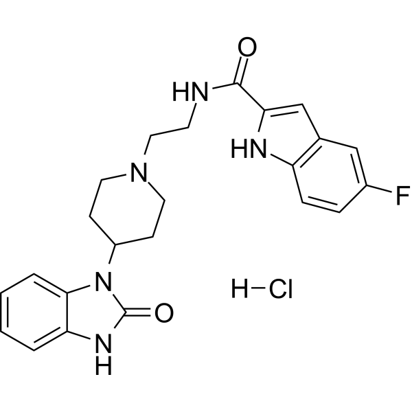
FIPI hydrochloride
Based on 5 publication(s) in Google Scholar
FIPI hydrochloride is a phospholipase D (PLD) inhibitor with an IC50 for PLD1 and PLD2 of about 25 nM. FIPI hydrochloride regulates cytoskeletal recombination, cell diffusion and chemotaxis. FIPI hydrochloride can be used in cancer research. In addition, FIPI hydrochloride can enhance the secretion and aggregation of platelet dense particles, inhibit thrombosis, reduce ischemic stroke infarct volume and improve nerve function.
For research use only. We do not sell to patients.
- CAS No.: 1781834-93-2
- Formula: C23H25ClFN5O2
- Molecular Weight:457.93
-
Storage:
Please store the product under the recommended conditions in the Certificate of Analysis.

To place orders, for customer services and technical support, please contact: MedChemExpress USA
Tel: 609-228-6898 E-mail: [email protected] [email protected]


-
 Biological Activity
-
Chemical Information
- Protocols
- Purity & Documentation
- References
-
 Help & FAQs


-
HY-100736
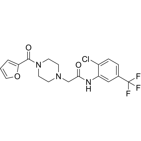
ML348APT1/LYPLA1 Inhibitor -
HY-70072
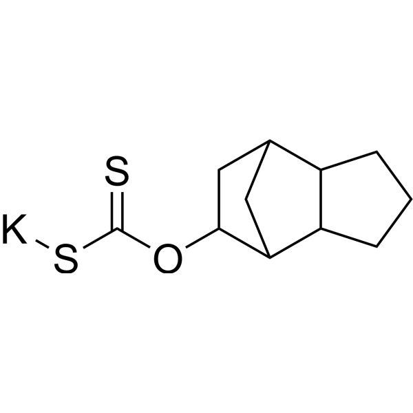
D609PC-PLC Inhibitor -
HY-116165
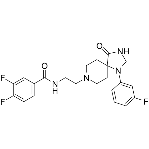
ML-298PLD2 Inhibitor -
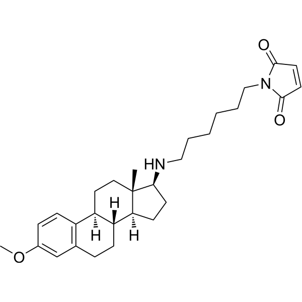
HY-13419
U-73122Phospholipase C Inhibitor -
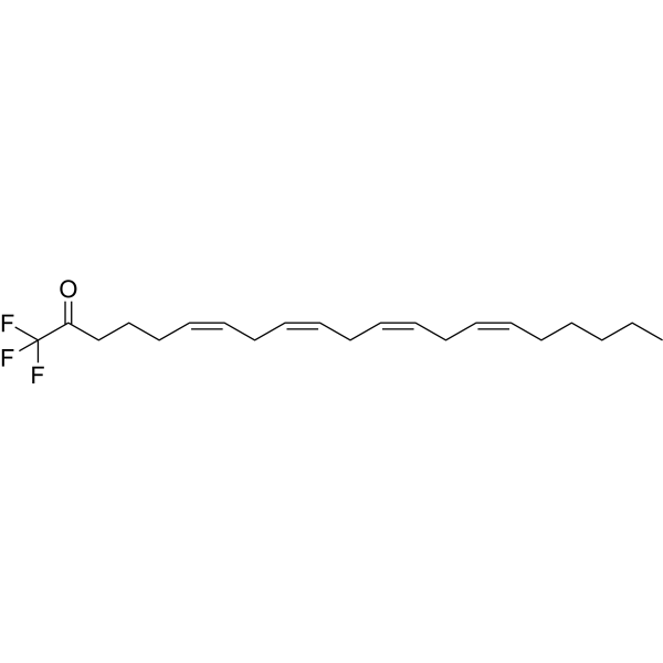
HY-108611
AACOCF3cPLA2 Inhibitor -
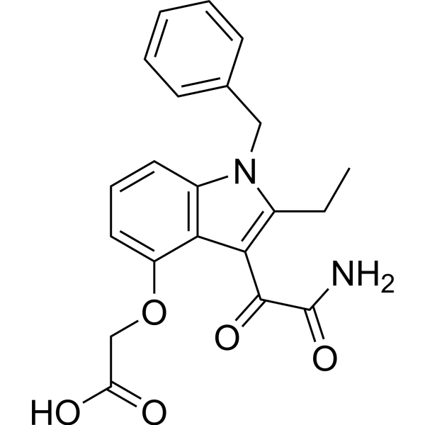
HY-13402
VarespladibsPLA2 Inhibitor -

HY-P0233
MelittinPLA2 Activator -
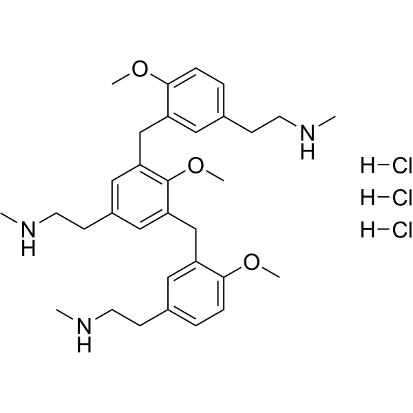
HY-130592
Compound 48/80 trihydrochlorideHistamine Releaser/Mast Cell Degranulator -
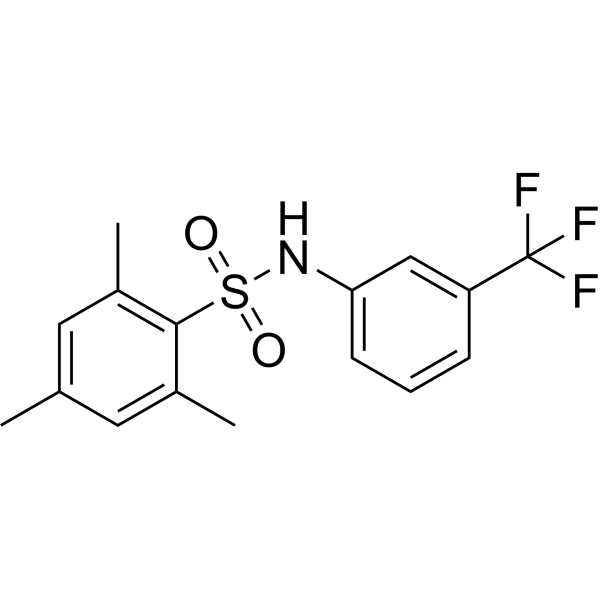
HY-19619
m-3M3FBSPhospholipase C Activator -
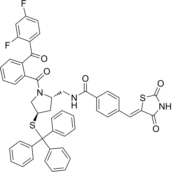
HY-111376
PyrrophenonePLA2 Inhibitor -
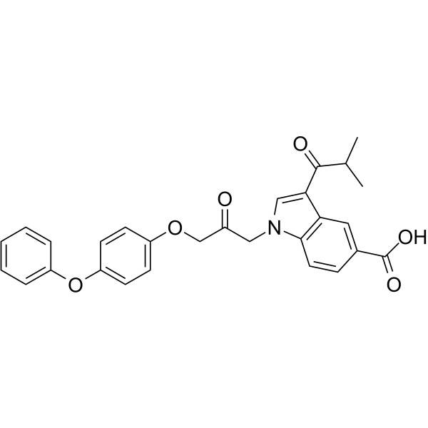
HY-10801
CAY10650cPLA2α Inhibitor -
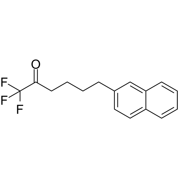
HY-115403
FKGK18GVIA iPLA2 Inhibitor
Publications Citing Use of MedChemExpress (MCE) FIPI hydrochloride
More Customer Validation & Images

-
Cell Proliferation/Viability Assay

-
Cell Migration/Invasion Assay

-
Cell Migration/Invasion Assay

-
Cell Migration/Invasion Assay

-
IF
All Phospholipase Isoforms
More

Biological Activity
Description
IC50 & Target
[1]|
PLD1 |
PLD2 |


Chemical Information
-
CAS No. 1781834-93-2
-
Molecular Weight 457.93
-
Formula C23H25ClFN5O2
-
SMILES
O=C(NCCN1CCC(CC1)N2C3=CC=CC=C3NC2=O)C4=CC5=C(C=CC(F)=C5)N4.[H]Cl
-
Synonyms
5-Fluoro-2-indolyl deschlorohalopemide hydrochloride
-
Shipping
Room temperature in continental US; may vary elsewhere.
-
Storage
Please store the product under the recommended conditions in the Certificate of Analysis.


Publications (5)
-
Journal Impact Factor
-
Most Recent
-
Anal Chem
Phospholipid-Tailored Titanium Carbide Nanosheets as a Novel Fluorescent Nanoprobe for Activity Assay and Imaging of Phospholipase D. [Abstract]2018 Jun 5;90(11):6742-6748. PMID: 29737155
FIPI hydrochloride purchased from MedChemExpress. Usage Cited in: Anal Chem. 2018 Jun 5;90(11):6742-6748. [Abstract]
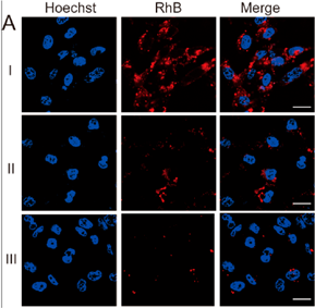
Typical CSLM images of MDA-MB-231 cells incubated with 50 μg/mL RhB-PL-Ti3C2 for 4 h at 37°C in the absence (I) and presence of 1.0 mg/mL FIPI (II), and CSLM images of MCF-7 cells (III) incubated with 50 μg/mL RhB-PL-Ti3C2 for 4 h at 37°C.
-
Biochem Pharmacol
Phospholipase D2 regulation by transcription factor FOXA1 promotes the drug resistance of vemurafenib in melanoma cells. [Abstract]2025 Dec;242(Pt 4):117434. PMID: 41110487 -
J Cell Sci
The protein kinase D1-mediated classical protein secretory pathway regulates the Ras oncogene-induced senescence response. [Abstract]2018 Mar 16;131(6):jcs207217. PMID: 29420297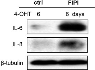
FIPI hydrochloride purchased from MedChemExpress. Usage Cited in: J Cell Sci. 2018 Mar 16;131(6):jcs207217. [Abstract]
ER:Ras IMR90 cells are treated with solvent or FIPI (250 nM, 2h).Western blotting are performed.
-
-
Nanoscale Res Lett
Preparation and Evaluation of Liposomes Co-Loaded with Doxorubicin, Phospholipase D Inhibitor 5-Fluoro-2-Indolyl Deschlorohalopemide (FIPI) and D-Alpha Tocopheryl Acid Succinate (α-TOS) for Anti-Metastasis. [Abstract]2019 Apr 18;14(1):138. PMID: 31001703
FIPI hydrochloride purchased from MedChemExpress. Usage Cited in: Nanoscale Res Lett. 2019 Apr 18;14(1):138. [Abstract]
Cell viability of MDA-MB-231 cells incubated with Blank-lip, FIPI-lip, DF-lip, DT-lip, and DFT-lip for 7 h or 24 h at a concentration of 2 μM FIPI, respectively.
FIPI hydrochloride purchased from MedChemExpress. Usage Cited in: Nanoscale Res Lett. 2019 Apr 18;14(1):138. [Abstract]
Inhibition effects on the cell motility in vitro. Images of MDA-MB-231 cells wound edge after incubation with control, Blank-lip, FIPI-lip, DF-lip, DT-lip, and DFT-lip for 48 h, respectively (magnification, × 100).
FIPI hydrochloride purchased from MedChemExpress. Usage Cited in: Nanoscale Res Lett. 2019 Apr 18;14(1):138. [Abstract]
Inhibition effects on cell migration. Images of MDA-MB-231 cell migration after incubation with control, Blank-lip, FIPI-lip, DF-lip, DT-lip, and DFT-lip for 7 h at a concentration of 2 μM FIPI.
FIPI hydrochloride purchased from MedChemExpress. Usage Cited in: Nanoscale Res Lett. 2019 Apr 18;14(1):138. [Abstract]
Inhibition effects on the cell invasion. Images of MDA-MB-231 cell invasion after incubation with control, Blank-lip, FIPI-lip, DF-lip, DT-lip, and DFT-lip for 24 h a concentration of 2 μM FIPI (magnification, × 100).

Protocols
-
Cell Cytotoxicity Assay
Cytotoxicity assays are usually based on the assessment of cell membrane damage, which can also be indirectly detected by measuring cell viability. Detection methods include MTT assay, CKK-8 assay, LDH assay and ATP assay, etc.
-
Cell migration
Cell migration is a method that plays an important role in wound healing, cell differentiation, embryonic development, etc.
-
Chemotaxis Gradient Chamber Assay 1
The chemotaxis gradient chamber assay is based on the principle of observing directional cell migration in response to a stable, linear or quasi-linear concentration gradient of a chemoattractant formed between two fluid reservoirs separated by a narrow observation chamber. Cells placed within the chamber respond to the gradient by polarized movement toward higher chemoattractant concentrations, allowing quantification of chemotactic behavior in real time under microscopy. The classic Zigmond chamber design enables simultaneous visualization of gradient formation and individual cell trajectories, making it suitable for studying leukocyte chemotaxis and other motile cell types in vitro.
-
Under-Agarose Cell Migration Assay
The under-agarose cell migration assay is a classical in vitro chemotaxis method designed to evaluate directed cell movement through a semi-solid agarose matrix toward soluble chemoattractant gradients, enabling visualization and quantification of leukocyte or motile cell migration in a confined 2D-like environment. In this system, cells and chemoattractants are placed in separate wells cut into an agarose gel, allowing diffusion-driven gradient formation that guides directional migration, which is typically assessed by measuring migration distance, cell morphology changes, and accumulation toward the chemoattractant source. This assay has been widely used to study neutrophil and leukocyte chemotaxis as a simple alternative to filter-based migration systems and allows direct microscopic observation of migrating cells under near-physiological confinement conditions.
-
Transwell/Boyden Chamber Migration Assay
The Transwell/Boyden chamber migration assay measures cell movement through a porous membrane separating an upper and lower chamber, usually after a chemoattractant gradient is established by placing cells in the upper chamber and chemoattractant-containing medium in the lower chamber. The readout is generated by quantifying cells that traverse the membrane and appear on the lower membrane surface or in the lower chamber, depending on whether the cell type is adherent or non-adherent. This assay reflects chemotactic or haptotactic migration rather than matrix invasion unless an extracellular-matrix barrier is added to the membrane.
-
Chemotaxis Gradient Chamber Assay 2
Chemotaxis gradient chamber assays measure directional cell migration in response to a soluble chemical gradient by imaging cells as they move across a defined observation region; the readout is generated from time-lapse cell trajectories, displacement toward the gradient, forward migration index, trajectory plots, rose/polar plots, and statistical tests of non-random directionality. The Dunn chamber is a direct-viewing glass chamber in which cells migrate across a bridge between control and chemoattractant wells, allowing observation of cells in a linear concentration gradient; related direct-viewing formats include the Insall chamber, which supports defined unidirectional gradients and high numerical-aperture microscopy, and the μ-Slide Chemotaxis chamber, which supports long-term live-cell imaging and gradient characterization with fluorescent dye.
-
Phalloidin F-actin cytoskeleton staining
Phalloidin F-actin staining detects polymerized filamentous actin in fixed and permeabilized specimens by using fluorescent phalloidin or phalloidin-derived phallotoxins that bind actin filaments and generate a fluorescence microscopy readout corresponding to F-actin organization, including stress fibers, cortical actin, filament bundles, and tissue-specific actin networks. Phalloidin stabilizes F-actin by reducing actin subunit dissociation from filament ends, and fluorescent phallotoxins were established as tools for visualizing actin-containing structures in eukaryotic cells.


Purity & Documentation
References
[1]. Su W, et al. 5-Fluoro-2-indolyl des-chlorohalopemide (FIPI), a phospholipase D pharmacological inhibitor that alters cell spreading and inhibits chemotaxis. Mol Pharmacol. 2009 Mar;75(3):437-46. [Content Brief]
[2]. Monovich L, et al. Optimization of halopemide for phospholipase D2 inhibition. Bioorg Med Chem Lett. 2007 Apr 15;17(8):2310-1. [Content Brief]
[3]. Claudia Dall'Armi, et al. The phospholipase D1 pathway modulates macroautophagy. Nat Commun. 2010:1:142. [Content Brief]

Calculators
Concentration (start) × Volume (start) = Concentration (final) × Volume (final)


FIPI hydrochloride Related Classifications
HY-12807A Related Classifications
 Powered by Bioz