



Flupentixol
Based on 2 publication(s) in Google Scholar
Flupentixol is an orally active D1/D2 dopamine receptor antagonist and new PI3K inhibitor (PI3Kα IC50=127 nM). Flupentixol shows anti-proliferative activity to cancer cells and induces apoptosis. Flupentixol can also be used in schizophrenia, anxiolytic and depressive research.
For research use only. We do not sell to patients.
- CAS No.: 2709-56-0
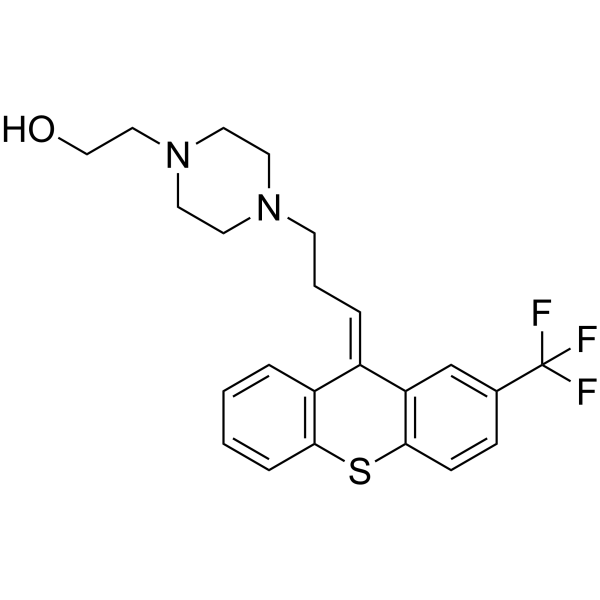
- Formula: C23H25F3N2OS
- Molecular Weight:434.52
-
Storage:
Please store the product under the recommended conditions in the Certificate of Analysis.

To place orders, for customer services and technical support, please contact: MedChemExpress USA
Tel: 609-228-6898 E-mail: [email protected] [email protected]


-
 Biological Activity
-
Chemical Information
- Protocols
- Purity & Documentation
- References
-
 Help & FAQs


Publications Citing Use of MedChemExpress (MCE) Flupentixol
MoreAll Dopamine Receptor Isoforms
More

Biological Activity
Description
IC50 & Target
[3]|
D1 Receptor |
D2 Receptor |
PI3Kα 127 nM (IC50) |
Cellular Effect
|
Cell Line

|
Type | Value | Description | References |
|---|---|---|---|---|
| Astrocyte | EC50 |
1.182 μM
Compound: 7
|
Antiproliferative activity against mouse astrocyte cells by MTT assay
Antiproliferative activity against mouse astrocyte cells by MTT assay
|
[PMID: 17417631] |
| HEK293 | IC50 |
89.5 μM
Compound: flupenthixol
|
Inhibition of 4-(4-(dimethylamino)styryl)-N-methylpyridinium uptake at human OCT1 expressed in HEK293 cells by confocal microscopy
Inhibition of 4-(4-(dimethylamino)styryl)-N-methylpyridinium uptake at human OCT1 expressed in HEK293 cells by confocal microscopy
|
[PMID: 18788725] |
| Medulloblastoma cell | EC50 |
0.187 μM
Compound: 7
|
Antiproliferative activity against mouse medulloblastoma cells harboring heterozygous ptch1 gene by MTT assay
Antiproliferative activity against mouse medulloblastoma cells harboring heterozygous ptch1 gene by MTT assay
|
[PMID: 17417631] |
In Vitro
Flupentixol (2.5-40 μM; 72 h) treatment inhibits the viability of lung cancer cells in a dose-dependent manner[3].
Flupentixol (2.5-40 μM; 24 h) induces apoptosis in lung cancer cells[3].
Flupentixol (2.5-15 μM; 24 h) inhibits p-AKT and Bcl-2 expression levels[3].
MedChemExpress (MCE) has not independently confirmed the accuracy of these methods. They are for reference only.
-
Cell Line:A549, H661, SK-SEM-1, and NCAL-H520 cells
-
Concentration:2.5, 5, 10, 20, or 40 μM
-
Incubation Time:72 hours
-
Result:Showed the IC50s of 5.708 μM and 6.374 μM for A549 and H661 cells, respectively.
-
Cell Line:A549 and H661 cells
-
Concentration:5, 10, 20 and 40 μM
-
Incubation Time:24 hours
-
Result:Increased the percentage of cells in early apoptosis compared with the negative control in both A549 and H661 (p< 0.05). Induced the cleavage of PARP and caspase-3 in a dose-dependent manner.
-
Cell Line:H661 and A549 cells
-
Concentration:2.5, 5, 10, and 15 μM
-
Incubation Time:24 hours
-
Result:Decreased AKT phosphorylation levels in a dose-dependent manner, decreased the expression levels of Bcl-2.
In Vivo
MedChemExpress (MCE) has not independently confirmed the accuracy of these methods. They are for reference only.
-
Animal Model:BALB/C nude mice injected with A549 cells[3]
-
Dosage:40 mg/kg
-
Administration:Intragastric injection; 40 mg/kg; once daily; 21 days
-
Result:Reduced tumor volumes compared to the vehicle control (p<0.05), reduced tumor weights by 64.1% (p<0.05).
Clinical Trial
| NCT Number | Sponsor | Condition | Start Date |
Phase
|
|---|---|---|---|---|
| NCT01329991 | 2011-05 | PHASE1 |


Chemical Information
-
CAS No. 2709-56-0
-
Molecular Weight 434.52
-
Formula C23H25F3N2OS
-
SMILES
OCCN1CCN(CC/C=C2C3=C(SC4=C/2C=CC=C4)C=CC(C(F)(F)F)=C3)CC1
-
Synonyms
Flupenthixol
-
Shipping
Room temperature in continental US; may vary elsewhere.
-
Storage
Please store the product under the recommended conditions in the Certificate of Analysis.


Publications (2)
-
Journal Impact Factor
-
Most Recent
-
Cell Rep Methods
RECOVER identifies synergistic drug combinations in vitro through sequential model optimization. [Abstract]2023 Oct 23;3(10):100599. PMID: 37797618 -
Insect Biochem Mol Biol
Molecular and pharmacological characterization of the dopamine receptors in the oriental fruit fly, Bactrocera dorsalis. [Abstract]2025 May:180:104312. PMID: 40245998


Purity & Documentation
References
[1]. Yonar D, et al. Effect of cis-(Z)-flupentixol on DPPC membranes in the presence and absence of cholesterol. Chem Phys Lipids. 2016 Jun;198:61-71. [Content Brief]
[2]. Ruhrmann S, et al. Efficacy of flupentixol and risperidone in chronic schizophrenia with predominantly negative symptoms. Prog Neuropsychopharmacol Biol Psychiatry. 2007 Jun 30;31(5):1012-22. [Content Brief]
[3]. Chao Dong, et al. The antipsychotic agent flupentixol is a new PI3K inhibitor and potential anticancer drug for lung cancer. Int J Biol Sci. 2019 Jun 2;15(7):1523-1532. [Content Brief]

Calculators
Concentration (start) × Volume (start) = Concentration (final) × Volume (final)


Flupentixol Related Classifications
HY-15856A Related Classifications
 Powered by Bioz