



Kartogenin
Based on 34 publication(s) in Google Scholar
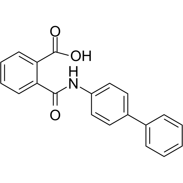
Kartogenin (KGN) is an inducer of chondrogenic tissue formation (EC50: 100 nM). Kartogenin induces chondrogenesis by binding to fibrin A, disrupting its interaction with the transcription factor core binding factor beta subunit (CBFβ), and by modulating the CBFβ-RUNX1 transcriptional program. Kartogenin also promotes tendon-bone junction (TBJ) wound healing by stimulating collagen synthesis. Kartogenin is widely used in cell-free therapy in the field of regeneration for cartilage regeneration and protection, tendon-bone healing, wound healing and limb development. Kartogenin promotes cartilage repair, coordinates limb development, and is also used in osteoarthritis (OA) research.
Nos produits utilisent uniquement pour la recherche. Nous ne vendons pas aux patients.
- Pureté : 99.05%
- CAS No.: 4727-31-5
- Formule: C20H15NO3
- Masse moléculaire:317.34
-
Stockage:Powder -20°C, 3 years , 4°C, 2 years ; In solvent -80°C, 6 months , -20°C, 1 month

To place orders, for customer services and technical support, please contact: MedChemExpress USA
Tel: 609-228-6898 E-mail: [email protected] [email protected]


-
 Activité biologique
-
Chemical Information
-
Solvant et solubilité
- Protocole
- Pureté et documentation
- Références
-
 Help & FAQs


-
HY-13013
SIS3Smad3 Inhibitor -
HY-100347
SRI-011381TGF-β/Smad Agonist -
HY-15108
PurmorphamineSmoothened Receptor Agonist -

HY-10219
RapamycinmTOR Inhibitor -

HY-14648
DexamethasoneGR Agonist -
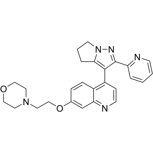
HY-12075
LY2109761TGF-βR I/II Inhibitor -

HY-Y1888
Corn oil -
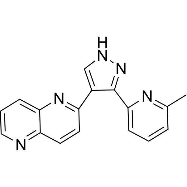
HY-13012
RepSoxTGFβR-1/ALK5 Inhibitor -

HY-10583
Y-27632 dihydrochlorideROCK Inhibitor -
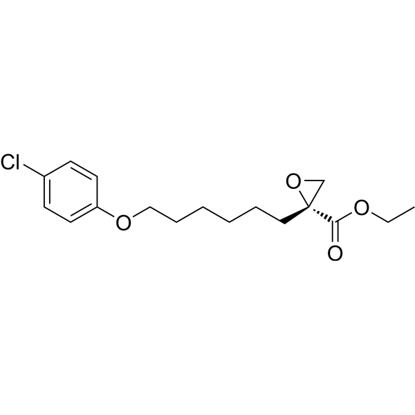
HY-50202
EtomoxirCPT1a Inhibitor -
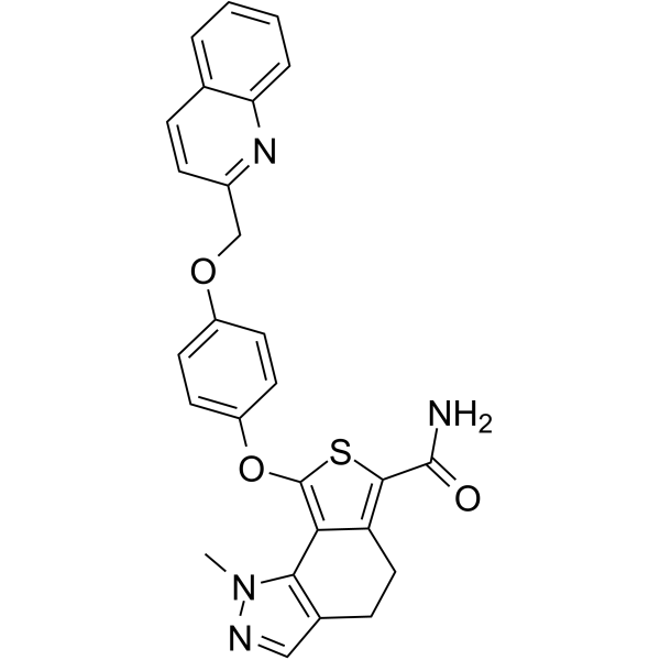
HY-15642
TD-198946Chondrogenic Agent -
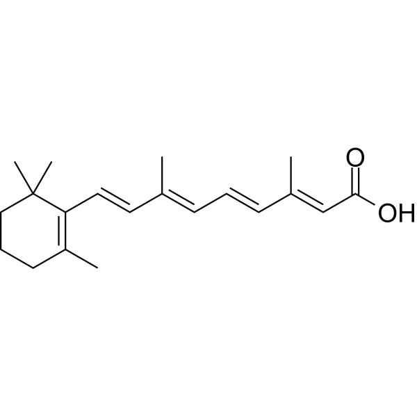
HY-14649
Retinoic acidRAR Agonist
Publications Citing Use of MedChemExpress (MCE) Kartogenin
More- Sci Bull. 2023 Sep 15;68(17):1904-1917. [Abstract]
- Adv Funct Mater. 2026 Feb 3.
- Adv Funct Mater. 2025 Dec 22.
- ACS Nano. 2025 Jun 10;19(22):20502-20515. [Abstract]
- J Mater Sci Technol. 2023 Sep 1.
- Theranostics. 2019 Sep 21;9(24):7108-7121. [Abstract]
- Biomaterials. 2022 Jun;285:121530. [Abstract]
- Biomaterials. 2021 Dec:279:121216. [Abstract]
- Chem Eng J. 2024 Aug 1.
- Chem Eng J. 1 March 2022, 133861.
- Chem Eng J. 400 (2020) 126004.
- Small. 2025 Sep 18:e07138. [Abstract]
- Small. 2022 Sep 3;e2202156. [Abstract]
- Adv Healthc Mater. 2023 Jul;12(18):e2203236. [Abstract]
- Sustainable Mater Technol. 2026 Mar 14.
- Mater Design. 2023 May 16, 112007.
- J Mater Chem B. 2026 Jul 1;14(25):7951-7968. [Abstract]
- Colloids Surf B Biointerfaces. 2020 Aug:192:111059. [Abstract]
- Int Immunopharmacol. 2026 Nov 1:188:117342.
- Int Immunopharmacol. 2026 Apr 15:175:116450. [Abstract]
- Int Immunopharmacol. 2025 Oct 10:163:115296. [Abstract]
- Stem Cell Rev Rep. 2022 Aug;18(6):2074-2087. [Abstract]
- Biomed Mater. 2026 May 28;21(3). [Abstract]
- Osteoarthr Cartil Open. 2023 May 12;5(3):100369. [Abstract]
- Anim Reprod Sci. 2021 Jun:229:106750. [Abstract]
- Orthop Surg. 2020 Jun;12(3):938-945. [Abstract]
- Res Sq. 2025 Jul 21.
- Res Sq. 2024 May 17.
- University of Oklahoma. 2024 May 10.
- Research Square Preprint. 2023 Apr 21.
- Authorea. 2023 Jan 27.
- Dis Markers. 2022 Aug 29:2022:6943630. [Abstract]
- Research Square Preprint. 2021 Sep.
- Patent. US20180263995A1.
 Customer Validation & Images

-
RT-PCR
-
WB
-
IHC


Activité biologique
Description
In Vitro
Kartogenin (100 nM; 72 h) induces chondrocyte nodule formation in primary hMSCs[1].
Kartogenin (10 nM-10 μM; 72 h) increases chondrocyte-specific gene expression in hMSCs[1].
Kartogenin (0.12-10 μM; 48 h) inhibits nitric oxide (NO) and glycosaminoglycan (GAG) release induced by cytokines in primary bovine articular chondrocytes[1].
Kartogenin (50-5000 nM; 2 weeks) induces the chondrogenetic differentiation of the BMSCs in a concentration-dependent manner[2].
MedChemExpress (MCE) has not independently confirmed the accuracy of these methods. They are for reference only.
In Vivo
MedChemExpress (MCE) has not independently confirmed the accuracy of these methods. They are for reference only.


Chemical Information
-
CAS No. 4727-31-5
-
Appearance Solid
-
Masse moléculaire 317.34
-
Formule C20H15NO3
-
Color White to off-white
-
SMILES
O=C(O)C1=CC=CC=C1C(NC2=CC=C(C3=CC=CC=C3)C=C2)=O
-
Synonyms
KGN
-
Livraison
Room temperature in continental US; may vary elsewhere.
-
Stockage
Powder -20°C 3 years 4°C 2 years In solvent -80°C 6 months -20°C 1 month


Publications (34)
-
Journal Impact Factor
-
Most Recent
-
Sci Bull
Enhanced osteochondral repair with hyaline cartilage formation using an extracellular matrix-inspired natural scaffold. [Abstract]2023 Sep 15;68(17):1904-1917. PMID: 37558534 -
-
-
ACS Nano
MXene-Based Cartilage-Adhesive Microspheres for Photothermal-Controlled Hydrophobic Drug Release and Mesenchymal Stem Cell Delivery in Osteoarthritis. [Abstract]2025 Jun 10;19(22):20502-20515. PMID: 40446309 -
-
Theranostics
Kartogenin hydrolysis product 4-aminobiphenyl distributes to cartilage and mediates cartilage regeneration. [Abstract]2019 Sep 21;9(24):7108-7121. PMID: 31695756
Kartogenin purchased from MedChemExpress. Usage Cited in: Theranostics. 2019 Sep 21;9(24):7108-7121. [Abstract]
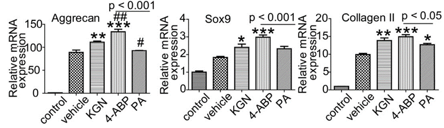
CSPC were isolated from cartilage and treated with vehicle or 10 μM Kartogenin (KGN), 4-ABP or phthalic acid (PA) in the chondrogenic differentiation medium. Aggrecan, Sox9 and type II collagen mRNA expression levels (determined by RT-qPCR) at day 21.
Kartogenin purchased from MedChemExpress. Usage Cited in: Theranostics. 2019 Sep 21;9(24):7108-7121. [Abstract]
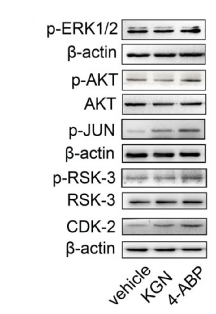
p-ERK1/2, p-AKT, p-JUN, p-RSK-3 and CDK-2 protein expression in UC-MSC treated with vehicle, Kartogenin (KGN) or 4-ABP (10 μM) for 3 days.
Kartogenin purchased from MedChemExpress. Usage Cited in: Theranostics. 2019 Sep 21;9(24):7108-7121. [Abstract]
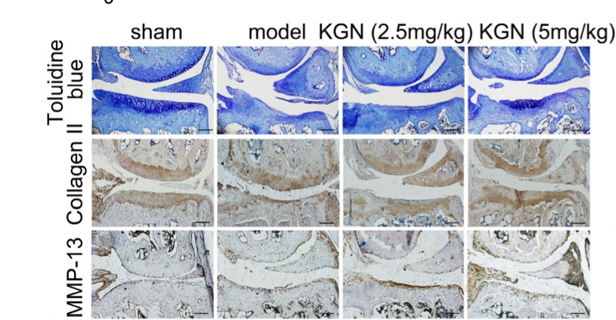
Oral Kartogenin (KGN) (2.5, 5 mg/kg) administration schedule in destabilization of medial meniscus (DMM)-induced osteoarthritis in STR/Ort mice. Representative images of toluidine blue and immunohistochemical staining of type II collagen and MMP-13 in articular cartilage.
-
Biomaterials
Injectable immunomodulation-based porous chitosan microspheres/HPCH hydrogel composites as a controlled drug delivery system for osteochondral regeneration. [Abstract]2022 Jun;285:121530. PMID: 35504181 -
Biomaterials
3D-bioprinted BMSC-laden biomimetic multiphasic scaffolds for efficient repair of osteochondral defects in an osteoarthritic rat model. [Abstract]2021 Dec:279:121216. PMID: 34739982 -
-
-
-
Small
2025 Sep 18:e07138. PMID: 40965039 -
Small
Injectable Double Positively Charged Hydrogel Microspheres for Targeting-Penetration-Phagocytosis. [Abstract]2022 Sep 3;e2202156. PMID: 36056898 -
Adv Healthc Mater
Sustained-Drug-Release, Strong, and Anti-Swelling Water-Lipid Biphasic Hydrogels Prepared via Digital Light Processing 3D Printing for Protection against Osteoarthritis: Demonstration in a Porcine Model. [Abstract]2023 Jul;12(18):e2203236. PMID: 36943891 -
-
-
J Mater Chem B
3D-printed bilayer hydrogel scaffolds incorporating HAP and KGN@Lip for osteochondral regeneration. [Abstract]2026 Jul 1;14(25):7951-7968. PMID: 42300767 -
Colloids Surf B Biointerfaces
Injectable in situ forming kartogenin-loaded chitosan hydrogel with tunable rheological properties for cartilage tissue engineering. [Abstract]2020 Aug:192:111059. PMID: 32380404 -
-
Int Immunopharmacol
Daucosterol alleviates osteoarthritis by targeting chondrocyte senescence via inhibiting of the JNK pathway. [Abstract]2026 Apr 15:175:116450. PMID: 41775106 -
Int Immunopharmacol
CORM-3 mitigates osteoarthritis by anti-inflammation and enhancing autophagy via inhibiting MAPK and mTOR pathways. [Abstract]2025 Oct 10:163:115296. PMID: 40749610 -
Stem Cell Rev Rep
Dual-specificity Tyrosine Phosphorylation-regulated Kinase Inhibitor ID-8 Promotes Human Somatic Cell Reprogramming by Activating PDK4 Expression. [Abstract]2022 Aug;18(6):2074-2087. PMID: 35080746 -
Biomed Mater
Chondrogenic differentiation of human periosteum-derived cells in spheroids, HAMA hydrogels, and bioprinted constructs: comparison of kartogenin and TGF- β 1. [Abstract]2026 May 28;21(3). PMID: 42102888 -
Osteoarthr Cartil Open
Comparison of multiple synthetic chondroinductive factors in pellet culture against a TGF-β positive control. [Abstract]2023 May 12;5(3):100369. PMID: 37252634 -
Anim Reprod Sci
Effects of TG interaction factor 1 on synthesis of estradiol and progesterone in granulosa cells of goats through SMAD2/3-SP1 signaling pathway. [Abstract]2021 Jun:229:106750. PMID: 33940561 -
Orthop Surg
Synergistic Effects of Kartogenin and Transforming Growth Factor-β3 on Chondrogenesis of Human Umbilical Cord Mesenchymal Stem Cells In Vitro. [Abstract]2020 Jun;12(3):938-945. PMID: 32462800 -
-
-
-
-
-
Dis Markers
Kartogenin Induced Adipose-Derived Stem Cell Exosomes Enhance the Chondrogenic Differentiation Ability of Adipose-Derived Stem Cells. [Abstract]2022 Aug 29:2022:6943630. PMID: 36072901 -
-


Solvant et solubilité
In Vitro:
DMSO : ≥ 100 mg/mL (315.12 mM; Hygroscopic DMSO has a significant impact on the solubility of product, please use newly opened DMSO)
Ethanol : 10 mg/mL (31.51 mM; Need ultrasonic)
H2O : < 0.1 mg/mL (insoluble)
* "≥" means soluble, but saturation unknown.
Please refer to the solubility information to select the appropriate solvent. Once prepared, please aliquot and store the solution to prevent product inactivation from repeated freeze-thaw cycles.
Storage method and period of stock solution: -80°C, 6 months; -20°C, 1 month. When stored at -80°C, please use it within 6 months. When stored at -20°C, please use it within 1 month.
Please refer to the solubility information to select the appropriate solvent. Once prepared, please aliquot and store the solution to prevent product inactivation from repeated freeze-thaw cycles.
Storage method and period of stock solution: -80°C, 6 months; -20°C, 1 month. When stored at -80°C, please use it within 6 months. When stored at -20°C, please use it within 1 month.
Concentration (start) × Volume (start) = Concentration (final) × Volume (final)
In Vivo:
Select the appropriate dissolution method based on your experimental animal and administration route.
- For the following dissolution methods, please ensure to first prepare a clear stock solution using an In Vitro approach and then sequentially add co-solvents:
- To ensure reliable experimental results, the clarified stock solution can be appropriately stored based on storage conditions. As for the working solution for In Vivo experiments, it is recommended to prepare freshly and use it on the same day.
- The percentages shown for the solvents indicate their volumetric ratio in the final prepared solution. If precipitation or phase separation occurs during preparation, heat and/or sonication can be used to aid dissolution.
Add each solvent one by one: 10% DMSO 40% PEG300 5% Tween-80 45% Saline
Solubility: ≥ 2.5 mg/mL (7.88 mM); Clear solution
This protocol yields a clear solution of ≥ 2.5 mg/mL (saturation unknown).
Taking 1 mL working solution as an example, add 100 μL DMSO stock solution (25.0 mg/mL) to 400 μL PEG300, and mix evenly; then add 50 μL Tween-80 and mix evenly; then add 450 μL Saline to adjust the volume to 1 mL.
Preparation of Saline: Dissolve 0.9 g sodium chloride in ddH₂O and dilute to 100 mL to obtain a clear Saline solution.
Add each solvent one by one: 10% DMSO 90% (20% SBE-β-CD in Saline)
Solubility: ≥ 2.5 mg/mL (7.88 mM); Clear solution
This protocol yields a clear solution of ≥ 2.5 mg/mL (saturation unknown).
Taking 1 mL working solution as an example, add 100 μL DMSO stock solution (25.0 mg/mL) to 900 μL 20% SBE-β-CD in Saline, and mix evenly.
Preparation of 20% SBE-β-CD in Saline (4°C, storage for one week): 2 g SBE-β-CD powder is dissolved in 10 mL Saline, completely dissolve until clear.
In Vivo Dissolution Calculator
Please enter the basic information of animal experiments:
-
-
-
-
Recommended: Prepare an additional quantity of animals to account for potential losses during experiments.
Please enter your animal formula composition:
-
%DMSO +
Recommended: Keep the proportion of DMSO in working solution below 2% if your animal is weak.
-
%+
-
+%Tween-80 + +
-
%Saline +
The co-solvents required include: DMSO, . All of co-solvents are available by MedChemExpress (MCE). , Tween 80. All of co-solvents are available by MedChemExpress (MCE).
Working solution concentration: 0.22 mg/mL
Method for preparing stock solution: mg drug dissolved in μL DMSO. Stock solution concentration: mg/mL.
1. Take μL DMSO stock solution;
2. Add μL .
μL , mix evenly;
3. Then add μL Tween 80, mix evenly;
4. Then add μL
Please ensure that the stock solution in the first step is dissolved to a clear state, and add co-solvents in sequence. You can use ultrasonic heating (ultrasonic cleaner, recommended frequency 20-40 kHz), vortexing, etc. to assist dissolution.


Pureté et documentation
-
Fiche technique (280 KB)
-
SDS (598 KB)
- English - EN (598 KB)
- Français - FR (598 KB)
- Deutsch - DE (598 KB)
- Norwegian - NO (598 KB)
- Español - ES (598 KB)
- Swedish - SV (598 KB)
- Italian - IT (598 KB)
- Korean - KR (598 KB)
- Portuguese - PT (598 KB)
-
Instruction de manipulation (2659 KB)
Références
[1]. Johnson K, et, al. A stem cell-based approach to cartilage repair. Science. 2012 May 11;336(6082):717-21. [Content Brief]
[2]. Liu F, et, al. A novel kartogenin-platelet-rich plasma gel enhances chondrogenesis of bone marrow mesenchymal stem cells in vitro and promotes wounded meniscus healing in vivo. Stem Cell Res Ther. 2019 Jul 8;10(1):201. [Content Brief]


Complete Stock Solution Preparation Table
Please refer to the solubility information to select the appropriate solvent. Once prepared, please aliquot and store the solution to prevent product inactivation from repeated freeze-thaw cycles.
Storage method and period of stock solution: -80°C, 6 months; -20°C, 1 month. When stored at -80°C, please use it within 6 months. When stored at -20°C, please use it within 1 month.
| Optional Solvent | Concentration Solvent Mass | 1 mg | 5 mg | 10 mg | 25 mg |
|---|---|---|---|---|---|
| Ethanol / DMSO | 1 mM | 3.1512 mL | 15.7560 mL | 31.5119 mL | 78.7799 mL |
| 5 mM | 0.6302 mL | 3.1512 mL | 6.3024 mL | 15.7560 mL | |
| 10 mM | 0.3151 mL | 1.5756 mL | 3.1512 mL | 7.8780 mL | |
| 15 mM | 0.2101 mL | 1.0504 mL | 2.1008 mL | 5.2520 mL | |
| 20 mM | 0.1576 mL | 0.7878 mL | 1.5756 mL | 3.9390 mL | |
| 25 mM | 0.1260 mL | 0.6302 mL | 1.2605 mL | 3.1512 mL | |
| 30 mM | 0.1050 mL | 0.5252 mL | 1.0504 mL | 2.6260 mL | |
| DMSO | 40 mM | 0.0788 mL | 0.3939 mL | 0.7878 mL | 1.9695 mL |
| 50 mM | 0.0630 mL | 0.3151 mL | 0.6302 mL | 1.5756 mL | |
| 60 mM | 0.0525 mL | 0.2626 mL | 0.5252 mL | 1.3130 mL | |
| 80 mM | 0.0394 mL | 0.1969 mL | 0.3939 mL | 0.9847 mL | |
| 100 mM | 0.0315 mL | 0.1576 mL | 0.3151 mL | 0.7878 mL |


Kartogenin Related Classifications
HY-16268 Related Classifications
 Powered by Bioz