



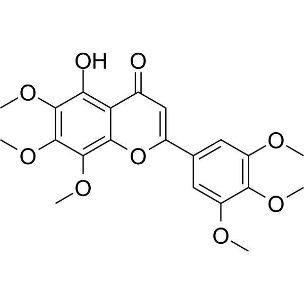
Gardenin A

Based on 1 Customer Validation
Gardenin A is an orally active and synthetic PMF analogue with the neurotrophic effect for neurite outgrowth and neuronal differentiation. Gardenin A promotes neuritogenesis via activating MAPK/ERK, PKC, and PKA, but not TrkA, CREB signaling pathways. Gardenin A also has sedative, anxiolytic, antidepressant, and anticonvulsant effects.
For research use only. We do not sell to patients.
- Purity : 97.61%
- CAS No.: 21187-73-5
- Formula: C21H22O9
- Molecular Weight:418.39
-
Storage:
-20°C, protect from light
* In solvent : -80°C, 6 months; -20°C, 1 month (protect from light)

To place orders, for customer services and technical support, please contact: MedChemExpress USA
Tel: 609-228-6898 E-mail: [email protected] [email protected]


-
 Biological Activity
-
Chemical Information
-
Solvent & Solubility
- Protocols
- Purity & Documentation
- References
-
 Help & FAQs


-
HY-W010907
Pamoic acid disodiumGPR35 Agonist -
HY-P4322
H-Ile-Lys-Val-Ala-Val-OHERK/Akt Activator -
HY-129566
Withanolide BNeuroprotective Agent -
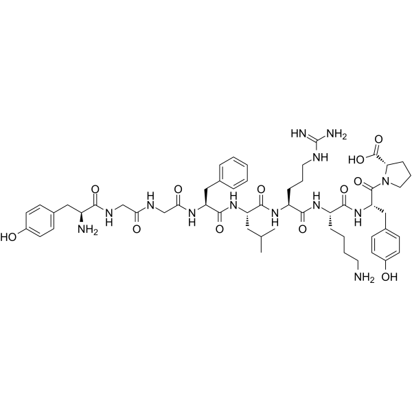
HY-P3513
β-Neo-EndorphinErk1/2 Activator -
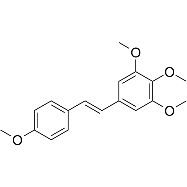
HY-137977
DMU-212Anticancer Compound -
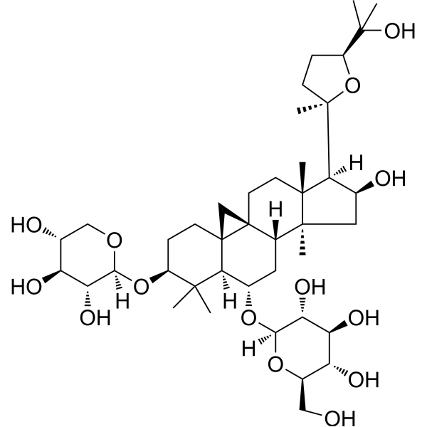
HY-N0431
Astragaloside IVMMP Inhibitor -
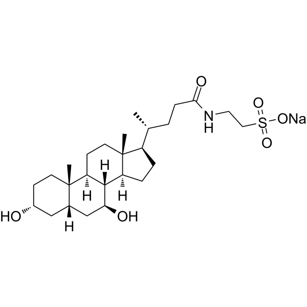
HY-19696A
Tauroursodeoxycholate sodiumEndoplasmic Reticulum Stress Inhibitor -

HY-N0431R
Astragaloside IV (Standard)MMP Inhibitor -
HY-W001174
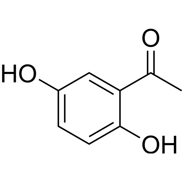
2,5-Dihydroxyacetophenone -
HY-15816A
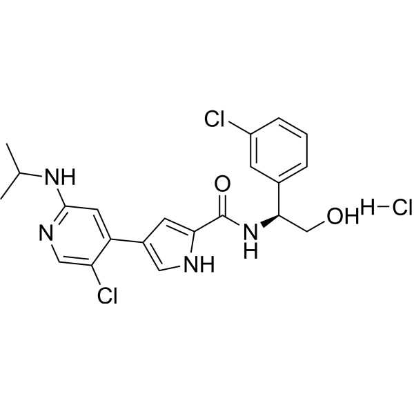
Ulixertinib hydrochlorideERK1/2 Inhibitor -
HY-108330
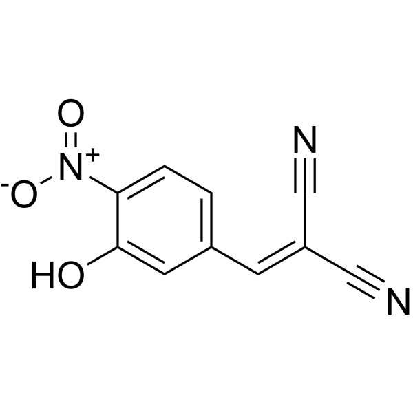
AG126Tyrosine Kinase Inhibitor -
HY-N1374
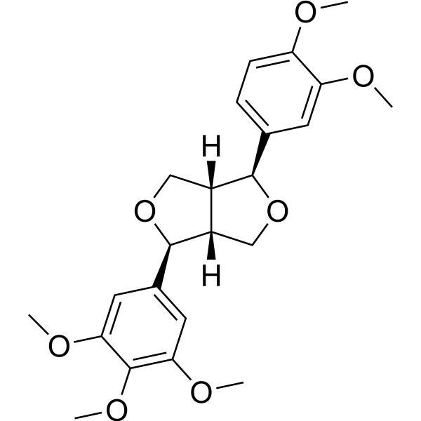
MagnolinERK Inhibitor


Biological Activity
Description
Cellular Effect
|
Cell Line

|
Type | Value | Description | References |
|---|---|---|---|---|
| HL-60 | IC50 |
>100 μM
Compound: XII
|
Antiproliferative activity against HL60 after 24 hrs
Antiproliferative activity against HL60 after 24 hrs
|
[PMID: 17391969] |
In Vitro
Gardenin A (10-20 μM; 48 h) potently induces neurite outgrowth in PC12 cells, and (10 μM; 24 h and 48 h) increases expression of neuronal differentiation and synapse formation marker proteins, growth-associated protein-43 (GAP-43), and synaptophysin[1].
Gardenin A (10 μM; 30-120 min) markedly induces the phosphorylation of both cyclic AMP response element-binding protein (CREB) and CRE-mediated transcription, which was suppressed through the administration of the inhibitor 2-naphthol AS-E phosphate (KG-501) or using CREB siRNA[1].
Gardenin A (10 μM; 15-120 min) increases ERK phosphorylation and PKA and PKC activities[1].
MedChemExpress (MCE) has not independently confirmed the accuracy of these methods. They are for reference only.
-
Cell Line:PC12 cells
-
Concentration:2, 5, 10, 20 μM
-
Incubation Time:48 hours
-
Result:showed cell growth supporting effect at higher concentrations (10-20 µM) and did not exert detectable 237 cytotoxicity on PC12 cells after 48 h incubation in low serum medium.
-
Cell Line:PC12 cells
-
Concentration:10 μM
-
Incubation Time:0, 15, 30, 60, and 120 min
-
Result:Significantly increased the phosphorylation of ERK1/2 (Thr202/Tyr204) at 15 min, subsequently reduced after 30 min.
Increased PKC and PKA activity peaked at 15 min. Increased CREB phosphorylation.
In Vivo
Gardenin A (1-10 mg/kg; p.o.; single dose) does not affect locomotor coordination in mice and delayed the onset of convulsions[2].
MedChemExpress (MCE) has not independently confirmed the accuracy of these methods. They are for reference only.
-
Animal Model:Male Balb/c mice (24-32 g)[2]
-
Dosage:1 mg/kg, 10 mg/kg, 25 mg/kg
-
Administration:Oral gavage; single dose
-
Result:Showed sedative effects at a dose of 25 mg/kg, which increased the duration of sleep but did not alter sleep onset.
- PC12 NGF-Induced Neuronal Differentiation Culture
- iPSC/hPSC-Derived Neuron Differentiation Culture
- SH-SY5Y Neuronal Differentiation Culture
- Human pluripotent stem cell neural induction and neuron differentiation
- SH-SY5Y neuronal-like differentiation
- Cell differentiation
- PC12 NGF-induced neuronal-like differentiation
- Directly Induced Neuron Culture


Chemical Information
-
CAS No. 21187-73-5
-
Appearance Solid
-
Molecular Weight 418.39
-
Formula C21H22O9
-
Color Off-white to light yellow
-
SMILES
O=C1C=C(C2=CC(OC)=C(OC)C(OC)=C2)OC3=C(OC)C(OC)=C(OC)C(O)=C13
-
Structure Classification
-
Initial Source
-
Shipping
Room temperature in continental US; may vary elsewhere.
-
Storage
-20°C, protect from light
* In solvent : -80°C, 6 months; -20°C, 1 month (protect from light)


Solvent & Solubility
In Vitro:
DMSO : 8.27 mg/mL (19.77 mM; Need ultrasonic and warming; Hygroscopic DMSO has a significant impact on the solubility of product, please use newly opened DMSO)
Please refer to the solubility information to select the appropriate solvent. Once prepared, please aliquot and store the solution to prevent product inactivation from repeated freeze-thaw cycles.
Storage method and period of stock solution: -80°C, 6 months; -20°C, 1 month (protect from light). When stored at -80°C, please use it within 6 months. When stored at -20°C, please use it within 1 month.
Please refer to the solubility information to select the appropriate solvent. Once prepared, please aliquot and store the solution to prevent product inactivation from repeated freeze-thaw cycles.
Storage method and period of stock solution: -80°C, 6 months; -20°C, 1 month (protect from light). When stored at -80°C, please use it within 6 months. When stored at -20°C, please use it within 1 month.
Concentration (start) × Volume (start) = Concentration (final) × Volume (final)


Purity & Documentation
-
Data Sheet (277 KB)
-
SDS (252 KB)
- English - EN (252 KB)
- Français - FR (252 KB)
- Deutsch - DE (252 KB)
- Norwegian - NO (252 KB)
- Español - ES (252 KB)
- Swedish - SV (252 KB)
- Italian - IT (252 KB)
- Korean - KR (252 KB)
- Portuguese - PT (252 KB)
-
Handling Instructions (2659 KB)
References
[1]. Chiu SP, et al. Neurotrophic action of 5-hydroxylated polymethoxyflavones: 5-demethylnobiletin and gardenin A stimulate neuritogenesis in PC12 cells. J Agric Food Chem. 2013 Oct 2;61(39):9453-63. [Content Brief]
[2]. Alonso-Castro AJ, et al. Evaluation of the neuropharmacological effects of Gardenin A in mice. Drug Dev Res. 2020 Aug;81(5):600-608. [Content Brief]


Complete Stock Solution Preparation Table
Please refer to the solubility information to select the appropriate solvent. Once prepared, please aliquot and store the solution to prevent product inactivation from repeated freeze-thaw cycles.
Storage method and period of stock solution: -80°C, 6 months; -20°C, 1 month (protect from light). When stored at -80°C, please use it within 6 months. When stored at -20°C, please use it within 1 month.
| Optional Solvent | Concentration Solvent Mass | 1 mg | 5 mg | 10 mg | 25 mg |
|---|---|---|---|---|---|
| DMSO | 1 mM | 2.3901 mL | 11.9506 mL | 23.9011 mL | 59.7529 mL |
| 5 mM | 0.4780 mL | 2.3901 mL | 4.7802 mL | 11.9506 mL | |
| 10 mM | 0.2390 mL | 1.1951 mL | 2.3901 mL | 5.9753 mL | |
| 15 mM | 0.1593 mL | 0.7967 mL | 1.5934 mL | 3.9835 mL |


Gardenin A Related Classifications
HY-N8303 Related Classifications
 Powered by Bioz