



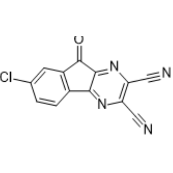
HBX 41108
Based on 4 publication(s) in Google Scholar
HBX 41108 is an inhibitor of ubiquitin-specific protease 7 (USP7) with an IC50 of 424 nM. HBX 41108 inhibits USP7-mediated p53 deubiquitination to stabilize p53 and inhibits cancer cell growth. BX 41108 can be used in cancer and diabetes research.
Nos produits utilisent uniquement pour la recherche. Nous ne vendons pas aux patients.
- Pureté : 98.91%
- CAS No.: 924296-39-9
- Formule: C13H3ClN4O
- Masse moléculaire:266.64
-
Stockage:
4°C, sealed storage, away from moisture
* In solvent : -80°C, 6 months; -20°C, 1 month (sealed storage, away from moisture)

To place orders, for customer services and technical support, please contact: MedChemExpress USA
Tel: 609-228-6898 E-mail: [email protected] [email protected]


-
 Activité biologique
-
Chemical Information
-
Solvant et solubilité
- Protocole
- Pureté et documentation
- Références
-
 Help & FAQs


-
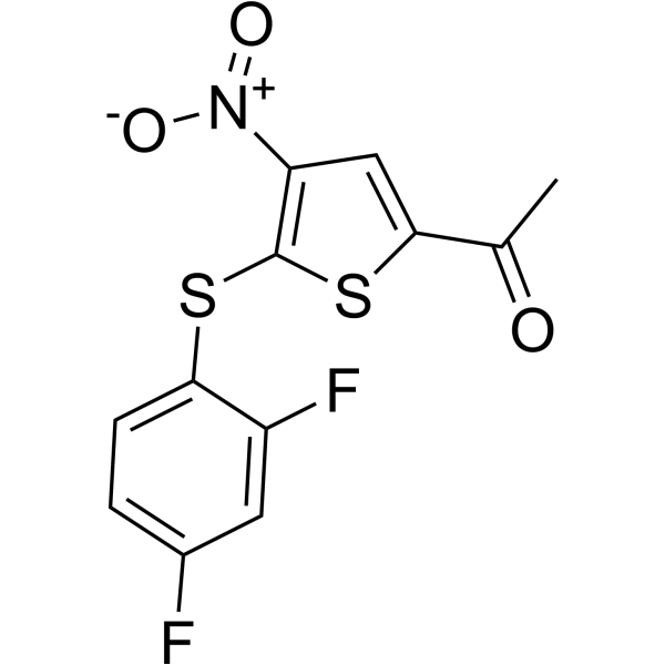
HY-13865
P 22077USP7/47 Inhibitor -
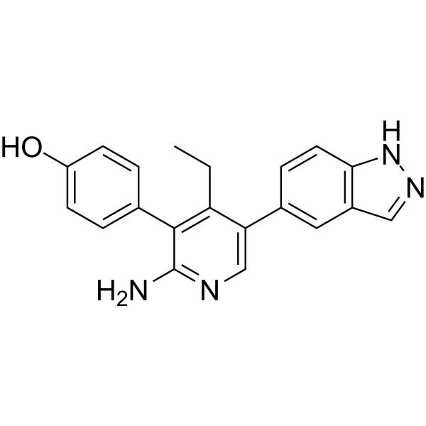
HY-112937
GNE-6640USP7 Inhibitor -
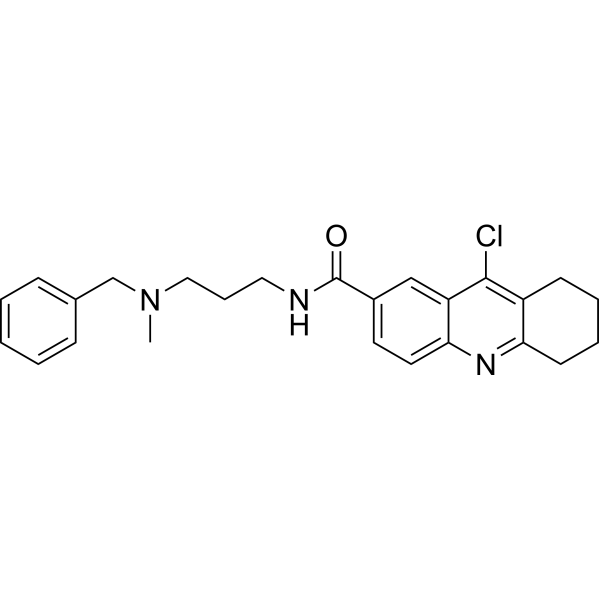
HY-17540
HBX 19818USP7 Inhibitor -
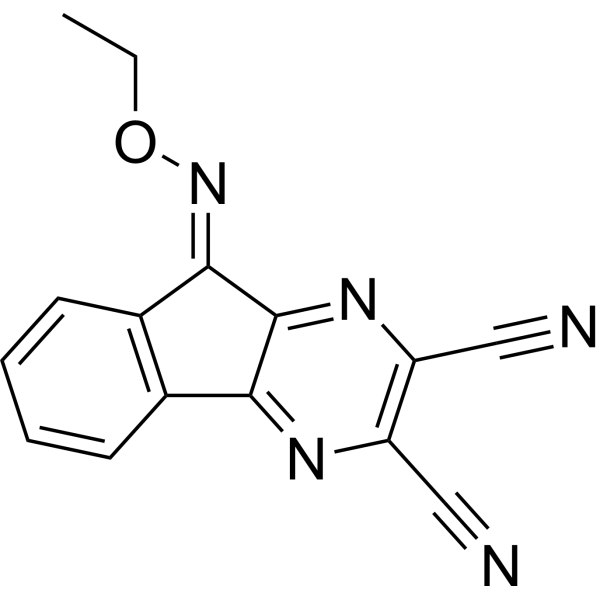
HY-50737A
DUB-IN-2Deubiquitinase Inhibitor -
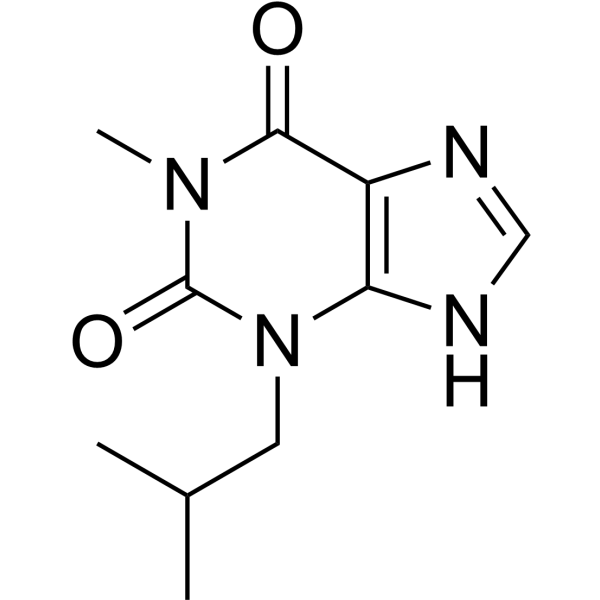
HY-12318
IBMXPDE Inhibitor -
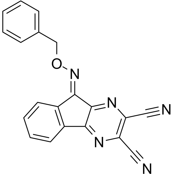
HY-50736
DUB-IN-1USP8 Inhibitor -
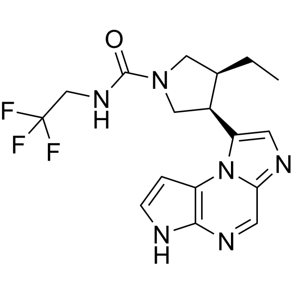
HY-19569
UpadacitinibJAK1 Inhibitor -
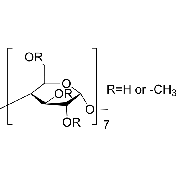
HY-101461
Methyl-β-cyclodextrinCholesterol-removing Agent -
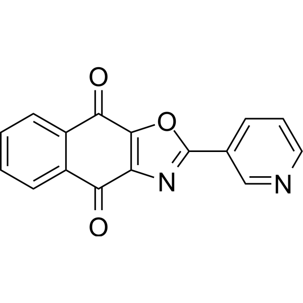
HY-80012
SJB3-019AUSP1 Inhibitor -
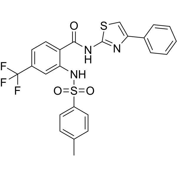
HY-100900
ML364USP2 Inhibitor -
HY-K1090
Cytotoxicity LDH Assay Kit -
HY-107986
GNE-6776USP7 Inhibitor
Publications Citing Use of MedChemExpress (MCE) HBX 41108
More

Activité biologique
Description
IC50 & Target
|
USP7 424 nM (IC50) |
hTPH2 |
In Vitro
HBX 41108 (0-3 μM, 24 h) inhibits the proliferation of tumor cells HCT-116, induces P53 dependent apoptosis and does not affect the activity of normal hepatocytes [1].
HBX 41108 (5 μM, 24 h) can inhibit cell cycle arrest and cell senescence induced by USP7 in HUVECs[3].
HBX 41108 (5-25 μM, 48 h) can enhances the hTPH2 promoter activity in RN46A cells[4].
MedChemExpress (MCE) has not independently confirmed the accuracy of these methods. They are for reference only.
-
Cell Line:HCT-116, NIH-3T3
-
Concentration:0-3 μM
-
Incubation Time:24 h
-
Result:HCT116 colon tumor cells were more sensitive to HBX 41108 (IC50 = 0.27 μmol/L) than normal diploid NIH-3T3 fibroblasts (p53 wild-type) with a 7-fold differential effect (IC50 = 1.77 μmol/L).
In Vivo
MedChemExpress (MCE) has not independently confirmed the accuracy of these methods. They are for reference only.


Chemical Information
-
CAS No. 924296-39-9
-
Appearance Solid
-
Masse moléculaire 266.64
-
Formule C13H3ClN4O
-
Color Light yellow to yellow
-
SMILES
N#CC1=C(C#N)N=C2C(C(C3=C2C=CC(Cl)=C3)=O)=N1
-
Livraison
Room temperature in continental US; may vary elsewhere.
-
Stockage
4°C, sealed storage, away from moisture
* In solvent : -80°C, 6 months; -20°C, 1 month (sealed storage, away from moisture)


Publications (4)
-
Journal Impact Factor
-
Most Recent
-
Cell Commun Signal
Inhibition of USP7 upregulates USP22 and activates its downstream cancer-related signaling pathways in human cancer cells. [Abstract]2023 Nov 9;21(1):319. PMID: 37946202 -
Cell Chem Biol
Development of potent and selective inhibitors targeting the papain-like protease of SARS-CoV-2. [Abstract]2021 Jun 17;28(6):855-865.e9. PMID: 33979649 -
Cell Rep
2024 Nov 4;43(11):114917. PMID: 39499616 -
Eur J Med Res
RHBDD1 induces cell cycle arrest in TP53-mutant NSCLC cells by promoting endoplasmic reticulum-associated degradation of p53 and DNA-PKcs. [Abstract]2025 Sep 26;30(1):859. PMID: 41013641


Solvant et solubilité
In Vitro:
DMSO : 250 mg/mL (937.59 mM; ultrasonic and warming and heat to 60°C; Hygroscopic DMSO has a significant impact on the solubility of product, please use newly opened DMSO)
Please refer to the solubility information to select the appropriate solvent. Once prepared, please aliquot and store the solution to prevent product inactivation from repeated freeze-thaw cycles.
Storage method and period of stock solution: -80°C, 6 months; -20°C, 1 month (sealed storage, away from moisture). When stored at -80°C, please use it within 6 months. When stored at -20°C, please use it within 1 month.
Please refer to the solubility information to select the appropriate solvent. Once prepared, please aliquot and store the solution to prevent product inactivation from repeated freeze-thaw cycles.
Storage method and period of stock solution: -80°C, 6 months; -20°C, 1 month (sealed storage, away from moisture). When stored at -80°C, please use it within 6 months. When stored at -20°C, please use it within 1 month.
Concentration (start) × Volume (start) = Concentration (final) × Volume (final)
In Vivo:
Select the appropriate dissolution method based on your experimental animal and administration route.
- For the following dissolution methods, please ensure to first prepare a clear stock solution using an In Vitro approach and then sequentially add co-solvents:
- To ensure reliable experimental results, the clarified stock solution can be appropriately stored based on storage conditions. As for the working solution for In Vivo experiments, it is recommended to prepare freshly and use it on the same day.
- The percentages shown for the solvents indicate their volumetric ratio in the final prepared solution. If precipitation or phase separation occurs during preparation, heat and/or sonication can be used to aid dissolution.
Add each solvent one by one: 10% DMSO 40% PEG300 5% Tween-80 45% Saline
Solubility: ≥ 1 mg/mL (3.75 mM); Clear solution
This protocol yields a clear solution of ≥ 1 mg/mL (saturation unknown).
Taking 1 mL working solution as an example, add 100 μL DMSO stock solution (10.0 mg/mL) to 400 μL PEG300, and mix evenly; then add 50 μL Tween-80 and mix evenly; then add 450 μL Saline to adjust the volume to 1 mL.
Preparation of Saline: Dissolve 0.9 g sodium chloride in ddH₂O and dilute to 100 mL to obtain a clear Saline solution.
In Vivo Dissolution Calculator
Please enter the basic information of animal experiments:
-
-
-
-
Recommended: Prepare an additional quantity of animals to account for potential losses during experiments.
Please enter your animal formula composition:
-
%DMSO +
Recommended: Keep the proportion of DMSO in working solution below 2% if your animal is weak.
-
%+
-
+%Tween-80 + +
-
%Saline +
The co-solvents required include: DMSO, . All of co-solvents are available by MedChemExpress (MCE). , Tween 80. All of co-solvents are available by MedChemExpress (MCE).
Working solution concentration: 0.22 mg/mL
Method for preparing stock solution: mg drug dissolved in μL DMSO. Stock solution concentration: mg/mL. * In solvent : -80°C, 6 months; -20°C, 1 month (sealed storage, away from moisture)
1. Take μL DMSO stock solution;
2. Add μL .
μL , mix evenly;
3. Then add μL Tween 80, mix evenly;
4. Then add μL
Please ensure that the stock solution in the first step is dissolved to a clear state, and add co-solvents in sequence. You can use ultrasonic heating (ultrasonic cleaner, recommended frequency 20-40 kHz), vortexing, etc. to assist dissolution.


Pureté et documentation
-
Fiche technique (278 KB)
-
SDS (252 KB)
- English - EN (252 KB)
- Français - FR (252 KB)
- Deutsch - DE (252 KB)
- Norwegian - NO (252 KB)
- Español - ES (252 KB)
- Swedish - SV (252 KB)
- Italian - IT (252 KB)
- Korean - KR (252 KB)
- Portuguese - PT (252 KB)
-
Instruction de manipulation (2659 KB)
Références
[1]. Colland F, et al. Small-molecule inhibitor of USP7/HAUSP ubiquitin protease stabilizes and activates p53 in cells. Mol Cancer Ther. 2009 Aug;8(8):2286-95. [Content Brief]
[2]. Colombo M, et al. Synthesis and biological evaluation of 9-oxo-9H-indeno[1,2-b]pyrazine-2,3-dicarbonitrile analogues as potential inhibitors of deubiquitinating enzymes. ChemMedChem. 2010 Apr 6;5(4):552-8. [Content Brief]
[3]. Li X, Wang T, et al. Inhibition of USP7 suppresses advanced glycation end-induced cell cycle arrest and senescence of human umbilical vein endothelial cells through ubiquitination of p53. Acta Biochim Biophys Sin (Shanghai). 2022 Mar 25;54(3):311-320. [Content Brief]
[4]. Nawa Y, et al. Functional characterization of the neuron-restrictive silencer element in the human tryptophan hydroxylase 2 gene expression. J Neurochem. 2017 Sep;142(6):827-840. [Content Brief]


Complete Stock Solution Preparation Table
Please refer to the solubility information to select the appropriate solvent. Once prepared, please aliquot and store the solution to prevent product inactivation from repeated freeze-thaw cycles.
Storage method and period of stock solution: -80°C, 6 months; -20°C, 1 month (sealed storage, away from moisture). When stored at -80°C, please use it within 6 months. When stored at -20°C, please use it within 1 month.
| Optional Solvent | Concentration Solvent Mass | 1 mg | 5 mg | 10 mg | 25 mg |
|---|---|---|---|---|---|
| DMSO | 1 mM | 3.7504 mL | 18.7519 mL | 37.5037 mL | 93.7594 mL |
| 5 mM | 0.7501 mL | 3.7504 mL | 7.5007 mL | 18.7519 mL | |
| 10 mM | 0.3750 mL | 1.8752 mL | 3.7504 mL | 9.3759 mL | |
| 15 mM | 0.2500 mL | 1.2501 mL | 2.5002 mL | 6.2506 mL | |
| 20 mM | 0.1875 mL | 0.9376 mL | 1.8752 mL | 4.6880 mL | |
| 25 mM | 0.1500 mL | 0.7501 mL | 1.5001 mL | 3.7504 mL | |
| 30 mM | 0.1250 mL | 0.6251 mL | 1.2501 mL | 3.1253 mL | |
| 40 mM | 0.0938 mL | 0.4688 mL | 0.9376 mL | 2.3440 mL | |
| 50 mM | 0.0750 mL | 0.3750 mL | 0.7501 mL | 1.8752 mL | |
| 60 mM | 0.0625 mL | 0.3125 mL | 0.6251 mL | 1.5627 mL | |
| 80 mM | 0.0469 mL | 0.2344 mL | 0.4688 mL | 1.1720 mL | |
| 100 mM | 0.0375 mL | 0.1875 mL | 0.3750 mL | 0.9376 mL |


HBX 41108 Related Classifications
HY-101666 Related Classifications
 Powered by Bioz