



Hirudin (54-65)

Based on 1 Customer Validation
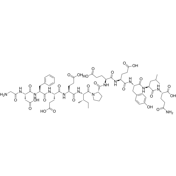
Hirudin (54-65) is a thrombin antagonist and YAP suppressor with anticoagulatory properties.Hirudin (54-65) blocks thrombin's anion binding site, acts on soluble and thrombus-bound thrombin.Hirudin (54-65) suppresses thrombin-induced profibrotic YAP activity, reduces YAP expression, nuclear translocation, and downstream effector signaling in vascular endothelial cells.Hirudin (54-65) ameliorates obstructive cholestasis, attenuates liver fibrosis symptoms, fibrosis-associated angiogenesis, and endothelial-to-mesenchymal transition.Hirudin (54-65) reduces liver inflammation and tissue hypoxia.Hirudin (54-65) promotes extracellular calcium influx through L-type calcium channels in canine coronary artery smooth muscle, mediates contraction.Hirudin (54-65) induces endothelium-independent contraction of canine coronary arterial segments; this response is not affected by indomethacin pretreatment.Hirudin (54-65) can be used for the research of liver obstructive cholestasis, liver fibrosis.
For research use only. We do not sell to patients.
- Purity : 99.20%
- CAS No.: 113274-56-9
- Formula: C66H93N13O25
- Molecular Weight:1468.52
-
Storage:
Sealed storage, away from moisture.
Powder -80°C, 2 years , -20°C, 1 year* In solvent : -80°C, 6 months; -20°C, 1 month (sealed storage, away from moisture)

To place orders, for customer services and technical support, please contact: MedChemExpress USA
Tel: 609-228-6898 E-mail: [email protected] [email protected]


-
 Biological Activity
-
Chemical Information
-
Solvent & Solubility
- Protocols
- Purity & Documentation
- References
-
 Help & FAQs


-
HY-10787
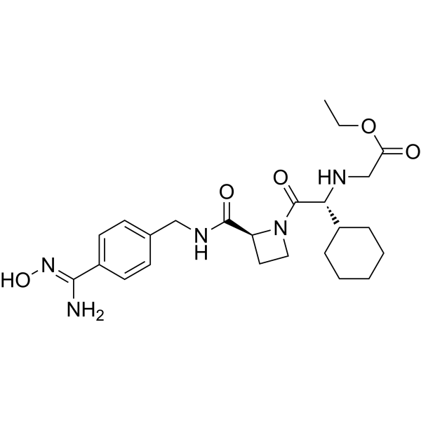
XimelagatranThrombin Inhibitor -
HY-B0375A
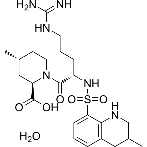
Argatroban monohydrateThrombin Inhibitor -
HY-P4803
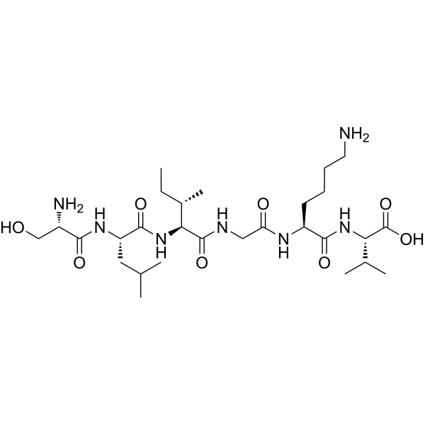
PAR-2 (1-6) (human)PAR2 Agonist -
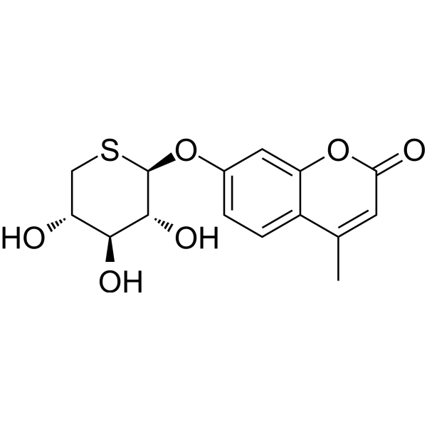
HY-10277
Odiparcilβ-D-xyloside Derivative -
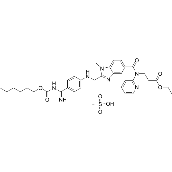
HY-10274A
Dabigatran etexilate mesylateAntithrombotic Agent -
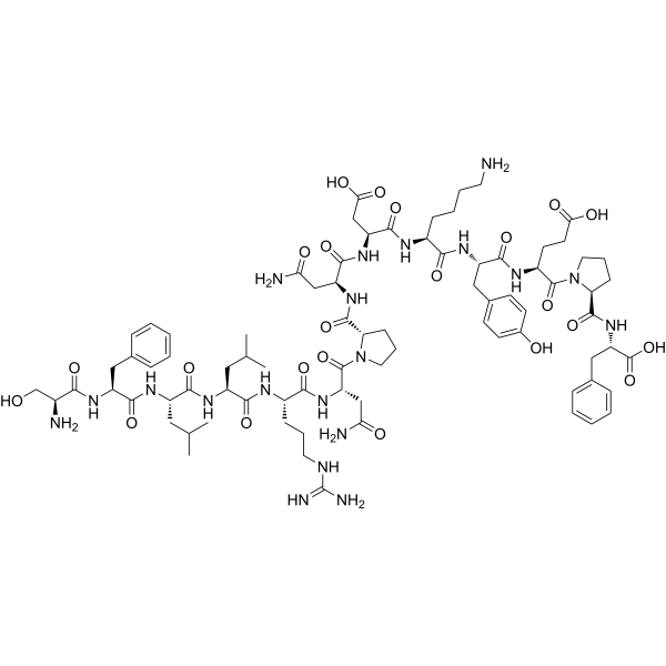
HY-P1000
TRAP-14Thrombin Receptor Agonist -

HY-10787R
Ximelagatran (Standard)Reference Standard -

HY-10274AR
Dabigatran etexilate mesylate (Standard)Antithrombotic Agent -

HY-B0375AR
Argatroban monohydrate (Standard)Thrombin Inhibitor -

HY-10277R
Odiparcil (Standard)Reference Standard -
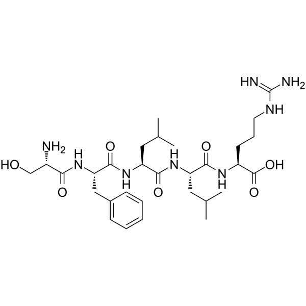
HY-P1536
Thrombin Receptor Activator for Peptide 5 (TRAP-5) -
HY-P4451
TRAP-5 amidePAR 1 Agonist


Biological Activity
Description
IC50 & Target
|
L-type calcium channel |
In Vitro
Hirudin (54-65) (10-10-10-6 mol/L) induces concentration-dependent, non-endothelium-dependent contraction in isolated canine left circumflex coronary artery segments in vitro. At the concentration of 10-6 mol/L, the maximum tension elevation reaches 33.6% in the endothelium-intact group and 31.8% in the endothelium-denuded group[2].
Hirudin (54-65) (10-8-10-6 mol/L) exerts a concentration-dependent contractile effect on canine left circumflex coronary artery segments with intact endothelium, and this effect is almost completely inhibited by pretreatment with 10-4 mol/L Verapamil (HY-14275) or Nifedipine (HY-B0284) for 60 min[2].
MedChemExpress (MCE) has not independently confirmed the accuracy of these methods. They are for reference only. Further protocols information, click here.


Chemical Information
-
CAS No. 113274-56-9
-
Appearance Solid
-
Molecular Weight 1468.52
-
Formula C66H93N13O25
-
Color White to off-white
-
Sequence
Gly-Asp-Phe-Glu-Glu-Ile-Pro-Glu-Glu-Tyr-Leu-Gln
-
Sequence Shortening
GDFEEIPEEYLQ
-
Shipping
Room temperature in continental US; may vary elsewhere.
-
Storage
Sealed storage, away from moisture
Powder -80°C 2 years -20°C 1 year * In solvent : -80°C, 6 months; -20°C, 1 month (sealed storage, away from moisture)


Solvent & Solubility
In Vitro:
DMSO : 100 mg/mL (68.10 mM; Need ultrasonic; Hygroscopic DMSO has a significant impact on the solubility of product, please use newly opened DMSO)
Please refer to the solubility information to select the appropriate solvent. Once prepared, please aliquot and store the solution to prevent product inactivation from repeated freeze-thaw cycles.
Storage method and period of stock solution: -80°C, 6 months; -20°C, 1 month (sealed storage, away from moisture). When stored at -80°C, please use it within 6 months. When stored at -20°C, please use it within 1 month.
Please refer to the solubility information to select the appropriate solvent. Once prepared, please aliquot and store the solution to prevent product inactivation from repeated freeze-thaw cycles.
Storage method and period of stock solution: -80°C, 6 months; -20°C, 1 month (sealed storage, away from moisture). When stored at -80°C, please use it within 6 months. When stored at -20°C, please use it within 1 month.
Concentration (start) × Volume (start) = Concentration (final) × Volume (final)

Protocols
-
Fibrosis/Collagen Morphometry
Fibrosis and collagen morphometry is based on the quantitative visualization of fibrillar collagen deposition in tissue sections using histochemical stains such as Sirius Red (Picrosirius Red) or Masson's trichrome, followed by image-based or polarization-enhanced analysis to estimate collagen proportional area as a surrogate of extracellular matrix accumulation during fibrotic remodeling. Sirius Red combined with polarized light microscopy enhances detection of collagen fibers due to birefringence properties, enabling more specific visualization of collagen type I and III fibrils compared to conventional bright-field histology, while whole-section or region-restricted digital morphometry reduces field-selection bias in fibrosis assessment. Alternative quantitative approaches include second harmonic generation (SHG) and two-photon excited fluorescence microscopy, which enable label-free detection of fibrillar collagen and have been validated against histological staining and biochemica
-
RNA extraction experimental
By lysing cells, releasing RNA, and removing impurities such as proteins and DNA, high-purity RNA products are finally obtained. The commonly used traditional method is the guanidine isothiocyanate/phenol/chloroform method (Trizol), which is suitable for a variety of animal materials including animal tissues, microorganisms, cultured cells, etc., and most plant materials.
-
Cytoplasmic-Nuclear Fractionated Protein Extraction
Cytoplasmic-nuclear fractionated protein extraction separates soluble cytoplasmic proteins from nuclear-enriched proteins by mild plasma-membrane permeabilization, differential centrifugation, washing of nuclei, and extraction of nuclear proteins for downstream immunoblotting or related molecular analysis. The readout is the relative abundance of a protein in cytoplasmic and nuclear fractions, commonly assessed by western blotting together with compartment markers such as tubulin or pyruvate kinase for cytoplasm and lamin, nucleoporin, hnRNP, H2AX, or Lamin B for nuclear fractions.
-
Collagen: Sirius Red Staining
Sirius Red or picrosirius red staining is a histochemical method for visualizing collagen-rich extracellular matrix in tissue sections, and collagen fibers are detected as red-stained structures under bright-field microscopy with enhanced birefringence under polarized light. Picrosirius red is useful for assessing total collagen organization, distribution, and fibrosis burden, but polarized color should not be interpreted as a definitive collagen type I versus type III readout because color is affected by fiber orientation, thickness, and packing.
-
Human pluripotent stem cell endothelial-cell differentiation
Human pluripotent stem cell endothelial differentiation is based on stepwise developmental patterning: early activation of WNT/GSK3β inhibition promotes mesodermal or vascular progenitor entry, followed by endothelial specification using VEGF-related signaling, BMP4, FGF2, Notch modulation, or cAMP depending on the published protocol. Endothelial differentiation is read out by acquisition of CD31, CD34, VE-cadherin/CD144, KDR/VEGFR2, vWF, Tie2, NOS3, acetylated LDL uptake, tube/network formation, barrier function, and in vivo vessel-forming capacity where tested.
-
Vascular/Branching Fractal Analysis
Vascular/branching fractal analysis quantifies the geometric complexity of vessel trees or vascular networks from segmented 2D images, commonly by converting vessels into binary and/or skeletonized maps and estimating fractal dimension using box-counting or related approaches. Fractal dimension is interpreted as an image-derived readout of vascular branching complexity, space filling, or density, and has been applied to retinal photographs, fluorescein angiography, OCT angiography, capillary perfusion maps, and in vitro Matrigel angiogenesis networks. The assay readout is generated from vessel-positive pixels after image preprocessing, vessel segmentation, binarization, and optional skeletonization; reported outputs include fractal dimension, vessel density, branchpoint density, endpoint density, vessel length density, tortuosity, and generation-based branching metrics when VESGEN-style analysis is used. The biological interpretation is limited to quantitative vascular patterning and s
-
Connective Tissue: Masson's Trichrome/Collagen Trichrome Staining
Masson’s Trichrome (collagen/trichrome staining) is a histological technique that differentially stains tissue compartments using sequential acidic dyes to distinguish collagen from muscle and cytoplasmic components based on dye affinity and tissue permeability differences, enabling visualization of fibrosis and connective tissue architecture in histological sections. The classical formulation typically uses Weigert's iron hematoxylin for nuclear staining, Biebrich scarlet-acid fuchsin for cytoplasm and muscle, and aniline blue (or light green variants) for collagen, producing a characteristic blue/green collagen signal contrasted against red cytoplasm and dark nuclei. The staining principle relies on selective displacement of smaller dye molecules by larger anionic dyes in collagen-rich regions under controlled acidified conditions, which enhances collagen-specific dye retention. This property makes the method widely used for fibrosis assessment in organs such as heart, liver, lung, a
-
Endothelial Tube Formation Assay
Endothelial tube formation assay evaluates the ability of endothelial cells to attach, migrate, align, and organize into capillary-like networks when cultured on gelled basement membrane extract or Matrigel; the readout is the morphology and quantity of tube-like networks, which reflects an in vitro endothelial morphogenesis step related to angiogenesis. Basement membrane extract/Matrigel provides laminin-rich extracellular matrix cues that support endothelial differentiation into capillary-like structures, but it can contain biologically active growth factors, so growth-factor-reduced matrix is preferred when testing defined angiogenic stimulators or inhibitors.
-
Research Protocol for Inflammation-related Diseases
The NLRP3 inflammasome is a cytosolic innate immune signaling platform that integrates priming signals and danger-signal activation to promote caspase-1 activation, maturation of IL-1β and IL-18, and gasdermin D-mediated pyroptotic cell death. The core experimental logic is to determine whether inflammatory disease phenotypes are driven by increased NLRP3 expression, ASC-containing inflammasome assembly, caspase-1 cleavage, GSDMD cleavage, and extracellular release of IL-1β/IL-18 rather than by nonspecific cell injury alone. The pathway is strongly linked to inflammation-related disease phenotypes because monosodium urate crystals activate NALP3/NLRP3 inflammasome signaling in gout-like crystal inflammation, cholesterol crystals activate NLRP3 inflammasomes in atherogenesis models, and DSS-induced intestinal inflammation has been reported to involve NLRP3 inflammasome activity. However, experimental colitis studies also show context-dependent protective effects of NLRP3 inflammasome co
-
Ca2+ Staining Technique
Ca2+ staining is an experimental technique that utilizes specific fluorescent probes (such as Fluo-4 AM, Fura-2, etc.) to qualitatively or quantitatively detect dynamic changes in intracellular Ca2+ concentrations; this is achieved by monitoring the changes in fluorescent signals generated when these probes bind to free intracellular calcium ions. The underlying principle relies primarily on the presence of chelating groups within the probe's molecular structure that possess high affinity for calcium ions.
-
Endothelial Cell Migration/Angiogenic Sprouting Assay
Endothelial cell migration and angiogenic sprouting assays are in vitro (and partially ex vivo-adapted) functional models that quantify the ability of endothelial cells to undergo coordinated migration, extracellular matrix invasion, and multicellular organization into capillary-like sprouts in response to pro-angiogenic stimuli such as VEGF, bFGF, or conditioned microenvironments. These assays are used to model early angiogenic events including tip-cell formation, directional migration, and lumen-like sprout extension, which collectively reflect angiogenic activation and vascular morphogenesis processes observed in vivo.
-
Liver Histomorphometry
Liver histomorphometry is a quantitative histological approach used to measure structural alterations in hepatic tissue, including parenchymal loss, steatosis, fibrosis, and vascular remodeling, by combining stained tissue section analysis with stereological or computerized image-based measurements. Classical morphometric frameworks quantify volume fractions of liver compartments and fibrotic regions using systematic sampling and image analysis, enabling objective comparison of pathological changes across experimental groups. These approaches are widely applied in liver cirrhosis and fibrosis studies to reduce subjectivity in histological scoring and improve reproducibility of tissue evaluation. Recent methodological advances integrate automated image analysis and radiomics-based extraction of histological features from standard liver stains (e. g. , H&E and fibrotic stains), enabling quantitative correlation between morphometric features and fibrosis stages in non-alcoholic fatty live


Purity & Documentation
-
Data Sheet (286 KB)
-
SDS (254 KB)
- English - EN (254 KB)
- Français - FR (254 KB)
- Deutsch - DE (254 KB)
- Norwegian - NO (254 KB)
- Español - ES (254 KB)
- Swedish - SV (254 KB)
- Italian - IT (254 KB)
- Korean - KR (254 KB)
- Portuguese - PT (254 KB)
-
Handling Instructions (2659 KB)
References
[1]. Yang X, et al. Targeting endothelial coagulation signaling ameliorates liver obstructive cholestasis and dysfunctional angiogenesis. Exp Biol Med (Maywood). 2023;248(14):1242-1253. [Content Brief]
[2]. Sorajja P, et al. Hirudin (desulfated, 54-65) contracts canine coronary arteries: extracellular calcium influx mediates hirudin-induced contractions. J Surg Res. 2004;121(1):38-41. [Content Brief]
[3]. Acquasaliente L, et al. From haemadin to haemanorm: Synthesis and characterization of full-length haemadin from the leech Haemadipsa sylvestris and of a novel bivalent, highly potent thrombin inhibitor (haemanorm). Protein Sci. 2023;32(12):e4825. [Content Brief]


Complete Stock Solution Preparation Table
Please refer to the solubility information to select the appropriate solvent. Once prepared, please aliquot and store the solution to prevent product inactivation from repeated freeze-thaw cycles.
Storage method and period of stock solution: -80°C, 6 months; -20°C, 1 month (sealed storage, away from moisture). When stored at -80°C, please use it within 6 months. When stored at -20°C, please use it within 1 month.
| Optional Solvent | Concentration Solvent Mass | 1 mg | 5 mg | 10 mg | 25 mg |
|---|---|---|---|---|---|
| DMSO | 1 mM | 0.6810 mL | 3.4048 mL | 6.8096 mL | 17.0239 mL |
| 5 mM | 0.1362 mL | 0.6810 mL | 1.3619 mL | 3.4048 mL | |
| 10 mM | 0.0681 mL | 0.3405 mL | 0.6810 mL | 1.7024 mL | |
| 15 mM | 0.0454 mL | 0.2270 mL | 0.4540 mL | 1.1349 mL | |
| 20 mM | 0.0340 mL | 0.1702 mL | 0.3405 mL | 0.8512 mL | |
| 25 mM | 0.0272 mL | 0.1362 mL | 0.2724 mL | 0.6810 mL | |
| 30 mM | 0.0227 mL | 0.1135 mL | 0.2270 mL | 0.5675 mL | |
| 40 mM | 0.0170 mL | 0.0851 mL | 0.1702 mL | 0.4256 mL | |
| 50 mM | 0.0136 mL | 0.0681 mL | 0.1362 mL | 0.3405 mL | |
| 60 mM | 0.0113 mL | 0.0567 mL | 0.1135 mL | 0.2837 mL |


Hirudin (54-65) Related Classifications
HY-P1636 Related Classifications
 Powered by Bioz
Keywords
- Hirudin (54-65)
- 113274-56-9
- Thrombin
- YAP
- Calcium Channel
- canine left circumflex coronary artery segments
- L-type calcium channels
- canine coronary artery smooth muscle
- obstructive cholestasis
- endothelial-to-mesenchymal transition
- thrombin
- vascular endothelial cells
- liver fibrosis
- verapamil
- Inhibitor
- inhibitor
- inhibit