



Anagrelide
Based on 3 publication(s) in Google Scholar
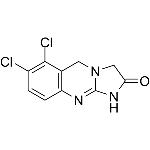
Anagrelide is a potent inhibitor of phosphodiesterase type III (PDE3) (IC50=36 nM). Anagrelide, an imidazoquinazoline derivative, acts as an inhibitor of platelet aggregation. Anagrelide inhibits bone marrow megakaryocytopoiesis. Anagrelide decreases gastrointestinal stromal tumor (GIST) cell proliferation and promotes their apoptosis in vitro. Anagrelide is a platelet-lowering agent and plays in the antithrombopoietic action.
For research use only. We do not sell to patients.
- Purity : 99.87%
- CAS No.: 68475-42-3
- Formula: C10H7Cl2N3O
- Molecular Weight:256.09
-
Storage:Powder -20°C, 3 years , 4°C, 2 years ; In solvent -80°C, 6 months , -20°C, 1 month

To place orders, for customer services and technical support, please contact: MedChemExpress USA
Tel: 609-228-6898 E-mail: [email protected] [email protected]


-
 Biological Activity
-
Chemical Information
-
Solvent & Solubility
- Protocols
- Purity & Documentation
- References
-
 Help & FAQs


-
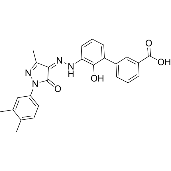
HY-15306
EltrombopagTPO Receptor Agonist -

HY-10182
LaduviglusibGSK-3 Inhibitor -
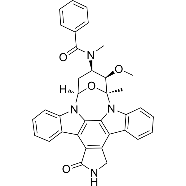
HY-10230
MidostaurinMulti-targeted Protein Kinase Inhibitor -

HY-114164
Thrombin (MW 37kDa)Procoagulant/Anticoagulant -
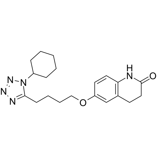
HY-17464
CilostazolPDE3 Inhibitor -
HY-15001
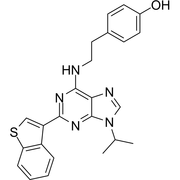
Stemregenin 1AhR Antagonist -
HY-122903
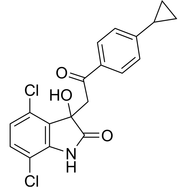
TK216ETS/EWS-FLI1 Inhibitor -
HY-N2193
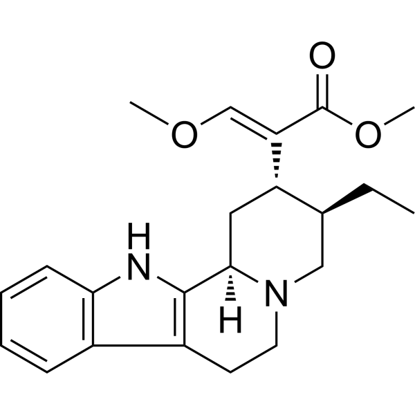
HirsutineApoptosis Inducer
Publications Citing Use of MedChemExpress (MCE) Anagrelide
More

Biological Activity
Description
IC50 & Target
PDEIII[1]
Cellular Effect
|
Cell Line

|
Type | Value | Description | References |
|---|---|---|---|---|
| HeLa | EC50 |
4 nM
Compound: 1
|
Antiproliferative activity against human HeLa cells assessed as reduction in cell growth
Antiproliferative activity against human HeLa cells assessed as reduction in cell growth
|
[PMID: 37077378] |
| HeLa | EC50 |
5.12 nM
Compound: 1
|
Antiproliferative activity against human HeLa cells assessed as reduction in cell viability measured after 72 hrs by MTT assay
Antiproliferative activity against human HeLa cells assessed as reduction in cell viability measured after 72 hrs by MTT assay
|
[PMID: 37077378] |
| HeLa | EC50 |
6.67 nM
Compound: 1
|
Cytotoxicity against human HeLa cells assessed as reduction in cell viability after 72 hrs by MTT assay
Cytotoxicity against human HeLa cells assessed as reduction in cell viability after 72 hrs by MTT assay
|
[PMID: 37077378] |
| Platelet | EC50 |
<1 μg/mL
Compound: 1
|
Antiplatelet aggregation activity in rabbit platelet-rich plasma assessed as reduction in ADP-induced aggregation incubated for 5 mins by aggregometry
Antiplatelet aggregation activity in rabbit platelet-rich plasma assessed as reduction in ADP-induced aggregation incubated for 5 mins by aggregometry
|
[PMID: 37077378] |
| Platelet | EC50 |
0.08 μM
Compound: 1
|
Antiplatelet aggregation activity in human rich plasma assessed as reduction in collagen-induced platelet aggregation
Antiplatelet aggregation activity in human rich plasma assessed as reduction in collagen-induced platelet aggregation
|
[PMID: 37077378] |
| Platelet | EC50 |
0.08 μM
Compound: 1
|
Antiplatelet aggregation activity in rhesus monkey platelet rich plasma assessed as reduction in collagen-induced platelet aggregation
Antiplatelet aggregation activity in rhesus monkey platelet rich plasma assessed as reduction in collagen-induced platelet aggregation
|
[PMID: 37077378] |
| Platelet | EC50 |
0.15 μM
Compound: 1
|
Antiplatelet aggregation activity in Sprague-Dawley rat platelet rich plasma assessed as reduction in collagen-induced platelet aggregation
Antiplatelet aggregation activity in Sprague-Dawley rat platelet rich plasma assessed as reduction in collagen-induced platelet aggregation
|
[PMID: 37077378] |
| Platelet | EC50 |
0.27 μM
Compound: 1
|
Antiplatelet aggregation activity in Sprague-Dawley rat platelet rich plasma assessed as reduction in ADP-induced platelet aggregation
Antiplatelet aggregation activity in Sprague-Dawley rat platelet rich plasma assessed as reduction in ADP-induced platelet aggregation
|
[PMID: 37077378] |
| Platelet | EC50 |
0.31 μM
Compound: 1
|
Antiplatelet activity in rabbit platelet rich plasma assessed as inhibition of collagen-induced platelet aggregation
Antiplatelet activity in rabbit platelet rich plasma assessed as inhibition of collagen-induced platelet aggregation
|
[PMID: 37077378] |
| Platelet | EC50 |
0.31 μM
Compound: 1
|
Antiplatelet aggregation activity in dog rich plasma assessed as reduction in ADP-induced platelet aggregation
Antiplatelet aggregation activity in dog rich plasma assessed as reduction in ADP-induced platelet aggregation
|
[PMID: 37077378] |
| Platelet | EC50 |
0.39 μM
Compound: 1
|
Antiplatelet activity in rabbit platelet rich plasma assessed as inhibition of arachidonic acid-induced platelet aggregation
Antiplatelet activity in rabbit platelet rich plasma assessed as inhibition of arachidonic acid-induced platelet aggregation
|
[PMID: 37077378] |
| Platelet | EC50 |
0.4 μM
Compound: 1
|
Antiplatelet aggregation activity in human rich plasma assessed as reduction in ADP-induced platelet aggregation
Antiplatelet aggregation activity in human rich plasma assessed as reduction in ADP-induced platelet aggregation
|
[PMID: 37077378] |
| Platelet | EC50 |
0.5 μM
Compound: 1
|
Antiplatelet aggregation activity in rhesus monkey platelet rich plasma assessed as reduction in ADP-induced platelet aggregation
Antiplatelet aggregation activity in rhesus monkey platelet rich plasma assessed as reduction in ADP-induced platelet aggregation
|
[PMID: 37077378] |
| Platelet | EC50 |
0.7 μM
Compound: 1
|
Antiplatelet activity in rabbit platelet rich plasma assessed as inhibition of thrombin-induced platelet aggregation
Antiplatelet activity in rabbit platelet rich plasma assessed as inhibition of thrombin-induced platelet aggregation
|
[PMID: 37077378] |
| Platelet | EC50 |
0.74 μM
Compound: 1
|
Antiplatelet activity in rabbit platelet rich plasma assessed as inhibition of antigen-antibody-induced platelet aggregation
Antiplatelet activity in rabbit platelet rich plasma assessed as inhibition of antigen-antibody-induced platelet aggregation
|
[PMID: 37077378] |
| Platelet | EC50 |
1.2 μM
Compound: 1
|
Antiplatelet aggregation activity in rabbit platelet rich plasma assessed as reduction in ADP-induced platelet aggregation
Antiplatelet aggregation activity in rabbit platelet rich plasma assessed as reduction in ADP-induced platelet aggregation
|
[PMID: 37077378] |
| Platelet | EC50 |
27 nM
Compound: 1
|
Antiplatelet activity against ADP-induced Wistar rat platelet aggregation assessed as inhibition of platelet aggregation
Antiplatelet activity against ADP-induced Wistar rat platelet aggregation assessed as inhibition of platelet aggregation
|
[PMID: 37077378] |
In Vitro
Anagrelide potently inhibits the development of marrow megakaryocytes (IC50=26 nM)[1].
Anagrelide (0.05, 0.3, 1 µM; 12-day) inhibits only megakaryocytic cell growth not non-megakaryocytic cells[2].
Anagrelide (0.1-10000 nM) induces a cytotoxic effect in the GIST882 cell line[3].
MedChemExpress (MCE) has not independently confirmed the accuracy of these methods. They are for reference only.
-
Cell Line:Megakaryocytic and non-megakaryocytic cells
-
Concentration:0.05, 0.3, 1 µM
-
Incubation Time:12-day
-
Result:Inhibited only megakaryocytic cell growth at every concentration tested
-
Cell Line:GIST882 and GIST48 cell line
-
Concentration:0.1, 1, 10, 100, 1000, 10000 nM
-
Incubation Time:
-
Result:Induced a cytotoxic effect in the GIST882 cell line (IC50= 16 nM), but was only weakly active in the GIST48 cell line.
In Vivo
MedChemExpress (MCE) has not independently confirmed the accuracy of these methods. They are for reference only.
-
Animal Model:Adult female athymic mice bearing GIST2B, GIST3, GIST9, GIST882 model[3]
-
Dosage:5 mg/kg
-
Administration:Twice daily; for 10 days
-
Result:Inhibited or reduced tumor growth in three (GIST2B, GIST9, GIST882) of these four models.
- Annexin V plus membrane-impermeant dye apoptosis staining
- BrdU Incorporation Assay
- Somatic Cell Culture
- CFSE Dye Dilution Proliferation Assay
- Dye-dilution cell tracking and proliferation staining
- CCK-8/WST-8 Cell Proliferation Assay
- Protocol for Cell Cycle
- Cell Counting-Based Growth Curve Assay
- Colony Formation (Clonogenic) Assay
- Cell Viability Determination by MTT Colorimetric Assay
- EdU Incorporation Assay (Click Chemistry-Based DNA Synthesis Measurement)
- Ki-67 Immunostaining Proliferation Assay
- Detection of Apoptosis
- TUNEL staining for apoptotic DNA fragmentation
- PCNA Immunodetection Proliferation Assay
- Protocol for Cell Counting and Cell Density Analysis
- Apoptosis Solutions
- MTT Cell Proliferation Assay
Clinical Trial
| NCT Number | Sponsor | Condition | Start Date |
Phase
|
|---|---|---|---|---|
| NCT01329991 | Plexxikon| | 2011-05 | PHASE1 |


Chemical Information
-
CAS No. 68475-42-3
-
Appearance Solid
-
Molecular Weight 256.09
-
Formula C10H7Cl2N3O
-
Color White to off-white
-
SMILES
O=C1NC2=NC3=C(C(Cl)=C(Cl)C=C3)CN2C1
-
Shipping
Room temperature in continental US; may vary elsewhere.
-
Storage
Powder -20°C 3 years 4°C 2 years In solvent -80°C 6 months -20°C 1 month


Publications (3)
-
Journal Impact Factor
-
Most Recent
-
Cell Metab
Imatinib and methazolamide ameliorate COVID-19-induced metabolic complications via elevating ACE2 enzymatic activity and inhibiting viral entry. [Abstract]2022 Mar 1;34(3):424-440.e7. PMID: 35150639 -
Anal Chem
Exposome-Scale Investigation of Cl-/Br-Containing Chemicals Using High-Resolution Mass Spectrometry, Multistage Machine Learning, and Cloud Computing. [Abstract]2025 Jun 3;97(21):11099-11109. PMID: 40401576 -
Exp Hematol
Purging myeloma cell contaminants and simultaneous expansion of peripheral blood mobilised stem cells. [Abstract]2024 Mar:131:104138. PMID: 38151170


Solvent & Solubility
In Vitro:
DMSO : 2.56 mg/mL (10.00 mM; ultrasonic and warming and heat to 60°C; Hygroscopic DMSO has a significant impact on the solubility of product, please use newly opened DMSO)
Please refer to the solubility information to select the appropriate solvent. Once prepared, please aliquot and store the solution to prevent product inactivation from repeated freeze-thaw cycles.
Storage method and period of stock solution: -80°C, 6 months; -20°C, 1 month. When stored at -80°C, please use it within 6 months. When stored at -20°C, please use it within 1 month.
Please refer to the solubility information to select the appropriate solvent. Once prepared, please aliquot and store the solution to prevent product inactivation from repeated freeze-thaw cycles.
Storage method and period of stock solution: -80°C, 6 months; -20°C, 1 month. When stored at -80°C, please use it within 6 months. When stored at -20°C, please use it within 1 month.
Concentration (start) × Volume (start) = Concentration (final) × Volume (final)


Purity & Documentation
-
Data Sheet (283 KB)
-
SDS (393 KB)
- English - EN (393 KB)
- Français - FR (393 KB)
- Deutsch - DE (393 KB)
- Norwegian - NO (393 KB)
- Español - ES (393 KB)
- Swedish - SV (393 KB)
- Italian - IT (393 KB)
- Korean - KR (393 KB)
- Portuguese - PT (393 KB)
-
Handling Instructions (2659 KB)
References
[1]. Guosu Wang, et al. Comparison of the biological activities of Anagrelide and its major metabolites in haematopoietic cell cultures. Br J Pharmacol. 2005 Oct;146(3):324-32. [Content Brief]
[2]. Y Hong, et al. Comparison between Anagrelide and hydroxycarbamide in their activities against haematopoietic progenitor cell growth and differentiation: selectivity of Anagrelide for the megakaryocytic lineage. Leukemia. 2006 Jun;20(6):1117-22. [Content Brief]
[3]. Olli-Pekka Pulkka, et al. Anagrelide for Gastrointestinal Stromal Tumor. Clin Cancer Res. 2019 Mar 1;25(5):1676-1687. [Content Brief]


Complete Stock Solution Preparation Table
Please refer to the solubility information to select the appropriate solvent. Once prepared, please aliquot and store the solution to prevent product inactivation from repeated freeze-thaw cycles.
Storage method and period of stock solution: -80°C, 6 months; -20°C, 1 month. When stored at -80°C, please use it within 6 months. When stored at -20°C, please use it within 1 month.
| Optional Solvent | Concentration Solvent Mass | 1 mg | 5 mg | 10 mg | 25 mg |
|---|---|---|---|---|---|
| DMSO | 1 mM | 3.9049 mL | 19.5244 mL | 39.0488 mL | 97.6219 mL |
| 5 mM | 0.7810 mL | 3.9049 mL | 7.8098 mL | 19.5244 mL |


Anagrelide Related Classifications
HY-B0523 Related Classifications
 Powered by Bioz