



SH-5
Based on 1 publication(s) in Google Scholar
SH-5 is a potent AKT inhibitor. SH-5 potentiates the apoptosis induced by tumor necrosis factor. SH-5 blocks NF-kB activation induced by TNF-a, lipopolysaccharide (HY-D1056), phorbol ester (HY-18739), and cigarette smoke.
For research use only. We do not sell to patients.
- CAS No.: 701976-54-7
- Formula: C29H59O10P
- Molecular Weight:598.75
-
Storage:
Please store the product under the recommended conditions in the Certificate of Analysis.


To place orders, for customer services and technical support, please contact: MedChemExpress USA
Tel: 609-228-6898 E-mail: [email protected] [email protected]


-
 Biological Activity
-
Chemical Information
- Protocols
- Purity & Documentation
- References
-
 Help & FAQs


-
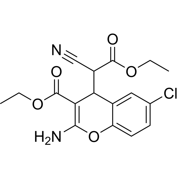
HY-18749
SC79Akt Activator -
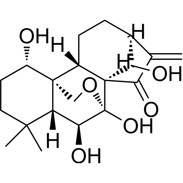
HY-N0004
OridoninBacterial Inhibitor -
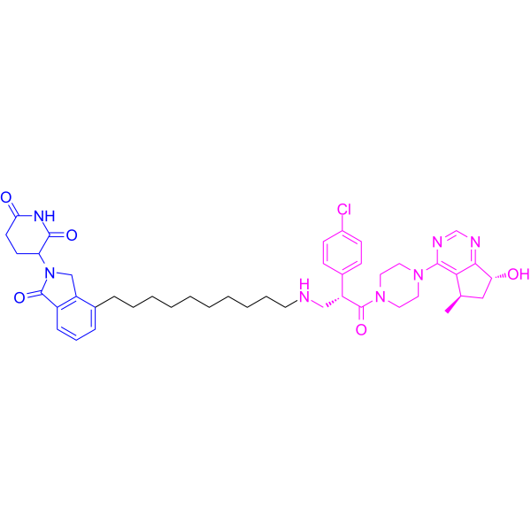
HY-133120
INY-03-041PROTAC Akt Degrader -
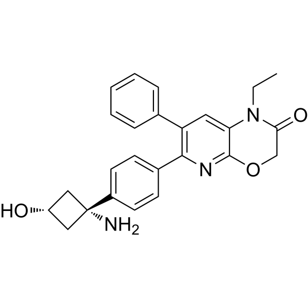
HY-151504
EngasertibAKT1/2 Inhibitor -
HY-50862
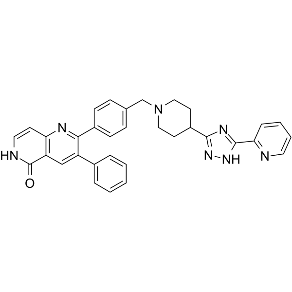
Akt1/Akt2-IN-1Akt Inhibitor -
HY-13254A
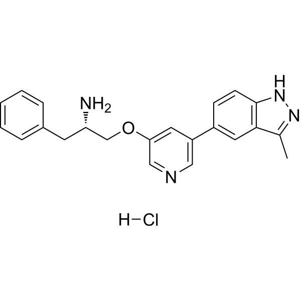
A-674563 hydrochlorideAkt1 Inhibitor -
HY-14971
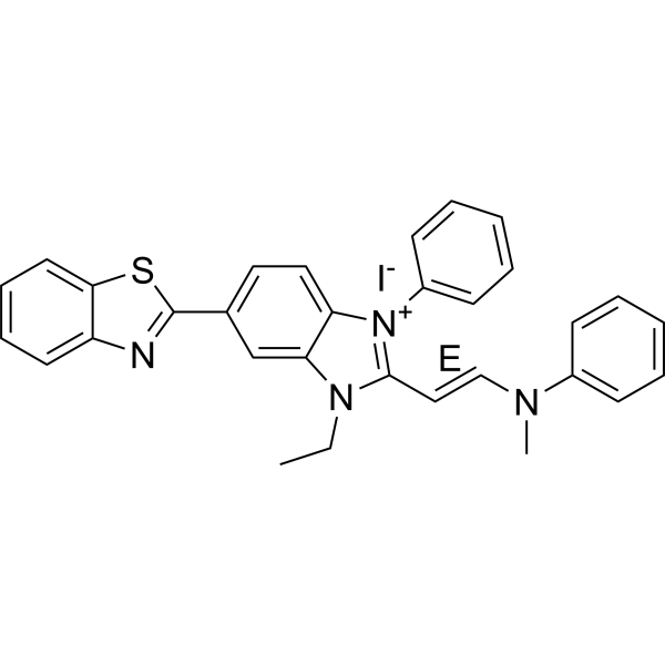
(E)-Akt inhibitor-IVPI3K-Akt Inhibitor -
HY-145282
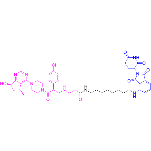
MS170PROTAC AKT Degrader -
HY-15186A
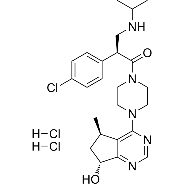
Ipatasertib dihydrochloridepan-Akt Inhibitor -
HY-110077
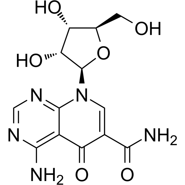
API-1Akt/PKB Inhibitor -
HY-16666
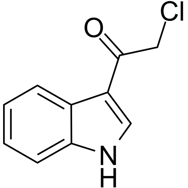
3CAIAkt Inhibitor
Publications Citing Use of MedChemExpress (MCE) SH-5
More

Biological Activity
Description
Cellular Effect
|
Cell Line

|
Type | Value | Description | References |
|---|---|---|---|---|
| 184B5 | GI50 |
10.35 μM
Compound: 2
|
Cytotoxicity against human 184B5 cells at 10 uM chloroquine by SRB assay
Cytotoxicity against human 184B5 cells at 10 uM chloroquine by SRB assay
|
[PMID: 18691894] |
| 184B5 | GI50 |
13.76 μM
Compound: 2
|
Cytotoxicity against human 184B5 cells by SRB assay
Cytotoxicity against human 184B5 cells by SRB assay
|
[PMID: 18691894] |
| 184B5 | GI50 |
8.98 μM
Compound: 2
|
Cytotoxicity against human 184B5 cells at 20 uM chloroquine by SRB assay
Cytotoxicity against human 184B5 cells at 20 uM chloroquine by SRB assay
|
[PMID: 18691894] |
| MCF7 | GI50 |
15.64 μM
Compound: 2
|
Cytotoxicity against human MCF7 cells in presence of 10 uM chloroquine by SRB assay
Cytotoxicity against human MCF7 cells in presence of 10 uM chloroquine by SRB assay
|
[PMID: 18691894] |
| MCF7 | GI50 |
16.01 μM
Compound: 2
|
Cytotoxicity against human MCF7 cells by SRB assay
Cytotoxicity against human MCF7 cells by SRB assay
|
[PMID: 18691894] |
| MCF7 | GI50 |
7.94 μM
Compound: 2
|
Cytotoxicity against human MCF7 cells in presence of 20 uM chloroquine by SRB assay
Cytotoxicity against human MCF7 cells in presence of 20 uM chloroquine by SRB assay
|
[PMID: 18691894] |
| MDA-MB-231 | GI50 |
14.67 μM
Compound: 2
|
Cytotoxicity against human MDA-MB-231 cells in presence of 10 uM chloroquine by SRB assay
Cytotoxicity against human MDA-MB-231 cells in presence of 10 uM chloroquine by SRB assay
|
[PMID: 18691894] |
| MDA-MB-231 | GI50 |
16.77 μM
Compound: 2
|
Cytotoxicity against human MDA-MB-231 cells by SRB assay
Cytotoxicity against human MDA-MB-231 cells by SRB assay
|
[PMID: 18691894] |
| MDA-MB-231 | GI50 |
9.39 μM
Compound: 2
|
Cytotoxicity against human MDA-MB-231 cells in presence of 20 uM chloroquine by SRB assay
Cytotoxicity against human MDA-MB-231 cells in presence of 20 uM chloroquine by SRB assay
|
[PMID: 18691894] |
| MDA-MB-468 | GI50 |
0.06 μM
Compound: 2
|
Cytotoxicity against human MDA-MB-468 cells in presence of 20 uM chloroquine by SRB assay
Cytotoxicity against human MDA-MB-468 cells in presence of 20 uM chloroquine by SRB assay
|
[PMID: 18691894] |
| MDA-MB-468 | GI50 |
19.26 μM
Compound: 2
|
Cytotoxicity against human MDA-MB-468 cells in presence of 10 uM chloroquine by SRB assay
Cytotoxicity against human MDA-MB-468 cells in presence of 10 uM chloroquine by SRB assay
|
[PMID: 18691894] |
| MDA-MB-468 | GI50 |
28.43 μM
Compound: 2
|
Cytotoxicity against human MDA-MB-468 cells by SRB assay
Cytotoxicity against human MDA-MB-468 cells by SRB assay
|
[PMID: 18691894] |
In Vitro
SH-5 (5 µM; 4+24 h) increases the cytotoxicity with 1 nM TNF in H1299 cells[1].
SH-5 (5 µM; 4+0, 6, 12, 18, 24 h) potentiates the apoptosis induced by tumor necrosis factor (TNF) in H1299 cells[1].
SH-5 (1, 3, 5 µM) inhibits TNF (1 nM; 24 h)-induced COX-2 gene expression in a dose-dependent manner[1].
SH-5 (5 µM; 4+0, 6, 12, 18, 24 h) decreases the protein expression of 1 nM TNF-induced IAP-1, XIAP, Bcl-Xl, Bcl-2, TRAF1, Survivin, COX-2, Cyclin D1, MMP-9 in a time-dependent manner in KBM-5 cells[1].
MedChemExpress (MCE) has not independently confirmed the accuracy of these methods. They are for reference only.
-
Cell Line:H1299 cells
-
Concentration:5 µM
-
Incubation Time:4+0, 6, 12, 18, 24 h
-
Result:Increased the protein expression of Cleaved-PARP in a time-dependent manner.


Chemical Information
-
CAS No. 701976-54-7
-
Molecular Weight 598.75
-
Formula C29H59O10P
-
SMILES
CCCCCCCCCCCCCCCCCCOC[C@@H](OC)COP(O[C@@H]1[C@@H](C[C@H]([C@@H]([C@H]1O)O)O)OC)(O)=O
-
Shipping
Room temperature in continental US; may vary elsewhere.
-
Storage
Please store the product under the recommended conditions in the Certificate of Analysis.


Publications (1)
-
Journal Impact Factor
-
Most Recent


Purity & Documentation
-
Data Sheet (263 KB)
-
SDS (252 KB)
- Français - FR (252 KB)
- Deutsch - DE (252 KB)
- Norwegian - NO (252 KB)
- Español - ES (252 KB)
- Swedish - SV (252 KB)
- Italian - IT (252 KB)
- Korean - KR (252 KB)
- Portuguese - PT (252 KB)
-
Handling Instructions (2659 KB)
References

Calculators
Concentration (start) × Volume (start) = Concentration (final) × Volume (final)


SH-5 Related Classifications
HY-100702 Related Classifications
 Powered by Bioz