



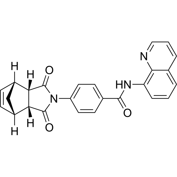
IWR-1 (IWR-1-endo) (GMP) is the IWR-1 (HY-12238) produced by using GMP guidelines. GMP small molecules work appropriately as an auxiliary reagent for cell therapy manufacture. IWR-1 (IWR-1-endo) is a tankyrase inhibitor targeting Wnt/β-catenin (IC50 = 180 nM). IWR-1 compromises critical steps of the canonical Wnt signaling, namely translocation of β-catenin to the nucleus and subsequent TCF/LEF activation and expression of Wnt/β-catenin downstream targets. IWR-1 promotes β-catenin phosphorylation by promoting stability of Axin-scaffolded destruction complexes. IWR-1 can be studied in research for anti-tumor purposes, and diseases such as osteosarcoma, colorectal cancer and psoriasis.
For research use only. We do not sell to patients.
- CAS No.: 1127442-82-3
- Formula: C25H19N3O3
- Molecular Weight:409.44
-
Storage:
Please store the product under the recommended conditions in the Certificate of Analysis.


To place orders, for customer services and technical support, please contact: MedChemExpress USA
Tel: 609-228-6898 E-mail: [email protected] [email protected]


-
 Biological Activity
-
Chemical Information
- Protocols
- Purity & Documentation
- References
-
 Help & FAQs


-
HY-12238
IWR-1Wnt Signaling Inhibitor -
HY-13912
IWP-2Wnt Production Inhibitor -
HY-14428
ICG-001Apoptosis Inducer -
HY-12879
IWP-4Wnt/CK1δ/ε Inhibitor -
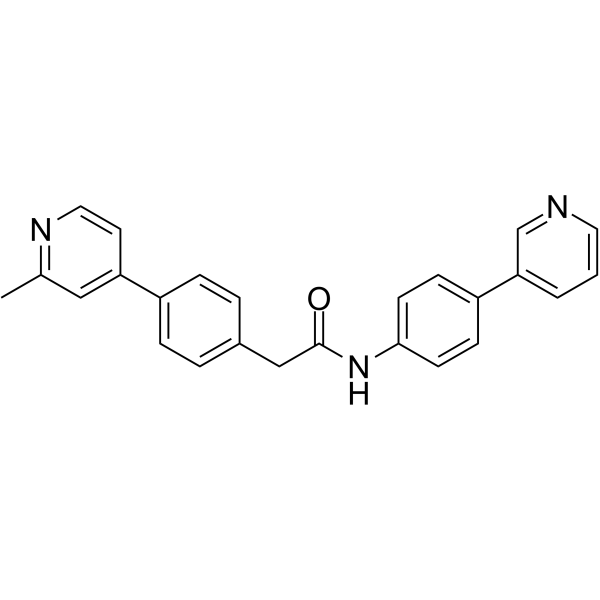
HY-15659
Wnt-C59PORCN Inhibitor -
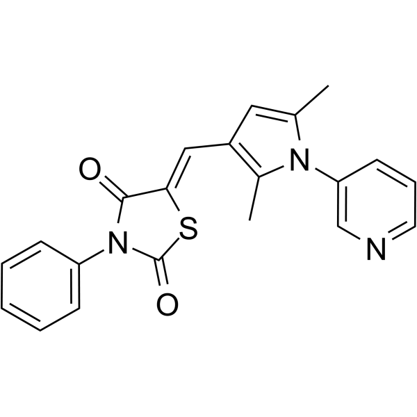
HY-16665
iCRT 14Wnt Inhibitor -
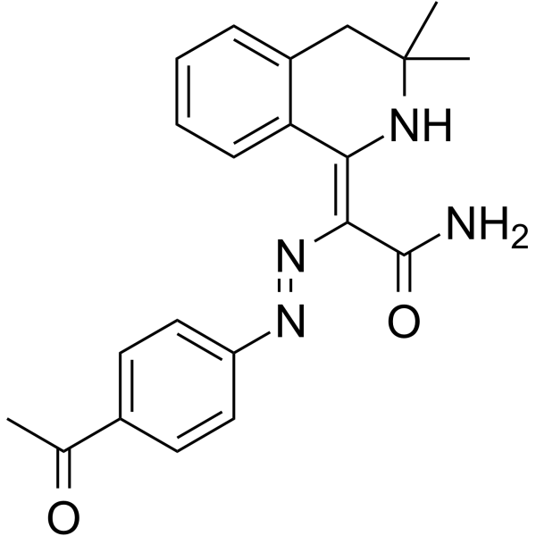
HY-10593
IQ 1Wnt/β-catenin/CBP Signaling Sustainer -
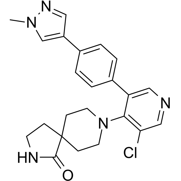
HY-12681
CCT251545WNT Pathway Inhibitor -
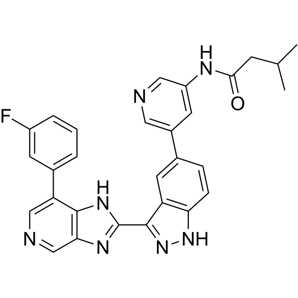
HY-109049
AdavivintWnt Pathway Inhibitor -
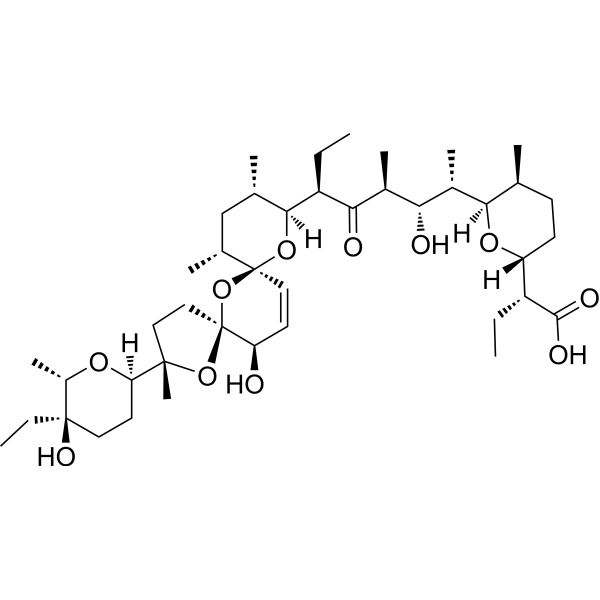
HY-15597
SalinomycinPotassium Ionophore Antibiotic -
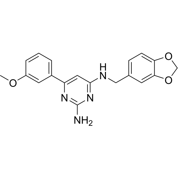
HY-19987
BML-284Wnt Signaling Activator -
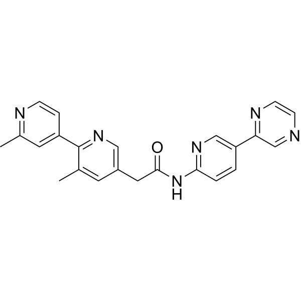
HY-17545
LGK974PORCN Inhibitor


Biological Activity
Description
Cellular Effect
|
Cell Line

|
Type | Value | Description | References |
|---|---|---|---|---|
| A549 | GI50 |
>100 μM
Compound: IWR1
|
Antiproliferative activity against human A549 cells after 72 hrs by MTT assay
Antiproliferative activity against human A549 cells after 72 hrs by MTT assay
|
[PMID: 24950489] |
| HEK293 | IC50 |
136 μM
Compound: 1, IWR1
|
Inhibition of Tankyrase in human HEK293 cells assessed as inhibition of Wnt pathway by Wnt3a-induced STF assay
Inhibition of Tankyrase in human HEK293 cells assessed as inhibition of Wnt pathway by Wnt3a-induced STF assay
|
[PMID: 23316926] |
| HEK-293T | IC50 |
26 nM
Compound: 1, IWR-1
|
Inhibition of beta-casein-dependent canonical Wnt3 pathway in human HEK293T cells by luciferase reporter gene assay
Inhibition of beta-casein-dependent canonical Wnt3 pathway in human HEK293T cells by luciferase reporter gene assay
|
[PMID: 22191557] |
| HepG2 | GI50 |
95.4 μM
Compound: IWR1
|
Antiproliferative activity against human HepG2 cells after 72 hrs by MTT assay
Antiproliferative activity against human HepG2 cells after 72 hrs by MTT assay
|
[PMID: 24950489] |
| HT-29 | GI50 |
>100 μM
Compound: IWR1
|
Antiproliferative activity against human HT-29 cells after 72 hrs by MTT assay
Antiproliferative activity against human HT-29 cells after 72 hrs by MTT assay
|
[PMID: 24950489] |
| HT-29 | IC50 |
24.4 μM
Compound: IWR1
|
Inhibition of Wnt signaling in human HT29 cells assessed as inhibition of beta-catenin-mediated Tcf/Lef transcriptional activity after 24 hrs by dual luciferase reporter gene assay relative to control
Inhibition of Wnt signaling in human HT29 cells assessed as inhibition of beta-catenin-mediated Tcf/Lef transcriptional activity after 24 hrs by dual luciferase reporter gene assay relative to control
|
[PMID: 24950489] |
| HT-29 | IC50 |
9.52 μM
Compound: IWR-1
|
Cytotoxicity against human HT-29 cells assessed as cell viability at 1.563 to 50 uM measured after 72 hrs by plate reader method
Cytotoxicity against human HT-29 cells assessed as cell viability at 1.563 to 50 uM measured after 72 hrs by plate reader method
|
[PMID: 34062253] |
| LoVo | GI50 |
63.1 μM
Compound: IWR1
|
Antiproliferative activity against human LoVo cells after 72 hrs by MTT assay
Antiproliferative activity against human LoVo cells after 72 hrs by MTT assay
|
[PMID: 24950489] |
| SW480 | EC50 |
2.5 μM
Compound: 1, IWR1
|
Inhibition of tankyrase in human SW480 cells assessed as accumulation of axin2 after 24 hrs by Hoechst dye-based method
Inhibition of tankyrase in human SW480 cells assessed as accumulation of axin2 after 24 hrs by Hoechst dye-based method
|
[PMID: 23701517] |
| SW480 | IC50 |
0.25 μM
Compound: 1, IWR1
|
Inhibition of tankyrase in human SW480 cells assessed as degradation of beta catenin after 40 to 48 hrs
Inhibition of tankyrase in human SW480 cells assessed as degradation of beta catenin after 40 to 48 hrs
|
[PMID: 23701517] |
In Vitro
IWR-1 (GMP) is cytotoxic for osteosarcoma cancer stem-like cells (CSCs)[1].
IWR-1 (GMP) suppresses cell migration, invasion, and matrix metalloproteinase activities of colorectal cancer cell lines[2].
IWR-1 (GMP) (2.5-10 μM, 48-96 h) is effective in reducing spheres’ viability in a concentration- and time- dependent manner in parental and spheres from MG-63 and MNNG-HOS cell lines[1].
IWR-1 (GMP) (10 μM, 96 h) increases the number of TUNEL-positive cells, reaching 4.65- and 15.83-fold differences relative to control at 96 h and promoted the activation of caspases 3/7 reaching 2.15- and 1.27-fold in MG-63 and MNNG-HOS spheres[1].
IWR-1 (GMP) (10 μM, 48 h) induces a cell cycle arrest in the G2/M phase in spheres derived from MG-63 and MNNG-HOS cell lines and increases the percentage of cells in the S phase slightly[1].
IWR-1 (GMP) (10 μM, 48 h) inhibits secondary sphere-forming efficacy by approximately 53% and 55% of the first-generation 7-day old spheres in MG-6t4 and MNNG-HOS cells[1].
IWR-1 (GMP) (5-50 μM, 24-48 h) decreases the proliferation of HCT116 cells in a dose- and time-dependent manner[2].
IWR-1 (GMP) (5-50 μM, 24-48 h) inhibits TNF-α-stimulated migration in HCT116 and HT29 cells[2].
MedChemExpress (MCE) has not independently confirmed the accuracy of these methods. They are for reference only. Further protocols information, click here.
-
Cell Line:MG-63 and MNNG-HOS spheres and parent cells
-
Concentration:2.5, 5, 7.5, 10 μM
-
Incubation Time:48 and 96 h
-
Result:Reduced cell viability when concentration is higher than 5 μM.
Elicited more than 70% reduction of cell viability in spheres derived from the two cell lines at 96 h with 10 μM.
Had minimal effect on parental cells since the Wnt/β-catenin signaling is absent in these cells. Increased the susceptibility of spheres towards Doxorubicin (HY-15142) when treated in combination with increasing concentrations of Doxorubicin (0.01-100 μM).
-
Cell Line:MG-63 and MNNG-HOS spheres and parent cells
-
Concentration:10 μM
-
Incubation Time:96 h
-
Result:Led to an upregulation of Bak and a downregulation of Bcl-2 (key proteins involved in mitochondrial-dependent apoptosis), which contributes to a Bak/Bcl-2 ratio >1 that prone the cells to undergo apoptosis.
-
Cell Line:MG-63 and MNNG-HOS spheres and parent cells
-
Concentration:10 μM
-
Incubation Time:96 h
-
Result:Led to an upregulation of Bak and a downregulation of Bcl-2 (key proteins involved in mitochondrial-dependent apoptosis), which contributes to a Bak/Bcl-2 ratio >1 that prone the cells to undergo apoptosis.
Results in a higher β-catenin nuclear/cytoplasmic ratio in spheres, indicating an increased Wnt/β-catenin pathway activation in osteosarcoma spheres.
Stabilized Axin2 protein levels.
Diminished the protein expression levels of Cyclin D1 in both parental cells and spheres.
-
Cell Line:HCT116 cells
-
Concentration:5, 10, 20, 50 μM
-
Incubation Time:0, 4, 8, 24, 48 h
-
Result:Increased the levels of the epithelial marker E-cadherin whilst decreased the mesenchymal markers N-cadherin, Vimentin, and Snail dose- and time-dependently.
Inhibited the EMT process in HCT116 cells effectively.
Decreased β-catenin expression and inhibited the EMT-like expressional changes whereby decreasing N-cadherin and Snail and increasing E-cadherin expressions, even in the presence of TNF-α (HY-P1860) (10 ng/mL for 24 h)-induced EMT stimulation.
Decreased the phosphorylation of Akt in a concentration- and time-dependent manner.
Decreased the surviving expression in a concentration- and time-dependent manner, thereby promoting tumor proliferation directly or indirectly through regulating cancer cell homeostasis.
Reduced MMP activities only when surviving was suppressed.
In Vivo
IWR-1 (GMP) (10 mg/kg, s.c., on days 1, 3, 5) ameliorates IL-36γ (2 μg/mouse on days 1, 3, 5)-mediated exacerbation of psoriatic skin lesions in an Imiquimod (IMQ) (HY-B0180)-induced psoriasis-like mice model[3].
MedChemExpress (MCE) has not independently confirmed the accuracy of these methods. They are for reference only.
-
Animal Model:Immunocompromised nude mice (6-week old female Swiss nude) were injected sub-cutaneously the pGL4-transfected MNNG-HOS cells (2×10⁶ cells/100 mL PBS)[1]
-
Dosage:5 mg/kg
-
Administration:Intratumorally, each 2 d for 12 d
-
Result:Resulted in slower tumor growth rate and reduction in tumor size by 73% and 71% in comparison to control and to Doxorubicin-treated (8 mg/kg, i.p., each 4 d for 12 d) groups.
Enhanced the therapeutic efficacy of Doxorubicin shown by a greater reduction of tumor burden at the end of the treatment in opposite to Doxorubicin alone.
-
Animal Model:Balb/c mice (aged 6-8 weeks) with shaven back and treated with a daily topical dose of IMQ cream[3]
-
Dosage:10 mg/kg
-
Administration:Subcutaneous injection (s.c.) on days 1, 3, 5
-
Result:Increased epidermal thickening with keratinocyte thickness.
Significantly diminished the effect of IMQ on hyperplasia in the IMQ+IWR-1 group.
Ameliorated the pathological changes in IL-36γ-induced psoriasiform skin lesion.
Reversed IL-36γ-mediated upregulation of inflammatory factors (IL-17 A and IFN-γ) in psoriatic lesions.
Reversed IL-36γ-mediated upregulation of β-catenin and DKK1 expression.


Chemical Information
-
CAS No. 1127442-82-3
-
Molecular Weight 409.44
-
Formula C25H19N3O3
-
SMILES
O=C(NC1=C2N=CC=CC2=CC=C1)C3=CC=C(N(C([C@]4([H])[C@](C5)([H])C=C[C@]5([H])[C@]64[H])=O)C6=O)C=C3
-
Synonyms
endo-IWR 1 (GMP); IWR-1-endo (GMP)
-
Shipping
Room temperature in continental US; may vary elsewhere.
-
Storage
Please store the product under the recommended conditions in the Certificate of Analysis.

Protocols
-
Kinase activity and phosphorylation assays
Kinase activity assays measure the ability of kinases to transfer phosphate groups from ATP to specific substrates, while phosphorylation assays detect the presence and levels of phosphorylated proteins. Common methods include radiolabeled ATP incorporation (e. g. ,), ADP release detection via bioluminescence (e. g. ,[3]), enzyme-linked immunosorbent assays (ELISA) for phospho-specific epitopes (e. g. ,[6]), and microtiter-based formats for high-throughput screening (e. g. ,[8]). The ADP-Glo assay quantifies kinase activity by measuring ADP produced during phosphorylation using a luciferase-based system. Radiometric assays involve autoradiography or scintillation counting after incorporation of 32P-labeled ATP into substrate proteins. ELISA-based approaches rely on phospho-specific antibodies to detect activated kinases in cell lysates or purified samples.
-
RNA extraction experimental
By lysing cells, releasing RNA, and removing impurities such as proteins and DNA, high-purity RNA products are finally obtained. The commonly used traditional method is the guanidine isothiocyanate/phenol/chloroform method (Trizol), which is suitable for a variety of animal materials including animal tissues, microorganisms, cultured cells, etc., and most plant materials.
-
Western Blot
Western blotting (WB) is a commonly used experimental method in molecular biology, biochemistry, and immunogenetics for identifying and quantifying target proteins. It combines gel electrophoresis with immunoassay, enabling researchers to analyze protein expression, post-translational modifications, and molecular weight.
-
Cell Cytotoxicity Assay
Cytotoxicity assays are usually based on the assessment of cell membrane damage, which can also be indirectly detected by measuring cell viability. Detection methods include MTT assay, CKK-8 assay, LDH assay and ATP assay, etc.
-
Imiquimod-Induced Psoriasiform Dermatitis
Imiquimod (IMQ)-induced psoriasiform dermatitis is a widely used murine model in which topical application of IMQ, a Toll-like receptor 7 (TLR7) agonist, triggers innate immune activation in the skin and induces a psoriasis-like inflammatory cascade characterized by epidermal hyperplasia, immune cell infiltration, and cytokine production dominated by the IL-23/IL-17 axis. This inflammatory response is mediated through activation of dendritic cells and downstream induction of IL-23, IL-17A, IL-22, and related pro-inflammatory mediators, recapitulating key features of human plaque psoriasis and enabling mechanistic and therapeutic studies. The model is commonly induced using Aldara (5% IMQ cream) applied topically to murine skin, resulting in rapid onset of erythema, scaling, and thickening that can be quantified as disease severity indices and validated histologically.
-
Protocol for Kinase activity and phosphorylation assays
Kinase activity assays measure transfer of phosphate from ATP to a protein or peptide substrate, generating phosphorylated substrate, ADP, or incorporated radiolabeled phosphate as the readout; phosphorylation assays measure site-specific phosphorylation in cells or tissues as a proxy for kinase-pathway activation, inhibition, or substrate regulation. Phosphorylation can be detected by phospho-specific Western blot, immunoprecipitation kinase assay, phospho-immunofluorescence, phospho-flow cytometry, luminescent ADP detection, radiolabeled ATP incorporation, or reporter-based pathway assays, and these readouts can be applied to cancer cells, primary neurons, mouse tumors, organoids, inflammatory macrophages, ferroptosis studies, and mitophagy studies when the kinase target is biologically relevant.


Purity & Documentation
References
[1]. Sara R. Martins-Neves, et al., IWR-1, a tankyrase inhibitor, attenuates Wnt/β-catenin signaling in cancer stem-like cells and inhibits in vivo the growth of a subcutaneous human osteosarcoma xenograft, Cancer Letters, Volume 414, 2018, Pages 1-15, ISSN 0304-3835. [Content Brief]
[3]. Wang, W. M., et al., IWR-1 attenuates the promotional effect of IL-36γ in a mouse model of psoriasis. BMC immunology, 25(1), 78. [Content Brief]
[4]. Chen, B., Dodge, M.E., Tang, W., et al. Small molecule-mediated disruption of Wnt-dependent signaling in tissue regeneration and cancer. Nat. Chem. Biol. 5(2), 100-107 (2009). [Content Brief]

Calculators
Concentration (start) × Volume (start) = Concentration (final) × Volume (final)


IWR-1 (GMP) Related Classifications
HY-12238G Related Classifications
 Powered by Bioz