



フィチン酸
Based on 1 publication(s) in Google Scholar
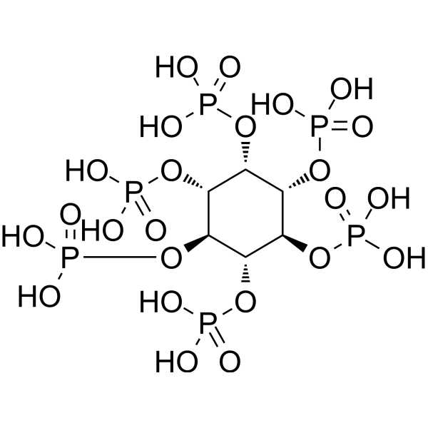
Phytic acid (myo-Inositol; hexakis dihydrogen phosphate; Inositol hexaphosphate) is an orally active compound. Phytic acid can be derived from the seeds of legumes. Phytic acid is a [PO4]3- storage depot and precursor for other inositol phosphates and pyrophosphates. Phytic acid attenuates Aβ oligomers and upregulates autophagy protein. Phytic acid can be used in cardiovascular disease, metabolic disease, nervous system disease and cancer research.
商品は「研究用試薬」です。人や動物の医療用・臨床診断用・食品用の製品ではありません。
研究用途以外に使用した場合、当社は一切の責任を負いかねます。
- 純度 : 99.60%
- CAS 番号: 83-86-3
- 分子式: C6H18O24P6
- 分子量:660.04
-
保管条件:
Solution, -20°C, 2 years

To place orders, for customer services and technical support, please contact: MedChemExpress USA
Tel: 609-228-6898 E-mail: [email protected] [email protected]


-
 生物活性
生物活性
-
化学情報
-
溶剤 & 溶解度
- プロトコル
- 純度とドキュメンテーション
- 参考文献
-
 Help & FAQs




MedChemExpress(MCE)の使用を引用している文献 Phytic acid (50% w/w in water)
MoreEndogenous Metabolite アイソフォーム固有の製品をすべて表示
More

生物活性
製品説明
IC50 & Target
Xanthine oxidase[3]
体外実験
Phytic acid (0.05%-0.4% (wt/wt), 5 min) and sodium chloride show marked synergistic bactericidal effects against nonadapted and acid-adapted Escherichia coli O157:H7 strains[2].
Phytic acid (1-5 mM, 2-3 h) decreases the integrity of Caco-2 cell monolayers by modulating the TJ proteins' localization and down-regulating the expression levels of TJ proteins, and does not cause cytotoxicity to Caco-2 cells[3].
Phytic acid (48-72 h) provides complete protection against amyloid precursor protein-C-terminal fragment-induced cytotoxicity by attenuating levels of increased intracellular calcium, hydrogen peroxide, superoxide, Aβ oligomers, and moderately upregulated the expression of autophagy (beclin-1) protein in MC65 cells[5].
Phytic acid (≥1 mM) stimulates suicidal human erythrocyte death[6].
MedChemExpress (MCE) has not independently confirmed the accuracy of these methods. They are for reference only. Further protocols information, click here.
-
Cell Line:Caco-2
-
Concentration:1, 2, 2.5, 3.5, 5 mM
-
Incubation Time:3 h
-
Result:Showed over 90% of the cells were viable when IP6 concentrations of 1-5 mM were used during the experiments.
体内実験
Phytic acid (2%, drinking water, 6 months) improves Alzheimer's pathology (AP180, beclin-1, LC3B, sirtuin 1) in Tg2576 mice[5].
Phytic acid (20 mg/kg, i.p., pretreatment at 5 h before coronary artery occlusion) suppresses ischemia-induced hydroxyl radical generation in rat myocardium[7].
Phytic acid (100 mg/kg/d, gavage, five days a week, 8 weeks) shows protective effects against CCl4-induced liver fibrosis in mice[8].
Phytic acid (4% of rodent chow, diet, 30 days) reduces lipogenic enzyme activities in diabetic rats[9].
Phytic acid (1-2 g/100 g, in water, 13 weeks) shows anticarcinogenic effect in colon carcinoma rats, with 36% (1 g/100 g Phytic acid) and 42% (2 g/100 g Phytic acid) reduction in ACF, respectively[10].
MedChemExpress (MCE) has not independently confirmed the accuracy of these methods. They are for reference only.
-
Animal Model:Piglets with an initial BW of 7.40 kg[4]
-
Dosage:2% (Phytic acid/food)
-
Administration:diet, 21 days
-
Result:Did not affect the blood plasma glucose and Na concentrations.
Reduced total potential difference and total short-circuit current.
Did not affect the blood plasma glucose and Na concentrations.
Did not affect the SGLT1 protein expression.
臨床実験
| NCT Number | Sponsor | Condition | Start Date |
Phase

|
|---|---|---|---|---|
| NCT01329991 | Plexxikon| | 2011-05 | PHASE1 |


化学情報
-
CAS 番号 83-86-3
-
性状 Liquid
-
分子量 660.04
-
分子式 C6H18O24P6
-
Color Colorless to light yellow
-
SMILES
O=P(O)(O)O[C@H]([C@H]([C@H](OP(O)(O)=O)[C@H]1OP(O)(O)=O)OP(O)(O)=O)[C@H]([C@H]1OP(O)(O)=O)OP(O)(O)=O
-
別名
Phytic acid; Inositol hexaphosphate; SNF472 (free acid)
-
Structure Classification
-
Initial Source
-
輸送条件
Room temperature in continental US; may vary elsewhere.
-
保管条件
Solution, -20°C, 2 years


Publications (1)
-
Journal Impact Factor
-
Most Recent


溶剤 & 溶解度
体外:
H2O : 300 mg/mL (454.52 mM; Need ultrasonic)

プロトコル
-
Autophagy
Autophagy is a process in which eukaryotic cells use lysosomes to degrade their own cytoplasmic proteins and damaged organelles under the regulation of autophagy related gene (Atg). Microtubule-associated proteins light chain 3 (LC3) is recognized as autophagy marker, which transfers from cytoplasmic LC3 (LC3-I) to membrane type (LC3-II). LC3-II/I ratio could be detected by Western Blot and fluorescence microscopy.
-
Research Protocol for Cardiovascular Diseases
Cardiovascular disease can be modeled as maladaptive cardiac remodeling, where ischemic injury or pressure overload activates inflammatory signaling, fibroblast activation, extracellular-matrix deposition, cardiomyocyte hypertrophy, vascular remodeling, and progressive ventricular dysfunction. The TGF-β/SMAD axis is a central profibrotic pathway after myocardial injury and pressure overload, while innate immune and cytokine pathways regulate leukocyte recruitment, scar formation, and adverse remodeling. Key unresolved questions include which inflammatory signals are reparative versus harmful, when fibrosis is protective versus maladaptive, and whether pathway inhibition improves function without weakening necessary infarct healing or compensatory remodeling.
-
Lysosome and acidic-vesicle live-cell staining
Lysosome and acidic-vesicle live-cell staining detects acidic intracellular compartments by using membrane-permeant acidotropic probes that accumulate in low-pH vesicles, including lysosomes, late endosomes, autolysosomes, and acidic phagosomes. LysoTracker staining is commonly used as an intensity-based readout of acidic lysosomal compartment abundance or enlargement, while acridine orange produces green fluorescence in less concentrated compartments and red fluorescence after concentration-dependent accumulation in acidic vesicular organelles. Loss or reduction of acridine-orange red signal can be used as a readout of lysosomal membrane permeabilization or reduced acidic-vesicle integrity. This protocol is designed for live cultured cells and can be adapted for fluorescence microscopy, high-content imaging, plate-reader readout, or flow cytometry when the selected literature supports the readout. Because these dyes report acidotropic accumulation rather than lysosome identity alone,
-
Macroautophagy Solutions
Macroautophagy is a conserved lysosome-dependent degradation pathway in which cytoplasmic material is sequestered into double-membrane autophagosomes and delivered to lysosomes for degradation and recycling. The pathway supports cellular homeostasis during nutrient limitation, organelle stress, protein-aggregate accumulation, infection, differentiation, and tissue remodeling by coupling cargo sequestration, autophagosome maturation, lysosomal fusion, and degradation of cargo-derived macromolecules. The core molecular sequence includes initiation by nutrient- and stress-regulated autophagy machinery, autophagosome nucleation, LC3/ATG8-family conjugation to autophagosomal membranes, cargo selection through receptors such as SQSTM1/p62, autophagosome-lysosome fusion, and lysosomal degradation. LC3 was identified as a mammalian homolog of yeast Atg8 that localizes to autophagosomal membranes after processing, and p62/SQSTM1 was shown to connect ubiquitinated cargo with autophagic degradati
-
Research Protocol for Metabolic Diseases
AMP-activated protein kinase, AMPK, is a conserved cellular energy sensor that responds to reduced cellular energy status and coordinates metabolism by increasing ATP-generating catabolic pathways while suppressing ATP-consuming anabolic processes. In metabolic disease research, the AMPK pathway is experimentally relevant because it regulates hepatic lipid synthesis, fatty acid oxidation, glucose production, skeletal-muscle glucose disposal, mTORC1-linked biosynthesis, autophagy, mitochondrial homeostasis, and whole-body energy balance. The central pathway logic is that energy stress, metformin, exercise-like stimulation, or direct AMPK activators increase AMPKα Thr172 phosphorylation and downstream substrate phosphorylation, including ACC and RAPTOR. Phosphorylation of ACC suppresses lipogenesis and supports fatty acid oxidation, whereas phosphorylation of RAPTOR suppresses mTORC1 signaling and links cellular energy status to growth and protein synthesis control. The pathway is linked
-
Alzheimer’s Disease Modeling
Alzheimer’s Disease (AD) is a neurodegenerative disorder characterized by a progressive decline in cognitive functions and loss of specific types of neurons and synapses. Alzheimer's symptoms can be simulated in mice by injecting drugs (such as Aβ) or genetically modified.


純度とドキュメンテーション
-
データシート (289 KB)
-
SDS (477 KB)
- English - EN (477 KB)
- Français - FR (477 KB)
- Deutsch - DE (477 KB)
- Norwegian - NO (477 KB)
- Español - ES (477 KB)
- Swedish - SV (477 KB)
- Italian - IT (477 KB)
- Korean - KR (477 KB)
- Portuguese - PT (477 KB)
-
取扱説明書 (2659 KB)
参考文献
[1]. Shears SB, et al.Assessing the omnipotence of inositol hexakisphosphate.Cell Signal. 2001 Mar;13(3):151-8. [Content Brief]
[2]. Kim NH, et al. Phytic Acid and Sodium Chloride Show Marked Synergistic Bactericidal Effects against Nonadapted and Acid-Adapted Escherichia coli O157:H7 Strains. Appl Environ Microbiol. 2015 Dec 4;82(4):1040-1049. [Content Brief]
[3]. Fu Q, et al. The effect of phytic acid on tight junctions in the human intestinal Caco-2 cell line and its mechanism. Eur J Pharm Sci. 2015 Dec 1;80:1-8. [Content Brief]
[4]. Woyengo TA, et al. Effect of dietary phytic acid on performance and nutrient uptake in the small intestine of piglets. J Anim Sci. 2012 Feb;90(2):543-9. [Content Brief]
[5]. Anekonda TS, et al. Phytic acid as a potential treatment for alzheimer's pathology: evidence from animal and in vitro models. J Alzheimers Dis. 2011;23(1):21-35. [Content Brief]
[6]. Eberhard M, et al. Effect of phytic acid on suicidal erythrocyte death. J Agric Food Chem. 2010 Feb 10;58(3):2028-33. [Content Brief]
[7]. Obata T, et al. Phytic acid suppresses ischemia-induced hydroxyl radical generation in rat myocardium. Eur J Pharmacol. 2016 Mar 5;774:20-4. [Content Brief]
[9]. Omoruyi FO, et al. The potential benefits and adverse effects of phytic Acid supplement in streptozotocin-induced diabetic rats. Adv Pharmacol Sci. 2013;2013:172494. [Content Brief]

Calculators
濃度 (開始) × 体積 (開始) = 濃度 (終了) × 体積 (終了)


Complete Stock Solution Preparation Table
Please refer to the solubility information to select the appropriate solvent. Once prepared, please aliquot and store the solution to prevent product inactivation from repeated freeze-thaw cycles.
Storage method and period of stock solution: -80°C, 6 months; -20°C, 1 month. When stored at -80°C, please use it within 6 months. When stored at -20°C, please use it within 1 month.
| Optional Solvent | Concentration Solvent Mass | 1 mg | 5 mg | 10 mg | 25 mg |
|---|---|---|---|---|---|
| H2O | 1 mM | 1.5151 mL | 7.5753 mL | 15.1506 mL | 37.8765 mL |
| 5 mM | 0.3030 mL | 1.5151 mL | 3.0301 mL | 7.5753 mL | |
| 10 mM | 0.1515 mL | 0.7575 mL | 1.5151 mL | 3.7876 mL | |
| 15 mM | 0.1010 mL | 0.5050 mL | 1.0100 mL | 2.5251 mL | |
| 20 mM | 0.0758 mL | 0.3788 mL | 0.7575 mL | 1.8938 mL | |
| 25 mM | 0.0606 mL | 0.3030 mL | 0.6060 mL | 1.5151 mL | |
| 30 mM | 0.0505 mL | 0.2525 mL | 0.5050 mL | 1.2625 mL | |
| 40 mM | 0.0379 mL | 0.1894 mL | 0.3788 mL | 0.9469 mL | |
| 50 mM | 0.0303 mL | 0.1515 mL | 0.3030 mL | 0.7575 mL | |
| 60 mM | 0.0253 mL | 0.1263 mL | 0.2525 mL | 0.6313 mL | |
| 80 mM | 0.0189 mL | 0.0947 mL | 0.1894 mL | 0.4735 mL | |
| 100 mM | 0.0152 mL | 0.0758 mL | 0.1515 mL | 0.3788 mL |
* Note: If you choose water as the stock solution, please dilute it to the working solution, then filter and sterilize it with a 0.22 μm filter before use.


Phytic acid (50% w/w in water) 関連分類
HY-N0814 関連分類
 Powered by Bioz