



AAPK-25

Based on 1 Customer Validation
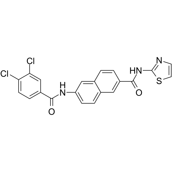
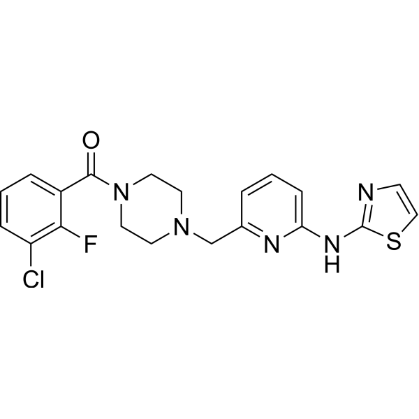
AAPK-25 is a potent and selective Aurora/PLK dual inhibitor with anti-tumor activity, which can cause mitotic delay and arrest cells in a prometaphase, reflecting by the biomarker histone H3Ser10 phosphorylation and followed by a surge in apoptosis. AAPK-25 targets Aurora-A, -B, and -C with Kd values ranging from 23-289 nM, as well as PLK-1, -2, and -3 with Kd values ranging from 55-456 nM.
商品は「研究用試薬」です。人や動物の医療用・臨床診断用・食品用の製品ではありません。
研究用途以外に使用した場合、当社は一切の責任を負いかねます。
- 純度 : 98.09%
- CAS 番号: 2247919-28-2
- 分子式: C21H13Cl2N3O2S
- 分子量:442.32
-
保管条件:
4°C, protect from light
* In solvent : -80°C, 6 months; -20°C, 1 month (protect from light)

To place orders, for customer services and technical support, please contact: MedChemExpress USA
Tel: 609-228-6898 E-mail: [email protected] [email protected]


-
 生物活性
生物活性
-
化学情報
-
溶剤 & 溶解度
- プロトコル
- 純度とドキュメンテーション
- 参考文献
-
 Help & FAQs


-
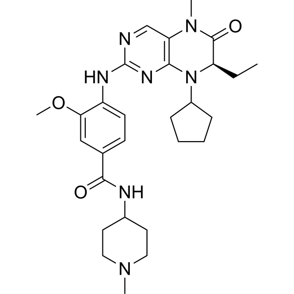
HY-50698
BI 2536PLK1 Inhibitor -
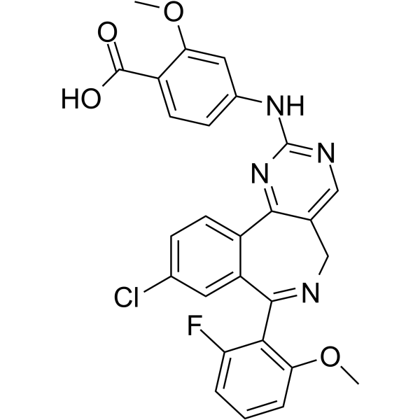
HY-10971
AlisertibAurora A Kinase Inhibitor -
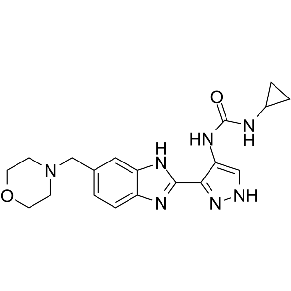
HY-50514
AT9283Aurora/JAK Inhibitor -
HY-12054
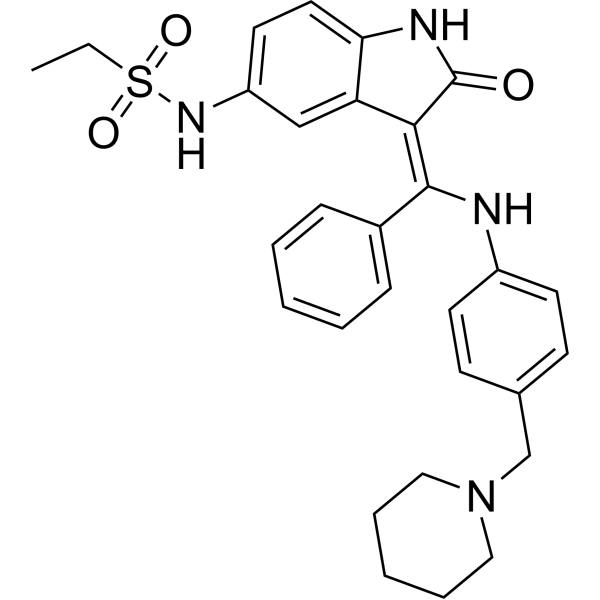
HesperadinAurora B Inhibitor -
HY-10128
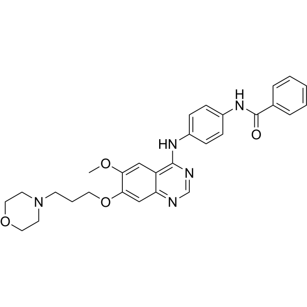
ZM-447439Aurora A/B Inhibitor -
HY-10987A
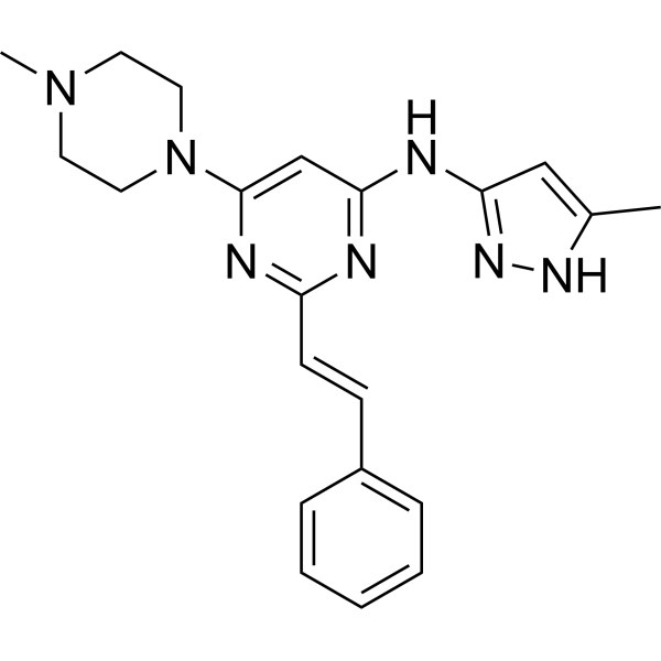
ENMD-2076 -
HY-10161
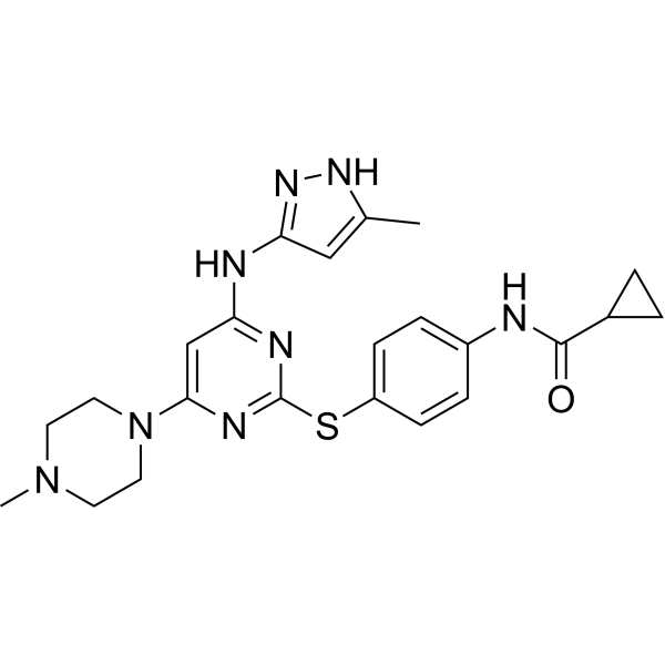
TozasertibAurora A/B/C Inhibitor -
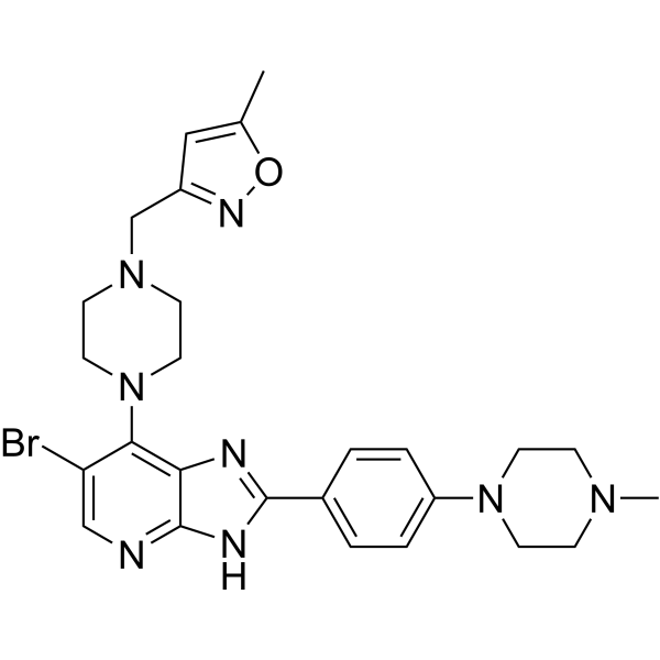
HY-10804
CCT 137690Apoptosis Inducer -
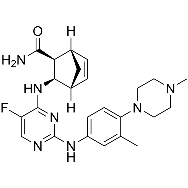
HY-13072
CenisertibMulti-Kinase Inhibitor -
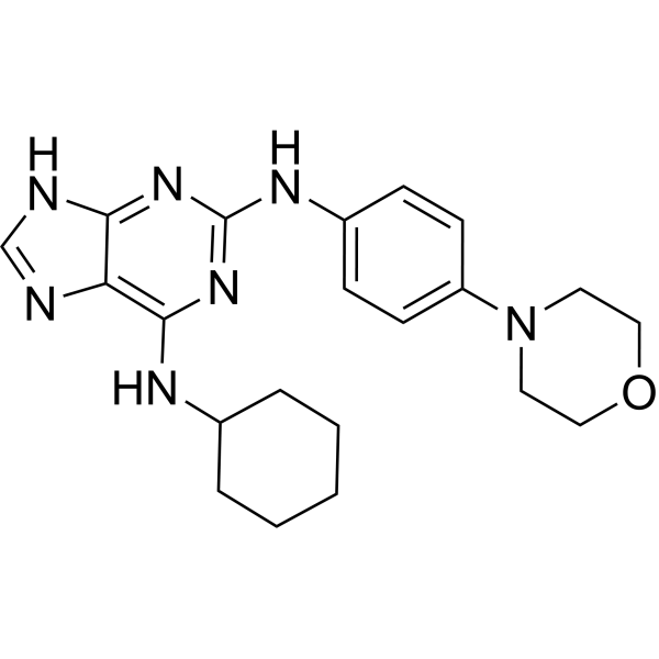
HY-14711
ReversineAutophagy Inducer -
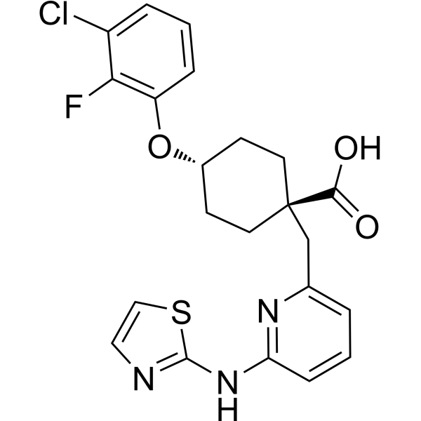
HY-13252
MK-5108Aurora Kinase Inhibitor -
HY-13819
MK-8745Apoptosis Inducer
Aurora Kinase アイソフォーム固有の製品をすべて表示
More

生物活性
製品説明
IC50 & Target
Kd: 23 nM (Aurora-A), 78 nM (Aurora-B), 289 nM (Aurora-C), 55 nM (PLK-1), 272 nM (PLK-2), 456 nM (PLK-3), 5.32 μM (ERK), 7.11 μM (PI3K), 8.02 μM (CDK)[1]
Cellular Effect
|
Cell Line

|
Type | Value | Description | References |
|---|---|---|---|---|
| A549 | IC50 |
11.6 μM
Compound: AAPK-25
|
Antiproliferative activity against human A549 cells measured after 48 hrs by MTT assay
Antiproliferative activity against human A549 cells measured after 48 hrs by MTT assay
|
[PMID: 31381325] |
| Calu-6 | IC50 |
5.3 μM
Compound: AAPK-25
|
Antiproliferative activity against human Calu6 cells measured after 48 hrs by MTT assay
Antiproliferative activity against human Calu6 cells measured after 48 hrs by MTT assay
|
[PMID: 31381325] |
| HCT-116 | IC50 |
0.4 μM
Compound: AAPK-25
|
Antiproliferative activity against human HCT116 cells measured after 48 hrs by MTT assay
Antiproliferative activity against human HCT116 cells measured after 48 hrs by MTT assay
|
[PMID: 31381325] |
| MCF7 | IC50 |
2.3 μM
Compound: AAPK-25
|
Antiproliferative activity against human MCF7 cells measured after 48 hrs by MTT assay
Antiproliferative activity against human MCF7 cells measured after 48 hrs by MTT assay
|
[PMID: 31381325] |
体外実験
AAPK-25 inhibits HCT-116, Calu6, A549 and MCF-7 cells growth with IC50s of 0.4, 5.3, 11.6, and 2.3 μM, respectively[1].
AAPK-25 induces apoptosis as a dose-dependent manner in HCT-116 cell line[1].
AAPK-25 has significantly increased histone H3Ser10 phosphorylation, indicating a markedly mitotic block[1].
AAPK-25 is in notably inhibition of the mitotic spindle checkpoint, which is mainly mediated by cell cycle signaling and mitotic pathways[1].
MedChemExpress (MCE) has not independently confirmed the accuracy of these methods. They are for reference only.
体内実験
MedChemExpress (MCE) has not independently confirmed the accuracy of these methods. They are for reference only.


化学情報
-
CAS 番号 2247919-28-2
-
性状 Solid
-
分子量 442.32
-
分子式 C21H13Cl2N3O2S
-
Color Off-white to light yellow
-
SMILES
ClC1=CC(C(NC2=CC=C(C=C(C(NC3=NC=CS3)=O)C=C4)C4=C2)=O)=CC=C1Cl
-
輸送条件
Room temperature in continental US; may vary elsewhere.
-
保管条件
4°C, protect from light
* In solvent : -80°C, 6 months; -20°C, 1 month (protect from light)


溶剤 & 溶解度
体外:
DMSO : 50 mg/mL (113.04 mM; Need ultrasonic; Hygroscopic DMSO has a significant impact on the solubility of product, please use newly opened DMSO)
Please refer to the solubility information to select the appropriate solvent. Once prepared, please aliquot and store the solution to prevent product inactivation from repeated freeze-thaw cycles.
Storage method and period of stock solution: -80°C, 6 months; -20°C, 1 month (protect from light). When stored at -80°C, please use it within 6 months. When stored at -20°C, please use it within 1 month.
Please refer to the solubility information to select the appropriate solvent. Once prepared, please aliquot and store the solution to prevent product inactivation from repeated freeze-thaw cycles.
Storage method and period of stock solution: -80°C, 6 months; -20°C, 1 month (protect from light). When stored at -80°C, please use it within 6 months. When stored at -20°C, please use it within 1 month.
濃度 (開始) × 体積 (開始) = 濃度 (終了) × 体積 (終了)


純度とドキュメンテーション
-
データシート (270 KB)
-
SDS (252 KB)
- English - EN (252 KB)
- Français - FR (252 KB)
- Deutsch - DE (252 KB)
- Norwegian - NO (252 KB)
- Español - ES (252 KB)
- Swedish - SV (252 KB)
- Italian - IT (252 KB)
- Korean - KR (252 KB)
- Portuguese - PT (252 KB)
-
取扱説明書 (2659 KB)
参考文献


Complete Stock Solution Preparation Table
Please refer to the solubility information to select the appropriate solvent. Once prepared, please aliquot and store the solution to prevent product inactivation from repeated freeze-thaw cycles.
Storage method and period of stock solution: -80°C, 6 months; -20°C, 1 month (protect from light). When stored at -80°C, please use it within 6 months. When stored at -20°C, please use it within 1 month.
| Optional Solvent | Concentration Solvent Mass | 1 mg | 5 mg | 10 mg | 25 mg |
|---|---|---|---|---|---|
| DMSO | 1 mM | 2.2608 mL | 11.3040 mL | 22.6081 mL | 56.5202 mL |
| 5 mM | 0.4522 mL | 2.2608 mL | 4.5216 mL | 11.3040 mL | |
| 10 mM | 0.2261 mL | 1.1304 mL | 2.2608 mL | 5.6520 mL | |
| 15 mM | 0.1507 mL | 0.7536 mL | 1.5072 mL | 3.7680 mL | |
| 20 mM | 0.1130 mL | 0.5652 mL | 1.1304 mL | 2.8260 mL | |
| 25 mM | 0.0904 mL | 0.4522 mL | 0.9043 mL | 2.2608 mL | |
| 30 mM | 0.0754 mL | 0.3768 mL | 0.7536 mL | 1.8840 mL | |
| 40 mM | 0.0565 mL | 0.2826 mL | 0.5652 mL | 1.4130 mL | |
| 50 mM | 0.0452 mL | 0.2261 mL | 0.4522 mL | 1.1304 mL | |
| 60 mM | 0.0377 mL | 0.1884 mL | 0.3768 mL | 0.9420 mL | |
| 80 mM | 0.0283 mL | 0.1413 mL | 0.2826 mL | 0.7065 mL | |
| 100 mM | 0.0226 mL | 0.1130 mL | 0.2261 mL | 0.5652 mL |


AAPK-25 関連分類
HY-126249 関連分類
 Powered by Bioz