



PB28
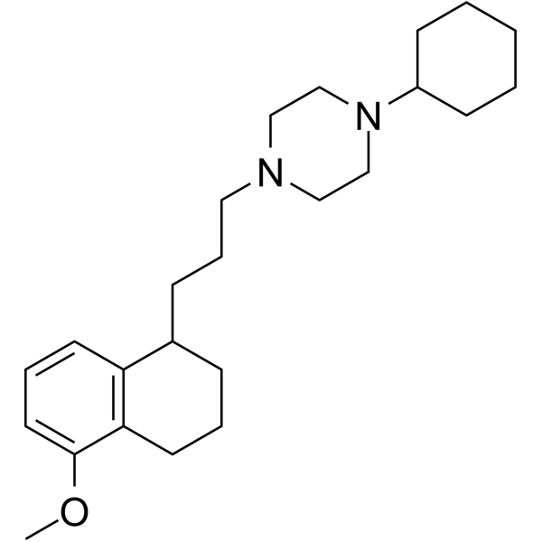
PB28 is a cyclohexylpiperazine derivative and a high affinity and selective sigma 2 (σ2) receptor agonist with a Ki of 0.68 nM. PB28 is also a σ1 antagonist with a Ki of 0.38 nM. PB28 is less affinity for other receptors. PB28 inhibits electrically evoked twitch in guinea pig bladder and ileum with EC50 values of 2.62 μM and 3.96 μM, respectively. PB28 can modulate SARS-CoV-2-human protein-protein interaction. PB28 induces caspase-independent apoptosis and has antitumor activity.
商品は「研究用試薬」です。人や動物の医療用・臨床診断用・食品用の製品ではありません。
研究用途以外に使用した場合、当社は一切の責任を負いかねます。
- CAS 番号: 172906-90-0
- 分子式: C24H38N2O
- 分子量:370.57
-
保管条件:
Please store the product under the recommended conditions in the Certificate of Analysis.

To place orders, for customer services and technical support, please contact: MedChemExpress USA
Tel: 609-228-6898 E-mail: [email protected] [email protected]


-
 生物活性
生物活性
-
化学情報
- プロトコル
- 純度とドキュメンテーション
- 参考文献
-
 Help & FAQs


Sigma Receptor アイソフォーム固有の製品をすべて表示
More

生物活性
製品説明
IC50 & Target
Ki: 0.68 nM (σ2 receptor); 0.38 nM (σ1 receptor)[4]
Cellular Effect
|
Cell Line

|
Type | Value | Description | References |
|---|---|---|---|---|
| Caco-2 | EC50 |
0.55 μM
Compound: PB-28
|
Inhibition of human Pgp mediated [3H]vinblastine transport in human Caco-2 cells
Inhibition of human Pgp mediated [3H]vinblastine transport in human Caco-2 cells
|
[PMID: 17936633] |
| MCF7 | EC50 |
27.4 μM
Compound: PB, PB28
|
Antiproliferative activity against human MCF7 cells after 48 hrs by MTT assay
Antiproliferative activity against human MCF7 cells after 48 hrs by MTT assay
|
[PMID: 23415062] |
| MCF7 | EC50 |
31.2 μM
Compound: PB, PB28
|
Antiproliferative activity against human MCF7 cells transfected with human sigma1 receptor after 48 hrs by MTT assay
Antiproliferative activity against human MCF7 cells transfected with human sigma1 receptor after 48 hrs by MTT assay
|
[PMID: 23415062] |
| MCF7 | IC50 |
1.2 nM
Compound: 1, PB28
|
Displacement of [3H]DTG from sigma 2 receptor in human MCF7 cells after 45 mins by FACS analysis
Displacement of [3H]DTG from sigma 2 receptor in human MCF7 cells after 45 mins by FACS analysis
|
[PMID: 24697311] |
| MCF7 | IC50 |
4.27 nM
Compound: 1, PB28
|
Displacement of 2-(6-{1-[3-(4-Cyclohexylpiperazin-1-yl)propyl]-1,2,3,4-tetrahydronaphthalen-5-yloxy}hexyl)-5-dimethylamino-1H-isoindole-1,3(2H)-dione from sigma 2 receptor in human MCF7 cells after 45 mins by FACS analysis
Displacement of 2-(6-{1-[3-(4-Cyclohexylpiperazin-1-yl)propyl]-1,2,3,4-tetrahydronaphthalen-5-yloxy}hexyl)-5-dimethylamino-1H-isoindole-1,3(2H)-dione from sigma 2 receptor in human MCF7 cells after 45 mins by FACS analysis
|
[PMID: 24697311] |
| PC-3 | EC50 |
64.2 μM
Compound: 36, PB-28
|
Cytotoxicity against human PC3 cells assessed as LDH release after 24 hrs
Cytotoxicity against human PC3 cells assessed as LDH release after 24 hrs
|
[PMID: 19053780] |
| RPMI-8226 | IC50 |
9.29 μM
Compound: 3, PB28
|
Cytotoxicity against human RPMI8226 cells assessed as cell viability after 72 hrs by MTT assay
Cytotoxicity against human RPMI8226 cells assessed as cell viability after 72 hrs by MTT assay
|
[PMID: 24331758] |
| SK-N-SH | EC50 |
9.04 μM
Compound: (+/-)-1, PB-28
|
Antiproliferative activity against human SK-N-SH cells after 48 hts by MTT assay
Antiproliferative activity against human SK-N-SH cells after 48 hts by MTT assay
|
[PMID: 21229979] |
| SK-N-SH | EC50 |
9.97 μM
Compound: 7, PB-28
|
Antiproliferative activity against human SK-N-SH cells after 48 hrs by MTT assay
Antiproliferative activity against human SK-N-SH cells after 48 hrs by MTT assay
|
[PMID: 19842660] |
体外実験
PB28 (15-25 nM; 24-48 hours; MCF7 and MCF7 ADR cells) treatment shows an accumulation in the G0-G1 phase for MCF7 and MCF7 ADR cells that are time and concentration independent[1].
PB28 has a higher σ2 receptor affinity expressed as Ki (0.28 nM and 0.17 nM in MCF7 and MCF7 ADR cells, respectively) than σ1 receptor affinity (13.0 nMand 10.0 nM, respectively)[1].
PB28 inhibits cell growth of MCF7 and MCF7 ADR cells with IC50s of 25 nM and 15 nM, respectively after 2-day treatment[1].
PB28 induces apoptosis through a caspase-independent pathway[1].
PB28 also reduces P-gp expression in a concentration- and time-dependent manner (approximately 60% in MCF7 and 90% in MCF7 ADR)[1].
PB28 displays antiproliferative and cytotoxic effects in both C6 rat glioma and SK-N-SH human neuroblastoma cell lines[1].
MedChemExpress (MCE) has not independently confirmed the accuracy of these methods. They are for reference only. Further protocols information, click here.
-
Cell Line:MCF7 and MCF7 ADR cells
-
Concentration:25 nM and 15 nM
-
Incubation Time:24 hours, 48 hours
-
Result:Showed an accumulation in the G0-G1 phase for MCF7 and MCF7 ADR cells that were time and concentration independent.
体内実験
MedChemExpress (MCE) has not independently confirmed the accuracy of these methods. They are for reference only.
-
Animal Model:C57BL/6 female mice (10 weeks old) injected with Panc02 cells[2]
-
Dosage:10.7 mg/mL
-
Administration:Intraperitoneal injection; daily; for two weeks
-
Result:Inhibited tumor growth in Panc02 tumor burden mice.


化学情報
-
CAS 番号 172906-90-0
-
分子量 370.57
-
分子式 C24H38N2O
-
SMILES
COC1=CC=CC2=C1CCCC2CCCN3CCN(C4CCCCC4)CC3
-
輸送条件
Room temperature in continental US; may vary elsewhere.
-
保管条件
Please store the product under the recommended conditions in the Certificate of Analysis.

プロトコル
-
Apoptosis
Apoptosis, also called programmed cell death, is generally characterized by distinct morphological characteristics.
-
TUNEL staining for apoptotic DNA fragmentation
TUNEL staining detects DNA strand breaks by using terminal deoxynucleotidyl transferase to add labeled nucleotides to exposed 3′-OH DNA termini, generating either microscopic staining in fixed cells or tissue sections, or fluorescence/cytometric signal in cell suspensions. TUNEL positivity reflects DNA fragmentation but should not be interpreted alone as definitive apoptosis, because TUNEL can also label necrotic, autolytic, mechanically damaged, or DNA-repair-associated DNA breaks.
-
Annexin V plus membrane-impermeant dye apoptosis staining
Annexin V-based apoptosis assays rely on the detection of phosphatidylserine (PS) externalization from the inner leaflet of the plasma membrane to the outer leaflet, an early biochemical hallmark of apoptosis. Fluorescently labeled Annexin V binds PS in a calcium-dependent manner, enabling identification of early apoptotic cells by flow cytometry or fluorescence microscopy. When combined with a membrane-impermeant DNA-binding dye (e. g. , propidium iodide), this approach allows discrimination between viable (Annexin V−/dye−), early apoptotic (Annexin V+/dye−), and late apoptotic or necrotic (Annexin V+/dye+) cell populations by assessing membrane integrity and PS exposure.
-
Pull-down
The pull-down assay is an in vitro technique used to detect physical interactions between two or more proteins and an invaluable tool for confirming a predicted protein-protein interaction or identifying novel interacting partners. This method typically involves the use of affinity purification with various wash and elution steps.
-
Immunoprecipitation
Immunoprecipitation (IP) is an experimental method that uses the principle of antibody specific binding to purify and enrich target proteins.
-
Protocol for Bimolecular Fluorescence Complementation (BiFC) Assay
Bimolecular fluorescence complementation detects protein-protein proximity in living or fixed cells by fusing two candidate interaction partners to nonfluorescent N- and C-terminal fragments of a fluorescent protein; when the partners interact or remain close enough, the fluorescent fragments complement, mature, and generate a fluorescent signal at the site of the protein complex. The BiFC readout is fluorescence intensity and subcellular localization of the reconstituted fluorophore, which reflects formation or stabilization of a protein complex rather than direct biochemical binding kinetics; BiFC is therefore useful for mapping where interactions occur in cancer cells, neurons, macrophages, organoid-derived cells, or drug-screening systems, but results should be validated by independent assays such as co-IP or Western blot. BiFC signal formation is delayed by fluorophore maturation and can stabilize otherwise transient complexes, so it is not a real-time reversible interaction assay
-
Apoptosis Solutions
Apoptosis is a regulated, generally non-lytic cell-death pathway that removes unwanted, damaged, infected, or abnormal cells through coordinated morphological changes, caspase activation, DNA fragmentation, and membrane remodeling. The intrinsic apoptosis pathway is controlled mainly by mitochondrial outer membrane permeabilization, BCL-2 family proteins, cytochrome c release, apoptosome formation, caspase-9 activation, and downstream executioner caspase-3/7 activation. The extrinsic apoptosis pathway is initiated by death receptors such as Fas, TNFR, and TRAIL receptors, which recruit adaptor proteins and activate caspase-8 before engaging executioner caspases or mitochondrial amplification through BID cleavage. Apoptosis is linked to many phenotypes, including cancer cell killing, tissue homeostasis, immune regulation, neurodegeneration, infection response, and treatment-induced cytotoxicity; unresolved questions include how apoptosis interacts with necroptosis, pyroptosis, ferroptos
-
Subcutaneous Cell-Line-Derived Xenograft
Subcutaneous cell-line-derived xenograft (CDX) models are established by implanting cultured human cancer cell lines into immunodeficient mice, where the injected cells form localized tumors that can be monitored in vivo as a measure of tumorigenic potential, growth kinetics, and treatment response. These models are widely used in oncology research because they allow reproducible tumor formation and enable comparative assessment of tumor growth between different cell lines or genetic manipulations in a controlled in vivo microenvironment. Subcutaneous implantation of cancer cells in immunodeficient mice is a standard approach for evaluating tumor growth behavior and therapeutic response across multiple cancer types, including prostate, esophageal, pancreatic, and colon cancer models.
-
Co-Immunoprecipitation
Co-immunoprecipitation technology can verify protein interaction based on the specific immune reaction between antibodies and antigens.
-
Protocol for Yeast Two-Hybrid (Y2H) Assay
The yeast two-hybrid assay detects binary protein-protein interactions by separating a transcription factor into a DNA-binding domain fused to a "bait" protein and a transcriptional activation domain fused to a "prey" protein; if bait and prey interact in yeast, the transcription factor is reconstituted and activates reporter genes such as HIS3, ADE2, lacZ, MEL1, or other selectable/readable reporters. The readout is yeast growth on selective medium and/or reporter activity, which reflects proximity-dependent transcriptional activation in the yeast nucleus rather than direct biochemical binding in the original mammalian, tumor, neuronal, macrophage, or organoid context. Because yeast two-hybrid can generate false positives and false negatives, interaction claims should be validated using independent assays such as co-immunoprecipitation, Western blot, immunofluorescence colocalization, BiFC, pull-down, or mammalian two-hybrid assays.


純度とドキュメンテーション
参考文献
[1]. Amalia Azzariti, et al. Cyclohexylpiperazine Derivative PB28, a sigma2 Agonist and sigma1 Antagonist Receptor, Inhibits Cell Growth, Modulates P-glycoprotein, and Synergizes With Anthracyclines in Breast Cancer. Mol Cancer Ther. 2006 Jul;5(7):1807-16. [Content Brief]
[2]. Maria Laura Pati, et al. Sigma-2 Receptor Agonist Derivatives of 1-Cyclohexyl-4-[3-(5-methoxy-1,2,3,4-tetrahydronaphthalen-1-yl)propyl]piperazine (PB28) Induce Cell Death via Mitochondrial Superoxide Production and Caspase Activation in Pancreatic Cancer. [Content Brief]
[3]. Nicola A Colabufo, et al. A New Method for Evaluating sigma(2) Ligand Activity in the Isolated Guinea-Pig Bladder. Naunyn Schmiedebergs Arch Pharmacol. 2003 Aug;368(2):106-12. [Content Brief]
[4]. Francesco Berardi, et al. Exploring the Importance of Piperazine N-atoms for sigma(2) Receptor Affinity and Activity in a Series of Analogs of 1-cyclohexyl-4-[3-(5-methoxy-1,2,3,4-tetrahydronaphthalen-1-yl)propyl]piperazine (PB28). J Med Chem. 2009 Dec 10 [Content Brief]
[5]. David E Gordon, et al. A SARS-CoV-2-Human Protein-Protein Interaction Map Reveals Drug Targets and Potential Drug-Repurposing. bioRxiv. 2020 Mar 22;2020.03.22.002386. [Content Brief]

Calculators
濃度 (開始) × 体積 (開始) = 濃度 (終了) × 体積 (終了)


PB28 関連分類
HY-108511A 関連分類
 Powered by Bioz