



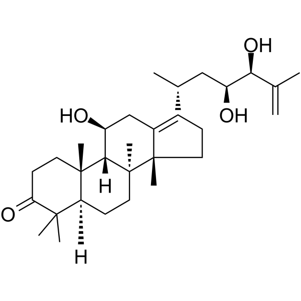
Alisol G

Based on 1 Customer Validation
Alisol G (25-Anhydroalisol A) is a human carboxylesterase 2 (hCES2) inhibitor with an IC50 of 3.85 μM. Alisol G exhibits cytotoxic activity against human cancer cells, antibacterial activity against Gram-positive strains, and anti-hepatitis B virus activity. Alisol G can be used in research related to lung cancer, breast cancer, prostate cancer, bacterial infections, and HBV infections.
연구목적의 판매만을 진행합니다. 환자를 대상으로 한 판매는 하지 않습니다.
- Purity : 98.02%
- CAS No.: 155521-46-3
- 화학식: C30H48O4
- 분자량:472.70
-
보관:Powder -20°C, 3 years , 4°C, 2 years ; In solvent -80°C, 6 months , -20°C, 1 month

To place orders, for customer services and technical support, please contact: MedChemExpress USA
Tel: 609-228-6898 E-mail: [email protected] [email protected]


-
 Biological Activity
-
Chemical Information
-
용액&용해도
- Protocol
- 순도&문서
- References
-
 Help & FAQs




Biological Activity
제품 설명
IC50 & Target
[1]|
hCES2 3.85 μM (IC50) |
Cellular Effect
|
Cell Line

|
Type | Value | Description | References |
|---|---|---|---|---|
| Erythrocyte | IC50 |
6.98 μM
Compound: 57d
|
Antiplasmodial activity against Plasmodium falciparum K1 infected in human erythrocyte assessed as inhibition of parasite growth incubated for 72 hrs by [3H]-hypoxanthine incorporation based liquid scintillation counting method
Antiplasmodial activity against Plasmodium falciparum K1 infected in human erythrocyte assessed as inhibition of parasite growth incubated for 72 hrs by [3H]-hypoxanthine incorporation based liquid scintillation counting method
|
[PMID: 35985254] |
| HepG2 | EC50 |
24.09 μM
Compound: 24
|
Transactivation of FXR (unknown origin) transfected in HepG2 cells co-expressing pBSEP/pGL4.74 incubated for 24 hrs by luciferase reporter gene assay
Transactivation of FXR (unknown origin) transfected in HepG2 cells co-expressing pBSEP/pGL4.74 incubated for 24 hrs by luciferase reporter gene assay
|
[PMID: 31494470] |
| HepG2 2.2.15 | IC50 |
0.6 μM
Compound: 23
|
Antiviral activity against Hepatitis B virus infected in human HepG2.2.15 cells assessed as decrease in HBV surface antigen secretion
Antiviral activity against Hepatitis B virus infected in human HepG2.2.15 cells assessed as decrease in HBV surface antigen secretion
|
[PMID: 24549242] |
In Vitro
Alisol G potently inhibits human carboxylesterase 2 with an IC50 of 3.85 μM; it exerts inhibitory effects on the HepG2 cell line with an IC50 of 11.0 μM; and it inhibits hepatitis B virus with an IC50 value of 17.6 μM[1].
Alisol G (24 h pre-culture, 5 days incubation) is completely converted into four novel oxidative metabolites by Penicillium janthinellum AS 3.510[1].
Alisol G (compound 10) (72 h) potently inhibits the viability of H460, MCF-7 and PC-3 human cancer cell lines, with IC50 values ranging from 11.5 μM to 16.3 μM[2].
Alisol G (24 h) inhibits the growth of two Gram-positive bacteria, Bacillus subtilis and Staphylococcus aureus, with an MIC of 12.5 mg/mL against both strains[2].
MedChemExpress (MCE) has not independently confirmed the accuracy of these methods. They are for reference only.


Chemical Information
-
CAS No. 155521-46-3
-
Appearance Solid
-
분자량 472.70
-
화학식 C30H48O4
-
Color White to off-white
-
SMILES
C[C@]([C@@]1(C2=C([C@H](C)C[C@H](O)[C@@H](O)C(C)=C)CC1)C)(CC[C@@]3([H])C4(C)C)[C@]([C@H](C2)O)([H])[C@]3(CCC4=O)C
-
Synonyms
Alisol-G; 25-Anhydroalisol A
-
Structure Classification
-
Initial Source
-
선적
Room temperature in continental US; may vary elsewhere.
-
보관
Powder -20°C 3 years 4°C 2 years In solvent -80°C 6 months -20°C 1 month


용액&용해도
In Vitro:
DMSO : ≥ 100 mg/mL (211.55 mM; Hygroscopic DMSO has a significant impact on the solubility of product, please use newly opened DMSO)
* "≥" means soluble, but saturation unknown.
Please refer to the solubility information to select the appropriate solvent. Once prepared, please aliquot and store the solution to prevent product inactivation from repeated freeze-thaw cycles.
Storage method and period of stock solution: -80°C, 6 months; -20°C, 1 month. When stored at -80°C, please use it within 6 months. When stored at -20°C, please use it within 1 month.
Please refer to the solubility information to select the appropriate solvent. Once prepared, please aliquot and store the solution to prevent product inactivation from repeated freeze-thaw cycles.
Storage method and period of stock solution: -80°C, 6 months; -20°C, 1 month. When stored at -80°C, please use it within 6 months. When stored at -20°C, please use it within 1 month.
Concentration (start) × Volume (start) = Concentration (final) × Volume (final)
In Vivo:
Select the appropriate dissolution method based on your experimental animal and administration route.
- For the following dissolution methods, please ensure to first prepare a clear stock solution using an In Vitro approach and then sequentially add co-solvents:
- To ensure reliable experimental results, the clarified stock solution can be appropriately stored based on storage conditions. As for the working solution for In Vivo experiments, it is recommended to prepare freshly and use it on the same day.
- The percentages shown for the solvents indicate their volumetric ratio in the final prepared solution. If precipitation or phase separation occurs during preparation, heat and/or sonication can be used to aid dissolution.
Add each solvent one by one: 10% DMSO 40% PEG300 5% Tween-80 45% Saline
Solubility: ≥ 2.5 mg/mL (5.29 mM); Clear solution
This protocol yields a clear solution of ≥ 2.5 mg/mL (saturation unknown).
Taking 1 mL working solution as an example, add 100 μL DMSO stock solution (25.0 mg/mL) to 400 μL PEG300, and mix evenly; then add 50 μL Tween-80 and mix evenly; then add 450 μL Saline to adjust the volume to 1 mL.
Preparation of Saline: Dissolve 0.9 g sodium chloride in ddH₂O and dilute to 100 mL to obtain a clear Saline solution.
In Vivo Dissolution Calculator
Please enter the basic information of animal experiments:
-
-
-
-
Recommended: Prepare an additional quantity of animals to account for potential losses during experiments.
Please enter your animal formula composition:
-
%DMSO +
Recommended: Keep the proportion of DMSO in working solution below 2% if your animal is weak.
-
%+
-
+%Tween-80 + +
-
%Saline +
The co-solvents required include: DMSO, . All of co-solvents are available by MedChemExpress (MCE). , Tween 80. All of co-solvents are available by MedChemExpress (MCE).
Working solution concentration: 0.22 mg/mL
Method for preparing stock solution: mg drug dissolved in μL DMSO. Stock solution concentration: mg/mL.
1. Take μL DMSO stock solution;
2. Add μL .
μL , mix evenly;
3. Then add μL Tween 80, mix evenly;
4. Then add μL
Please ensure that the stock solution in the first step is dissolved to a clear state, and add co-solvents in sequence. You can use ultrasonic heating (ultrasonic cleaner, recommended frequency 20-40 kHz), vortexing, etc. to assist dissolution.


순도&문서
-
Data Sheet (276 KB)
-
SDS (252 KB)
- English - EN (252 KB)
- Français - FR (252 KB)
- Deutsch - DE (252 KB)
- Norwegian - NO (252 KB)
- Español - ES (252 KB)
- Swedish - SV (252 KB)
- Italian - IT (252 KB)
- Korean - KR (252 KB)
- Portuguese - PT (252 KB)
-
Handling Instructions (2659 KB)
References


Complete Stock Solution Preparation Table
Please refer to the solubility information to select the appropriate solvent. Once prepared, please aliquot and store the solution to prevent product inactivation from repeated freeze-thaw cycles.
Storage method and period of stock solution: -80°C, 6 months; -20°C, 1 month. When stored at -80°C, please use it within 6 months. When stored at -20°C, please use it within 1 month.
| Optional Solvent | Concentration Solvent Mass | 1 mg | 5 mg | 10 mg | 25 mg |
|---|---|---|---|---|---|
| DMSO | 1 mM | 2.1155 mL | 10.5775 mL | 21.1551 mL | 52.8877 mL |
| 5 mM | 0.4231 mL | 2.1155 mL | 4.2310 mL | 10.5775 mL | |
| 10 mM | 0.2116 mL | 1.0578 mL | 2.1155 mL | 5.2888 mL | |
| 15 mM | 0.1410 mL | 0.7052 mL | 1.4103 mL | 3.5258 mL | |
| 20 mM | 0.1058 mL | 0.5289 mL | 1.0578 mL | 2.6444 mL | |
| 25 mM | 0.0846 mL | 0.4231 mL | 0.8462 mL | 2.1155 mL | |
| 30 mM | 0.0705 mL | 0.3526 mL | 0.7052 mL | 1.7629 mL | |
| 40 mM | 0.0529 mL | 0.2644 mL | 0.5289 mL | 1.3222 mL | |
| 50 mM | 0.0423 mL | 0.2116 mL | 0.4231 mL | 1.0578 mL | |
| 60 mM | 0.0353 mL | 0.1763 mL | 0.3526 mL | 0.8815 mL | |
| 80 mM | 0.0264 mL | 0.1322 mL | 0.2644 mL | 0.6611 mL | |
| 100 mM | 0.0212 mL | 0.1058 mL | 0.2116 mL | 0.5289 mL |


Alisol G Related Classifications
HY-N0855 Related Classifications
 Powered by Bioz