



PF-00356231
Based on 2 publication(s) in Google Scholar
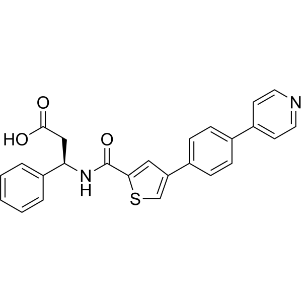
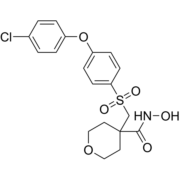
PF-00356231 hydrochloride is a specific, non-peptidic, non-zinc chelating ligand and inhibitor of matrix metalloproteinase MMP-12 (IC50=1.4 μM). PF-00356231 hydrochloride binds to MMP-12 and forms PF-00356231/MMP-12 complex. PF-00356231 hydrochloride shows potency against MMP-13, MMP-8, MMP-9, MMP-3 with IC50s of 0.00065, 1.7, 0.98, 0.39 μM, respectively.
연구목적의 판매만을 진행합니다. 환자를 대상으로 한 판매는 하지 않습니다.
- CAS No.: 766536-21-4
- 화학식: C25H20N2O3S
- 분자량:428.50
-
보관:
Please store the product under the recommended conditions in the Certificate of Analysis.

To place orders, for customer services and technical support, please contact: MedChemExpress USA
Tel: 609-228-6898 E-mail: [email protected] [email protected]


-
 Biological Activity
-
Chemical Information
- Protocol
- 순도&문서
- References
-
 Help & FAQs


-
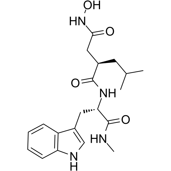
HY-15768
IlomastatMMP Inhibitor -
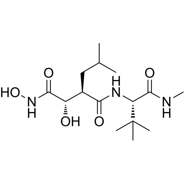
HY-12169
MarimastatMMP Inhibitor -
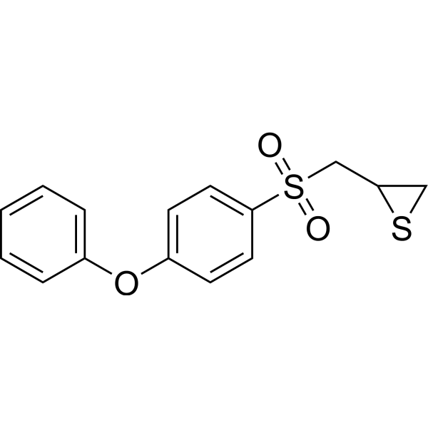
HY-12354
SB-3CTMMP2/9 Inhibitor -
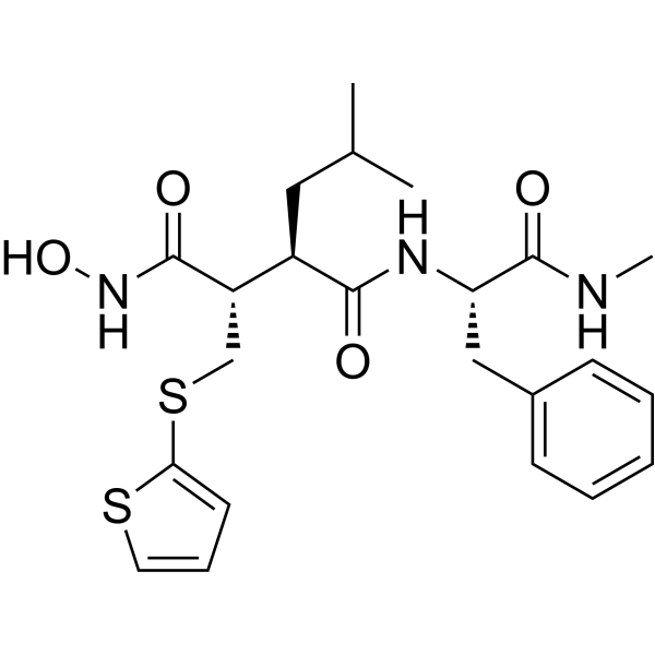
HY-13564
BatimastatMMP Inhibitor -
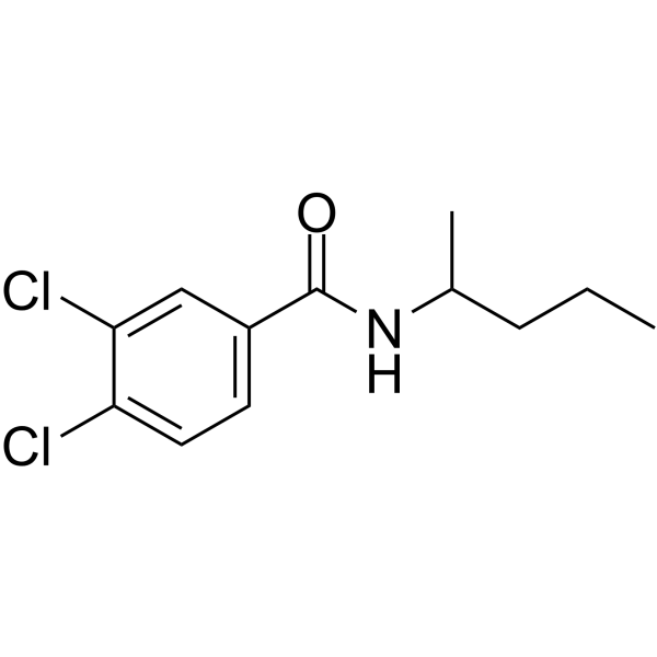
HY-15827
NSC 405020MMP14 Inhibitor -
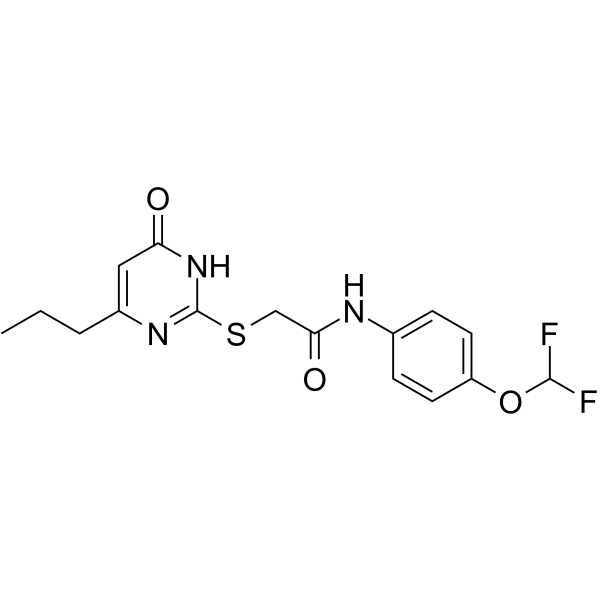
HY-135232
MMP-9-IN-1MMP-9 Inhibitor -
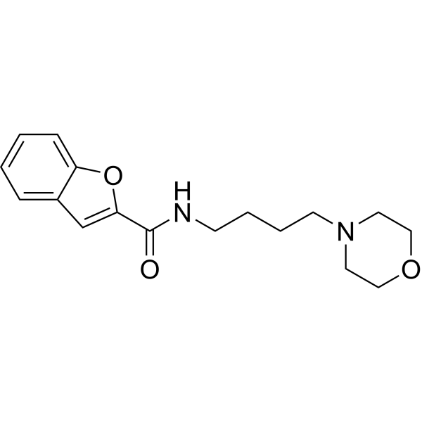
HY-100359
CL-82198MMP-13 Inhibitor -
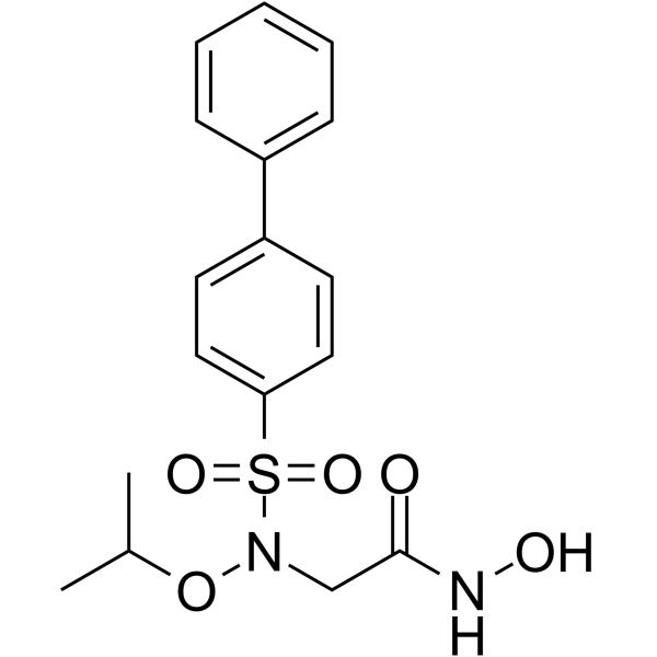
HY-103444
ARP-100MMP-2 Inhibitor -
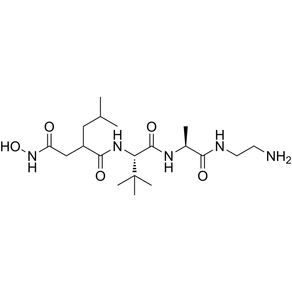
HY-100211
TAPI-2MMP/TACE/ADAM Inhibitor -
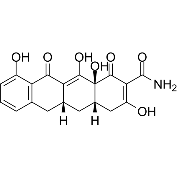
HY-13648
IncyclinideMMP Inhibitor -
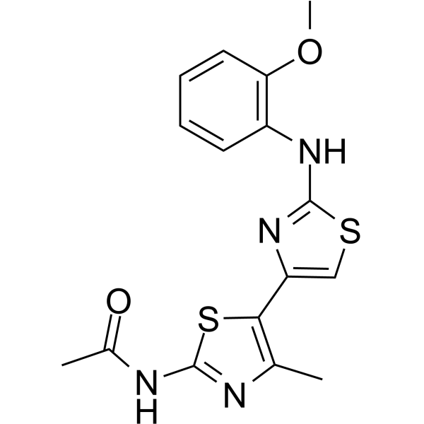
HY-103482
JNJ0966MMP-9 Inhibitor -
HY-10398
CTS-1027MMP Inhibitor
Publications Citing Use of MedChemExpress (MCE) PF-00356231
More

Biological Activity
제품 설명
In Vitro
PF-00356231 hydrochloride against MMP-12/13 can be affected significantly by the presence of acetohydroxamate (AH). PF-00356231 hydrochloride decreases the IC50 values of MMP-12 (0.014 μM) and MMP-13 (0.27 μM) in the presence AH[1].
MedChemExpress (MCE) has not independently confirmed the accuracy of these methods. They are for reference only. Further protocols information, click here.


Chemical Information
-
CAS No. 766536-21-4
-
분자량 428.50
-
화학식 C25H20N2O3S
-
SMILES
OC(C[C@H](C1=CC=CC=C1)NC(C2=CC(C3=CC=C(C4=CC=NC=C4)C=C3)=CS2)=O)=O
-
선적
Room temperature in continental US; may vary elsewhere.
-
보관
Please store the product under the recommended conditions in the Certificate of Analysis.


Publications (2)
-
Journal Impact Factor
-
Most Recent
-
JCI Insight
2025 Nov 24;10(22):e195743. PMID: 41277556 -
J Immunol Res
ADAM-10 Regulates MMP-12 during Lipopolysaccharide-Induced Inflammatory Response in Macrophages. [Abstract]2022 Sep 16;2022:3012218. PMID: 36157882

Protocol
-
Mitochondrial membrane-potential fluorescent assay
Mitochondrial membrane potential fluorescent assays estimate ΔΨm in living cells using lipophilic cationic dyes such as TMRM, TMRE, rhodamine 123, and JC-1, which accumulate in mitochondria according to membrane polarization; loss of signal after FCCP or CCCP treatment is interpreted as mitochondrial depolarization. TMRM/TMRE and rhodamine 123 are commonly used for semi-quantitative live-cell microscopy or flow cytometry, while JC-1 can report a shift from red aggregate fluorescence to green monomer fluorescence during depolarization; interpretation requires controls because dye concentration, quenching mode, cell type, dye efflux, and mitochondrial mass can affect fluorescence independently of ΔΨm.
-
Cell Cytotoxicity Assay
Cytotoxicity assays are usually based on the assessment of cell membrane damage, which can also be indirectly detected by measuring cell viability. Detection methods include MTT assay, CKK-8 assay, LDH assay and ATP assay, etc.
-
Mitochondrial membrane-potential and mitochondrial mass staining
Mitochondrial membrane potential staining measures the electrochemical polarization across the mitochondrial inner membrane in live cells using lipophilic cationic fluorescent probes; early rhodamine-based work showed that selective mitochondrial dye accumulation is lost when the mitochondrial transmembrane potential is dissipated. JC-1 reports mitochondrial polarization by shifting from green monomer fluorescence to red J-aggregate fluorescence as dye concentration increases within energized mitochondria; therefore, the red/green fluorescence ratio is used as a relative readout of mitochondrial membrane potential. TMRE or TMRM staining provides a single-channel relative readout because these cationic rhodamine esters accumulate in polarized mitochondria, and lower fluorescence indicates reduced mitochondrial polarization when acquisition and dye-loading conditions are controlled. Mitochondrial mass staining is commonly performed with MitoTracker Green FM or related MitoTracker dyes as


순도&문서
References

Calculators
Concentration (start) × Volume (start) = Concentration (final) × Volume (final)


PF-00356231 Related Classifications
HY-114091A Related Classifications
 Powered by Bioz