



Thioflavin T (solution)

Based on 1 Customer Validation
Thioflavin T (solution) is a cationic Benzothiazole dye that shows enhanced fluorescence upon binding to amyloid in tissue sections. Excitation max.: ~385 nm (free) ; ~450 nm (bound) ; Emission max.: ~445 nm (free) ; ~485 nm (bound) .
Solvent and Concentration: Sterile water: 10 mM
The 1 mL volume is defined as the base specification. All larger sizes correspond to incremental volumes of this base.
연구목적의 판매만을 진행합니다. 환자를 대상으로 한 판매는 하지 않습니다.
- CAS No.: 2390-54-7
- 화학식: C17H19ClN2S
- 분자량:318.86
-
보관:
Please store the product under the recommended conditions in the Certificate of Analysis.


To place orders, for customer services and technical support, please contact: MedChemExpress USA
Tel: 609-228-6898 E-mail: [email protected] [email protected]


-
 Biological Activity
-
Chemical Information
- Protocol
- 순도&문서
- References
-
 Help & FAQs


-
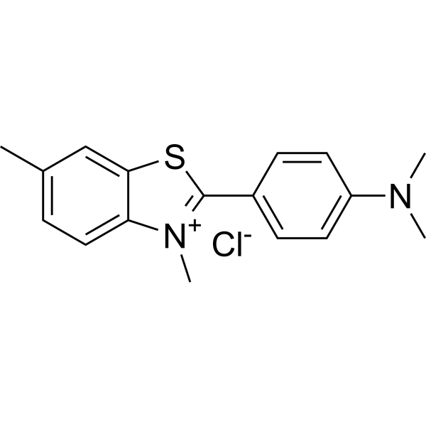
HY-D0218
Thioflavin Tβ-amyloid Dye -
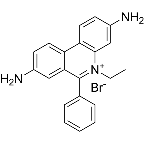
HY-D0021
EthD bromideFluorescent Tag -
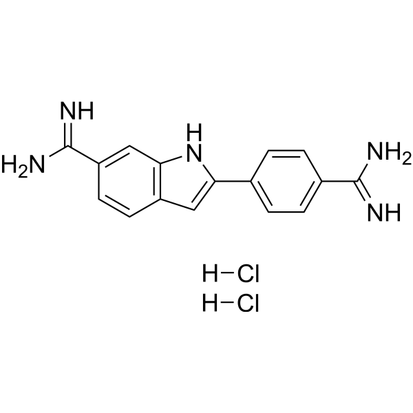
HY-D0814
DAPI dihydrochlorideFluorescent DNA Stain -
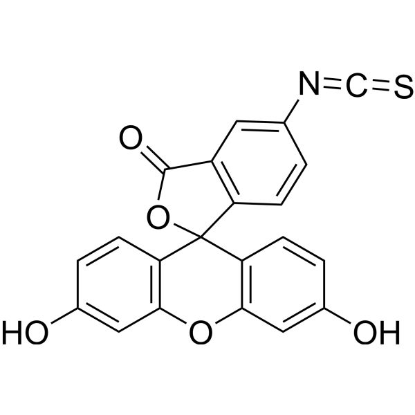
HY-66019
FITCFluorochrome -
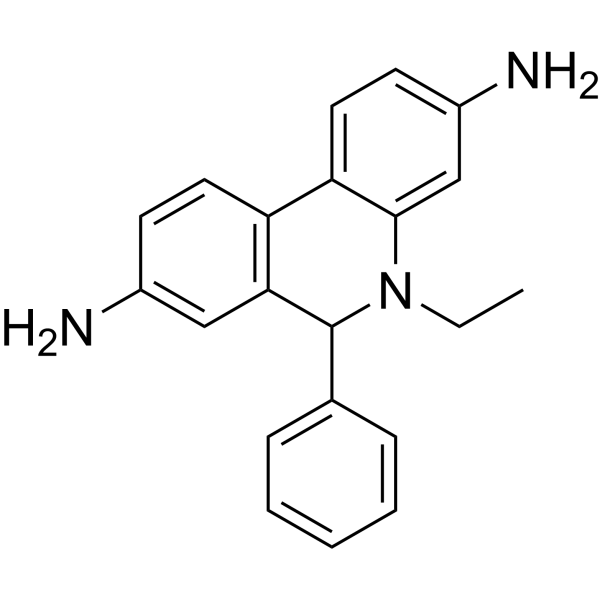
HY-D0079
DihydroethidiumFluorescent -

HY-DY1050
Ethidium bromide (solution)Fluorescent Tag -
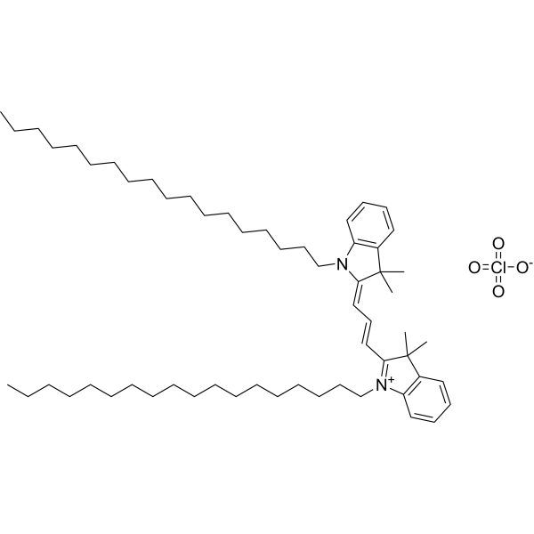
HY-D0083
DiIFluorescent Dye -
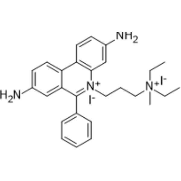
HY-D0815
Propidium IodideFluorochrome -

HY-DY1081
DAPI dihydrochloride (solution)Fluorescent DNA Stain -
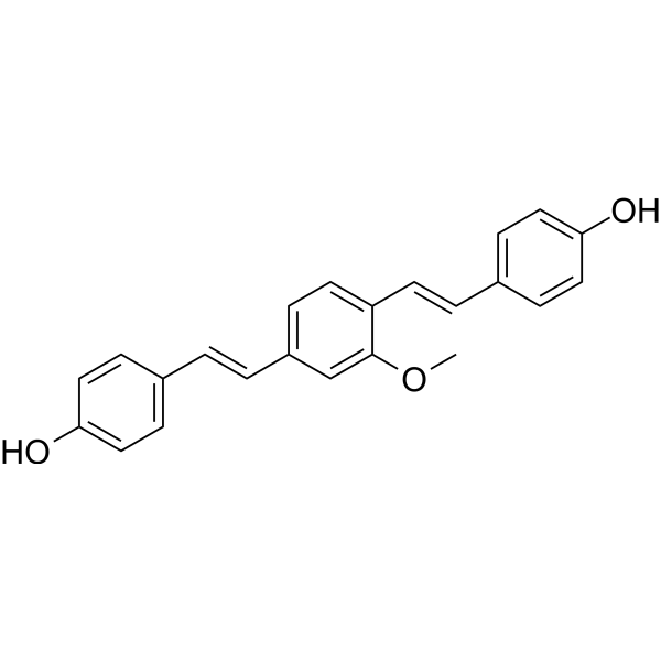
HY-103240
Methoxy-X04Fluorescent Dye -
HY-15902
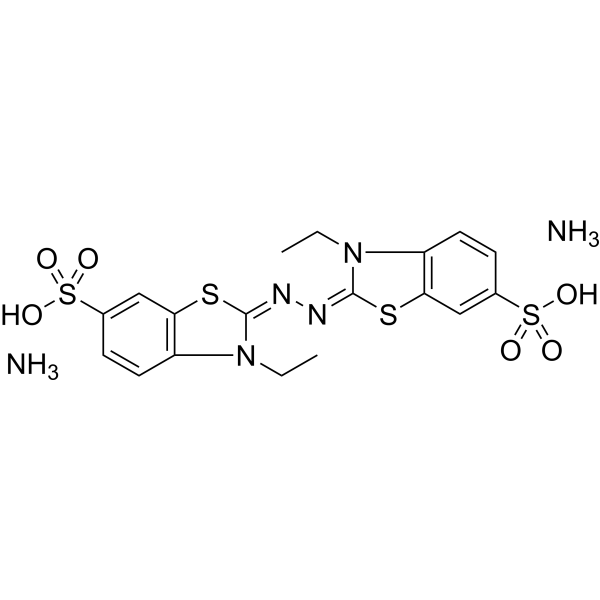
ABTS diammonium saltHRP Substrate -
HY-100041
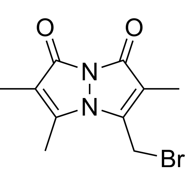
BromobimaneDye Reagent


Biological Activity
제품 설명
Solvent and Concentration: Sterile water: 10 mM
The 1 mL volume is defined as the base specification. All larger sizes correspond to incremental volumes of this base.
In Vitro
Thioflavin T is a fluorescent dye that can bind to β-fold-rich structures and is often used to measure protein fibrillation. Once bound, the emission spectrum of Thioflavin T will be red-shifted and the fluorescence intensity will increase, which can also be used to detect fibril formation. Here is a reference protocol for Aβ fibrils detection[2][3].
Guide (The following is our recommended protocol. This protocol is only a guide and should be modified according to your specific needs).
1. Prepare a mixed sample containing LMW Aβ42 (5.5 μM) , Thioflavin T solution (20 μM) , and NaCl (100 mM).
2. The mixtures were prepared in a 96-well half-area microplate. Seal the well plate and place it in a shaking incubator at 37°C for indicated time.
3. Shake for 20 seconds before testing. Set the excitation light to 450 nm and the emission light to 485 nm. Read the fluorescence value using a microplate reader.
MedChemExpress (MCE) has not independently confirmed the accuracy of these methods. They are for reference only.


Chemical Information
-
CAS No. 2390-54-7
-
Appearance Liquid (Density: 1.301 g/cm3)
-
분자량 318.86
-
화학식 C17H19ClN2S
-
Color Light yellow to yellow
-
SMILES
CC1=CC=C2[N+](C)=C(C3=CC=C(N(C)C)C=C3)SC2=C1.[Cl-]
-
선적
Room temperature in continental US; may vary elsewhere.
-
보관
Please store the product under the recommended conditions in the Certificate of Analysis.


순도&문서
-
Data Sheet (269 KB)
-
SDS (252 KB)
- English - EN (252 KB)
- Français - FR (252 KB)
- Deutsch - DE (252 KB)
- Norwegian - NO (252 KB)
- Español - ES (252 KB)
- Swedish - SV (252 KB)
- Italian - IT (252 KB)
- Korean - KR (252 KB)
- Portuguese - PT (252 KB)
-
Handling Instructions (2659 KB)
References
[1]. Nakano H, et al. Globular-shaped Aβ oligomers have diverse mechanisms for promoting Aβ aggregations with the facilitation of fibril elongation. Neurobiol Dis. 2025 Feb;205:106775. [Content Brief]
[2]. Muramatsu D, et al. ALZ-801 prevents amyloid β-protein assembly and reduces cytotoxicity: A preclinical experimental study. FASEB J. 2025 Feb 15;39(3):e70382. [Content Brief]
[3]. Khurana R, et al. Mechanism of thioflavin T binding to amyloid fibrils. J Struct Biol. 2005 Sep;151(3):229-38. [Content Brief]

Calculators
Concentration (start) × Volume (start) = Concentration (final) × Volume (final)


Thioflavin T (solution) Related Classifications
HY-DY1049 Related Classifications
 Powered by Bioz