



L-Asparagine-d3 hydrate
Based on 1 publication(s) in Google Scholar
L-Asparagine-d3 hydrate is the deuterium labeled L-Asparagine (HY-N0667). L-Asparagine is an essential amino acid for leukemic cells and a substrate for L-Asparaginase. L-Asparaginase is a potent anti-leukemic enzyme that promotes asparagine (Asn) and glutamine (Gln) depletion and inhibits protein biosynthesis in lymphoblasts. Removal of L-asparagine from plasma by L-Asparaginase results in inhibition of RNA and DNA synthesis and subsequent apoptosis. L-Asparaginase has cell-killing ability in vitro and in vivo, and selectively inhibits the growth of cancer cells with low asparagine synthetase (AASNS) expression. L-Asparagine can be used as a biomarker and sensor for the study of childhood acute lymphoblastic leukemia.
For research use only. We do not sell to patients.
- Purity : 99.64%
- CAS No.: 2483831-59-8
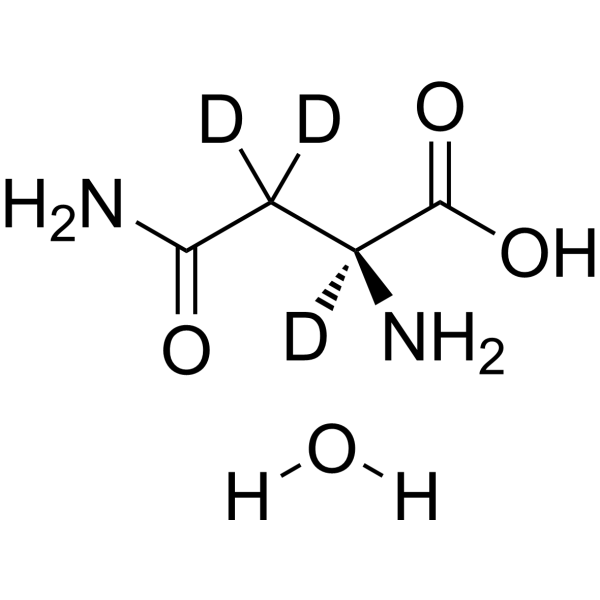
- Formula: C4H7D3N2O4
- Molecular Weight:153.15
-
Storage:Powder -20°C, 3 years , 4°C, 2 years ; In solvent -80°C, 6 months , -20°C, 1 month

To place orders, for customer services and technical support, please contact: MedChemExpress USA
Tel: 609-228-6898 E-mail: [email protected] [email protected]


-
 Biological Activity
-
Chemical Information
-
Solvent & Solubility
- Protocols
- Purity & Documentation
- References
-
 Help & FAQs


-
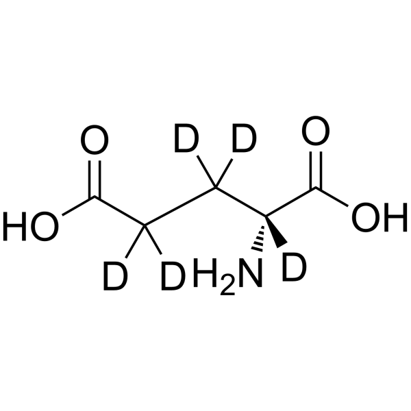
HY-100805S
D-Glutamic acid-d5Stable Isotope -
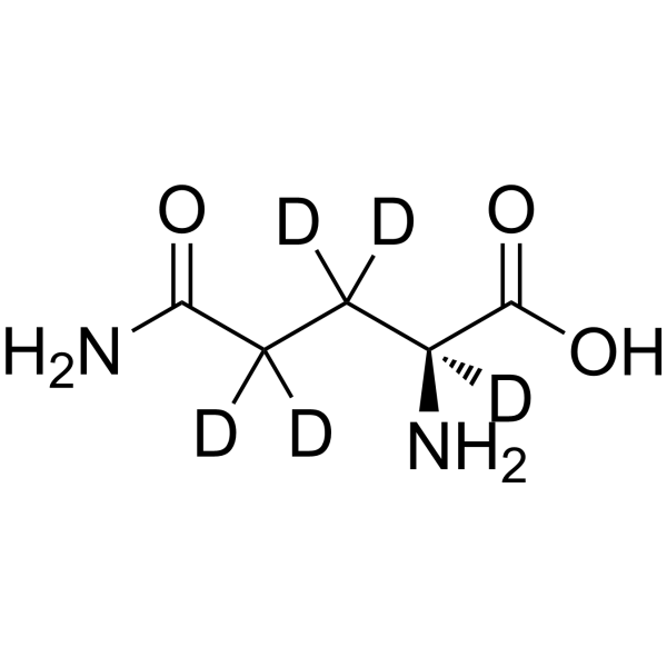
HY-N0390S2
L-Glutamine-d5Stable?Isotope -
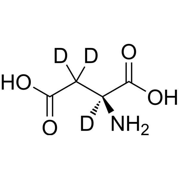
HY-N0666S8
L-Aspartic acid-d3Stable Isotope -
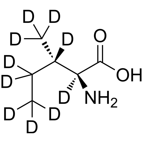
HY-N0771S3
L-Isoleucine-d10Stable Isotope -
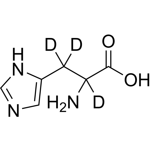
HY-W010209S2
DL-Histidine-d3Stable Isotope -
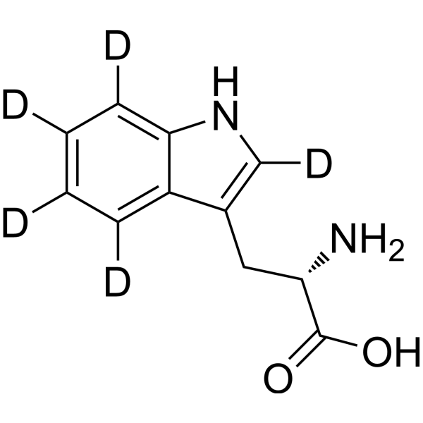
HY-N0623S
L-Tryptophan-d5Stable Isotope -
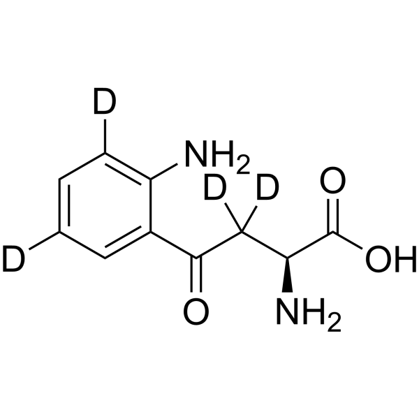
HY-104026S
L-Kynurenine-d4Stable Isotope -
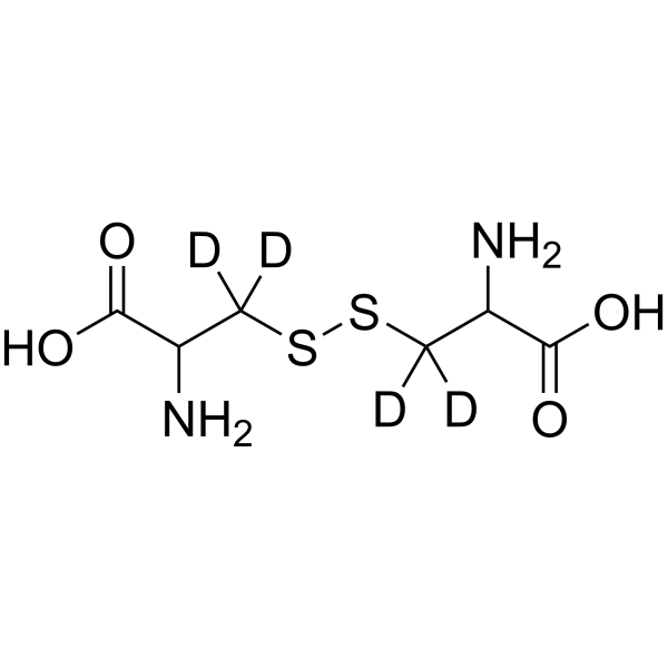
HY-W013940S
DL-Cystine-d4Stable Isotope -
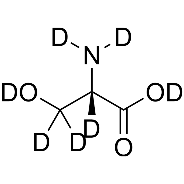
HY-N0650S11
L-Serine-d7Stable Isotope -
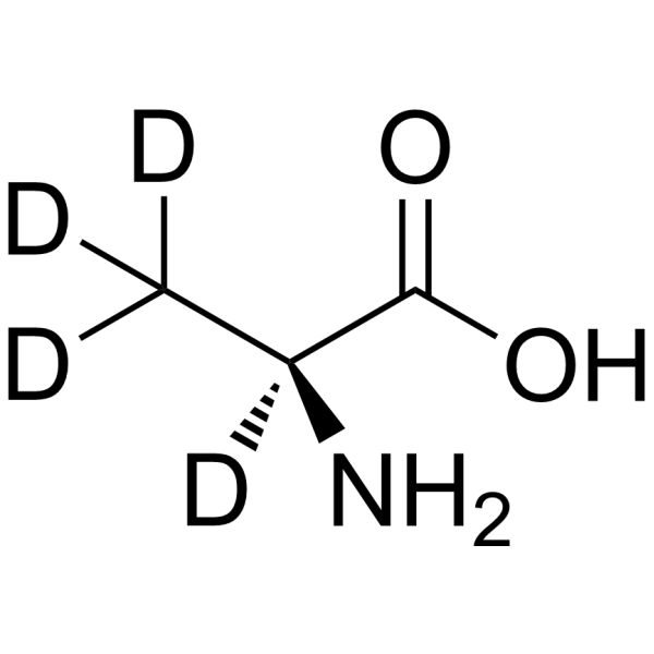
HY-N0229S3
L-Alanine-d4Stable Isotope -
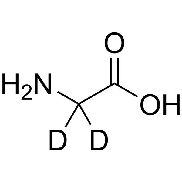
HY-Y0966S1
Glycine-d2Stable Isotope -
HY-N0623S5
L-Tryptophan-d8Stable Isotope
Publications Citing Use of MedChemExpress (MCE) L-Asparagine-d3 hydrate
MoreAll Endogenous Metabolite Isoforms
MoreAll DNA/RNA Synthesis Isoforms
More

Biological Activity
Description
In Vitro
Stable heavy isotopes of hydrogen, carbon, and other elements have been incorporated into drug molecules, largely as tracers for quantitation during the drug development process. Deuteration has gained attention because of its potential to affect the pharmacokinetic and metabolic profiles of drugs[1].
MedChemExpress (MCE) has not independently confirmed the accuracy of these methods. They are for reference only. Further protocols information, click here.
Application
1. This compound can be used as a tracer
2. This compound can be used as an internal standard for quantitative analysis by NMR, GC-MS, or LC-MS.


Chemical Information
-
CAS No. 2483831-59-8
-
Unlabeled CAS 70-47-3
-
Appearance Solid
-
Molecular Weight 153.15
-
Formula C4H7D3N2O4
-
Color White to off-white
-
SMILES
NC(C([2H])([2H])[C@@](N)([2H])C(O)=O)=O.[H]O[H]
-
Shipping
Room temperature in continental US; may vary elsewhere.
-
Storage
Powder -20°C 3 years 4°C 2 years In solvent -80°C 6 months -20°C 1 month


Publications (1)
-
Journal Impact Factor
-
Most Recent
-
Mol Cell
Asparagine sensing by TBK1 controls its phase separation to drive antiviral innate immune responses. [Abstract]2026 Feb 19;86(4):722-739.e8. PMID: 41653919


Solvent & Solubility
In Vitro:
H2O : 6.67 mg/mL (43.55 mM; Need ultrasonic)
Please refer to the solubility information to select the appropriate solvent. Once prepared, please aliquot and store the solution to prevent product inactivation from repeated freeze-thaw cycles.
Storage method and period of stock solution: -80°C, 6 months; -20°C, 1 month. When stored at -80°C, please use it within 6 months. When stored at -20°C, please use it within 1 month.
* Note: If you choose water as the stock solution, please dilute it to the working solution, then filter and sterilize it with a 0.22 μm filter before use.
Please refer to the solubility information to select the appropriate solvent. Once prepared, please aliquot and store the solution to prevent product inactivation from repeated freeze-thaw cycles.
Storage method and period of stock solution: -80°C, 6 months; -20°C, 1 month. When stored at -80°C, please use it within 6 months. When stored at -20°C, please use it within 1 month.
* Note: If you choose water as the stock solution, please dilute it to the working solution, then filter and sterilize it with a 0.22 μm filter before use.
Concentration (start) × Volume (start) = Concentration (final) × Volume (final)

Protocols
-
EdU Incorporation Assay (Click Chemistry-Based DNA Synthesis Measurement)
The EdU incorporation assay measures DNA synthesis by adding the thymidine analog 5-ethynyl-2′-deoxyuridine to cells or tissues, where it is incorporated into newly synthesized DNA during S phase. Incorporated EdU is detected by copper-catalyzed azide-alkyne cycloaddition, in which a fluorescent azide covalently reacts with the ethynyl group on EdU, allowing S-phase cells to be detected by fluorescence microscopy, flow cytometry, or high-content imaging. EdU detection does not require DNA denaturation or anti-BrdU antibody access, which preserves sample structure and improves compatibility with immunostaining and multiparameter cytometry compared with BrdU-based detection. EdU can be cytotoxic in a cell-type- and exposure-dependent manner, so pulse duration, concentration, and continuous-labeling designs should be validated for each cell type.
-
RNA extraction experimental
By lysing cells, releasing RNA, and removing impurities such as proteins and DNA, high-purity RNA products are finally obtained. The commonly used traditional method is the guanidine isothiocyanate/phenol/chloroform method (Trizol), which is suitable for a variety of animal materials including animal tissues, microorganisms, cultured cells, etc., and most plant materials.
-
Apoptosis
Apoptosis, also called programmed cell death, is generally characterized by distinct morphological characteristics.
-
TUNEL staining for apoptotic DNA fragmentation
TUNEL staining detects DNA strand breaks by using terminal deoxynucleotidyl transferase to add labeled nucleotides to exposed 3′-OH DNA termini, generating either microscopic staining in fixed cells or tissue sections, or fluorescence/cytometric signal in cell suspensions. TUNEL positivity reflects DNA fragmentation but should not be interpreted alone as definitive apoptosis, because TUNEL can also label necrotic, autolytic, mechanically damaged, or DNA-repair-associated DNA breaks.
-
Annexin V plus membrane-impermeant dye apoptosis staining
Annexin V-based apoptosis assays rely on the detection of phosphatidylserine (PS) externalization from the inner leaflet of the plasma membrane to the outer leaflet, an early biochemical hallmark of apoptosis. Fluorescently labeled Annexin V binds PS in a calcium-dependent manner, enabling identification of early apoptotic cells by flow cytometry or fluorescence microscopy. When combined with a membrane-impermeant DNA-binding dye (e. g. , propidium iodide), this approach allows discrimination between viable (Annexin V−/dye−), early apoptotic (Annexin V+/dye−), and late apoptotic or necrotic (Annexin V+/dye+) cell populations by assessing membrane integrity and PS exposure.
-
BrdU Incorporation Assay
Bromodeoxyuridine (BrdU) incorporation assay is based on the principle that BrdU, a thymidine analog, is incorporated into newly synthesized DNA during the S phase of the cell cycle, thereby serving as a marker of DNA replication and cellular proliferation. Incorporated BrdU can be detected using anti-BrdU antibodies following DNA denaturation, enabling visualization or quantification of proliferating cells through immunochemical detection methods such as immunofluorescence or immunohistochemistry.
-
Apoptosis Solutions
Apoptosis is a regulated, generally non-lytic cell-death pathway that removes unwanted, damaged, infected, or abnormal cells through coordinated morphological changes, caspase activation, DNA fragmentation, and membrane remodeling. The intrinsic apoptosis pathway is controlled mainly by mitochondrial outer membrane permeabilization, BCL-2 family proteins, cytochrome c release, apoptosome formation, caspase-9 activation, and downstream executioner caspase-3/7 activation. The extrinsic apoptosis pathway is initiated by death receptors such as Fas, TNFR, and TRAIL receptors, which recruit adaptor proteins and activate caspase-8 before engaging executioner caspases or mitochondrial amplification through BID cleavage. Apoptosis is linked to many phenotypes, including cancer cell killing, tissue homeostasis, immune regulation, neurodegeneration, infection response, and treatment-induced cytotoxicity; unresolved questions include how apoptosis interacts with necroptosis, pyroptosis, ferroptos


Purity & Documentation
-
Data Sheet (276 KB)
-
SDS (252 KB)
- English - EN (252 KB)
- Français - FR (252 KB)
- Deutsch - DE (252 KB)
- Norwegian - NO (252 KB)
- Español - ES (252 KB)
- Swedish - SV (252 KB)
- Italian - IT (252 KB)
- Korean - KR (252 KB)
- Portuguese - PT (252 KB)
-
Handling Instructions (2659 KB)
References
[1]. Russak EM, et al. Impact of Deuterium Substitution on the Pharmacokinetics of Pharmaceuticals. Ann Pharmacother. 2019;53(2):211-236. [Content Brief]
[2]. Batool T, et al. A Comprehensive Review on L-Asparaginase and Its Applications. Appl Biochem Biotechnol. 2016 Mar;178(5):900-23. [Content Brief]
[3]. Asselin BL, et al. In vitro and in vivo killing of acute lymphoblastic leukemia cells by L-asparaginase. Cancer Res. 1989 Aug 1;49(15):4363-8. [Content Brief]
[4]. Horvath TD, et al. Assessment of l-Asparaginase Pharmacodynamics in Mouse Models of Cancer. Metabolites. 2019 Jan 9;9(1):10. [Content Brief]
[5]. Kravtzoff R, et al. Erythrocytes as carriers for L-asparaginase. Methodological and mouse in-vivo studies. J Pharm Pharmacol. 1990 Jul;42(7):473-6. [Content Brief]
[6]. Ilaria M Michelozzi, et al. Acute myeloid leukaemia niche regulates response to L-asparaginase. Br J Haematol. 2019 Aug;186(3):420-430. doi: 10.1111/bjh.15920. [Content Brief]


Complete Stock Solution Preparation Table
Please refer to the solubility information to select the appropriate solvent. Once prepared, please aliquot and store the solution to prevent product inactivation from repeated freeze-thaw cycles.
Storage method and period of stock solution: -80°C, 6 months; -20°C, 1 month. When stored at -80°C, please use it within 6 months. When stored at -20°C, please use it within 1 month.
| Optional Solvent | Concentration Solvent Mass | 1 mg | 5 mg | 10 mg | 25 mg |
|---|---|---|---|---|---|
| H2O | 1 mM | 6.5295 mL | 32.6477 mL | 65.2955 mL | 163.2387 mL |
| 5 mM | 1.3059 mL | 6.5295 mL | 13.0591 mL | 32.6477 mL | |
| 10 mM | 0.6530 mL | 3.2648 mL | 6.5295 mL | 16.3239 mL | |
| 15 mM | 0.4353 mL | 2.1765 mL | 4.3530 mL | 10.8826 mL | |
| 20 mM | 0.3265 mL | 1.6324 mL | 3.2648 mL | 8.1619 mL | |
| 25 mM | 0.2612 mL | 1.3059 mL | 2.6118 mL | 6.5295 mL | |
| 30 mM | 0.2177 mL | 1.0883 mL | 2.1765 mL | 5.4413 mL | |
| 40 mM | 0.1632 mL | 0.8162 mL | 1.6324 mL | 4.0810 mL |
* Note: If you choose water as the stock solution, please dilute it to the working solution, then filter and sterilize it with a 0.22 μm filter before use.


L-Asparagine-d3 hydrate Related Classifications
HY-N0667S5 Related Classifications
 Powered by Bioz