



Lanuginosine

Based on 1 Customer Validation
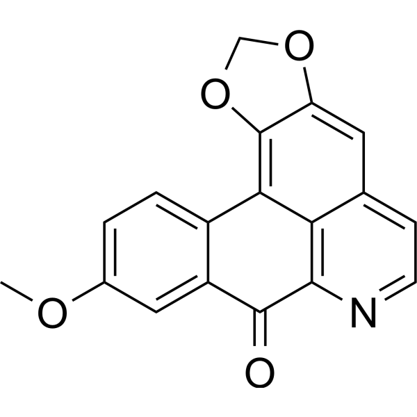
Lanuginosine is an alkaloid. Lanuginosine can be isolated from the stems of Xylopia laevigata (Annonaceae) and the leaves of Magnolia grandiflora. Lanuginosine induces Apoptosis. Lanuginosine inhibits AChE (IC50: 10.9 μM). Lanuginosine inhibits Aβ aggregation. Lanuginosine exhibits anticancer activity against hepatocellular carcinoma, human promyelocytic leukemia, human chronic myeloid leukemia, melanoma, and brain tumors. Lanuginosine can be used in the research of Alzheimer's disease.
For research use only. We do not sell to patients.
- Purity : 98.27%
- CAS No.: 23740-25-2
- Formula: C18H11NO4
- Molecular Weight:305.28
-
Storage:Powder -20°C, 3 years , 4°C, 2 years ; In solvent -80°C, 6 months , -20°C, 1 month

To place orders, for customer services and technical support, please contact: MedChemExpress USA
Tel: 609-228-6898 E-mail: [email protected] [email protected]


-
 Biological Activity
-
Chemical Information
-
Solvent & Solubility
- Protocols
- Purity & Documentation
- References
-
 Help & FAQs




Biological Activity
Description
IC50 & Target
10.9 μM (AChE)[1]
Cellular Effect
|
Cell Line

|
Type | Value | Description | References |
|---|---|---|---|---|
| B-cell | IC50 |
7.29 μM
Compound: 20
|
Inhibition of LPS-induced BALB/c mouse B cell proliferation
Inhibition of LPS-induced BALB/c mouse B cell proliferation
|
[PMID: 17081761] |
| HepG2 | IC50 |
0.8 μg/mL
Compound: atherospermidine
|
Cytotoxicity against human HepG2 cells
Cytotoxicity against human HepG2 cells
|
[PMID: 11575948] |
| HepG2 2.2.15 | IC50 |
2.2 μg/mL
Compound: atherospermidine
|
Cytotoxicity against human HepG2(2.2.15) cells
Cytotoxicity against human HepG2(2.2.15) cells
|
[PMID: 11575948] |
| T-cell | IC50 |
4.16 μM
Compound: 20
|
Inhibition of ConA-induced BALB/c mouse T cell proliferation
Inhibition of ConA-induced BALB/c mouse T cell proliferation
|
[PMID: 17081761] |
In Vitro
Lanuginosine (72 h) shows cytotoxic activity against B16-F10, HepG2, HL-60, and K562 tumor cell lines[1].
Lanuginosine (10-20 μg/mL; 24 h) induces apoptosis in HepG2 cells[1].
Lanuginosine is cytotoxic to U251 cells (a brain tumor cell line) with an IC50 of 4 μg/mL[2].
Lanuginosine (Compound 12) is an AChE inhibitor (IC50: 10.9 μM)[3].
MedChemExpress (MCE) has not independently confirmed the accuracy of these methods. They are for reference only. Further protocols information, click here.
-
Cell Line:HepG2
-
Concentration:10 μg/mL, 20 μg/mL
-
Incubation Time:24 h
-
Result:Increased apoptotic cells, and significantly increased necrotic cells (AO/EB staining).
Displayed apoptotic morphology (chromatin condensation and nuclear fragmentation).
Showed significant phosphatidylserine externalization.


Chemical Information
-
CAS No. 23740-25-2
-
Appearance Solid
-
Molecular Weight 305.28
-
Formula C18H11NO4
-
Color Yellow to brown
-
SMILES
O=C1C2=CC(OC)=CC=C2C3=C4OCOC4=CC5=CC=NC1=C53
-
Structure Classification
-
Shipping
Room temperature in continental US; may vary elsewhere.
-
Storage
Powder -20°C 3 years 4°C 2 years In solvent -80°C 6 months -20°C 1 month


Solvent & Solubility
In Vitro:
DMSO : 0.5 mg/mL (1.64 mM; ultrasonic and warming and adjust pH to 2 with 1 M HCl and heat to 60°C; Hygroscopic DMSO has a significant impact on the solubility of product, please use newly opened DMSO)
Please refer to the solubility information to select the appropriate solvent. Once prepared, please aliquot and store the solution to prevent product inactivation from repeated freeze-thaw cycles.
Storage method and period of stock solution: -80°C, 6 months; -20°C, 1 month. When stored at -80°C, please use it within 6 months. When stored at -20°C, please use it within 1 month.
Please refer to the solubility information to select the appropriate solvent. Once prepared, please aliquot and store the solution to prevent product inactivation from repeated freeze-thaw cycles.
Storage method and period of stock solution: -80°C, 6 months; -20°C, 1 month. When stored at -80°C, please use it within 6 months. When stored at -20°C, please use it within 1 month.
Concentration (start) × Volume (start) = Concentration (final) × Volume (final)

Protocols
-
Detection of Apoptosis
Apoptosis, also called programmed cell death, is generally characterized by distinct morphological characteristics.
-
TUNEL staining for apoptotic DNA fragmentation
TUNEL staining detects DNA strand breaks by using terminal deoxynucleotidyl transferase to add labeled nucleotides to exposed 3′-OH DNA termini, generating either microscopic staining in fixed cells or tissue sections, or fluorescence/cytometric signal in cell suspensions. TUNEL positivity reflects DNA fragmentation but should not be interpreted alone as definitive apoptosis, because TUNEL can also label necrotic, autolytic, mechanically damaged, or DNA-repair-associated DNA breaks.
-
Annexin V plus membrane-impermeant dye apoptosis staining
Annexin V-based apoptosis assays rely on the detection of phosphatidylserine (PS) externalization from the inner leaflet of the plasma membrane to the outer leaflet, an early biochemical hallmark of apoptosis. Fluorescently labeled Annexin V binds PS in a calcium-dependent manner, enabling identification of early apoptotic cells by flow cytometry or fluorescence microscopy. When combined with a membrane-impermeant DNA-binding dye (e. g. , propidium iodide), this approach allows discrimination between viable (Annexin V−/dye−), early apoptotic (Annexin V+/dye−), and late apoptotic or necrotic (Annexin V+/dye+) cell populations by assessing membrane integrity and PS exposure.
-
Apoptosis Solutions
Apoptosis is a regulated, generally non-lytic cell-death pathway that removes unwanted, damaged, infected, or abnormal cells through coordinated morphological changes, caspase activation, DNA fragmentation, and membrane remodeling. The intrinsic apoptosis pathway is controlled mainly by mitochondrial outer membrane permeabilization, BCL-2 family proteins, cytochrome c release, apoptosome formation, caspase-9 activation, and downstream executioner caspase-3/7 activation. The extrinsic apoptosis pathway is initiated by death receptors such as Fas, TNFR, and TRAIL receptors, which recruit adaptor proteins and activate caspase-8 before engaging executioner caspases or mitochondrial amplification through BID cleavage. Apoptosis is linked to many phenotypes, including cancer cell killing, tissue homeostasis, immune regulation, neurodegeneration, infection response, and treatment-induced cytotoxicity; unresolved questions include how apoptosis interacts with necroptosis, pyroptosis, ferroptos
-
Liver Cancer Modeling
Liver cancer can be classified into primary liver cancer and secondary liver cancer. Secondary liver cancer is the metastatic liver cancer. Primary liver cancer includes hepatocellular carcinoma (HCC), intrahepatic cholangiocarcinoma (ICC) and fibrolamellar HCC, of which HCC is the most common form, accounting for approximately 90% of primary liver cancers[1]. HCC mouse models include chemical agent-induced models, transplanted tumor models, and genetic engineered models.
-
Alzheimer’s Disease Modeling
Alzheimer’s Disease (AD) is a neurodegenerative disorder characterized by a progressive decline in cognitive functions and loss of specific types of neurons and synapses. Alzheimer's symptoms can be simulated in mice by injecting drugs (such as Aβ) or genetically modified.


Purity & Documentation
-
Data Sheet (278 KB)
-
SDS (251 KB)
- English - EN (251 KB)
- Français - FR (251 KB)
- Deutsch - DE (251 KB)
- Norwegian - NO (251 KB)
- Español - ES (251 KB)
- Swedish - SV (251 KB)
- Italian - IT (251 KB)
- Korean - KR (251 KB)
- Portuguese - PT (251 KB)
-
Handling Instructions (2659 KB)
References
[1]. Menezes LR, et al. Cytotoxic Alkaloids from the Stem of Xylopia laevigata. Molecules. 2016 Jul 8;21(7):890. [Content Brief]
[2]. Mohamed SM, et al. Cytotoxic and antiviral activities of aporphine alkaloids of Magnolia grandiflora L. Nat Prod Res. 2010 Sep;24(15):1395-402. [Content Brief]
[3]. Jiang K, et al. Bioactive Isoquinoline Alkaloids with Diverse Skeletons from Fissistigma polyanthum. J Nat Prod. 2023 Aug 24. [Content Brief]


Complete Stock Solution Preparation Table
Please refer to the solubility information to select the appropriate solvent. Once prepared, please aliquot and store the solution to prevent product inactivation from repeated freeze-thaw cycles.
Storage method and period of stock solution: -80°C, 6 months; -20°C, 1 month. When stored at -80°C, please use it within 6 months. When stored at -20°C, please use it within 1 month.
| Optional Solvent | Concentration Solvent Mass | 1 mg | 5 mg | 10 mg | 25 mg |
|---|---|---|---|---|---|
| DMSO | 1 mM | 3.2757 mL | 16.3784 mL | 32.7568 mL | 81.8920 mL |


Lanuginosine Related Classifications
HY-119398 Related Classifications
 Powered by Bioz