



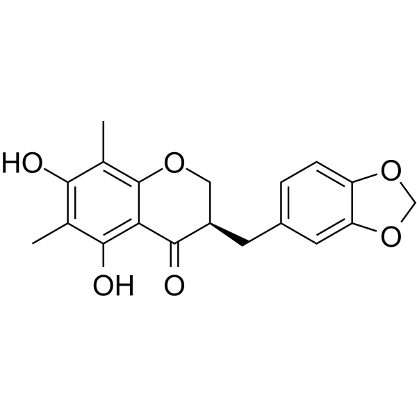
Methylophiopogonone A

Based on 1 Customer Validation
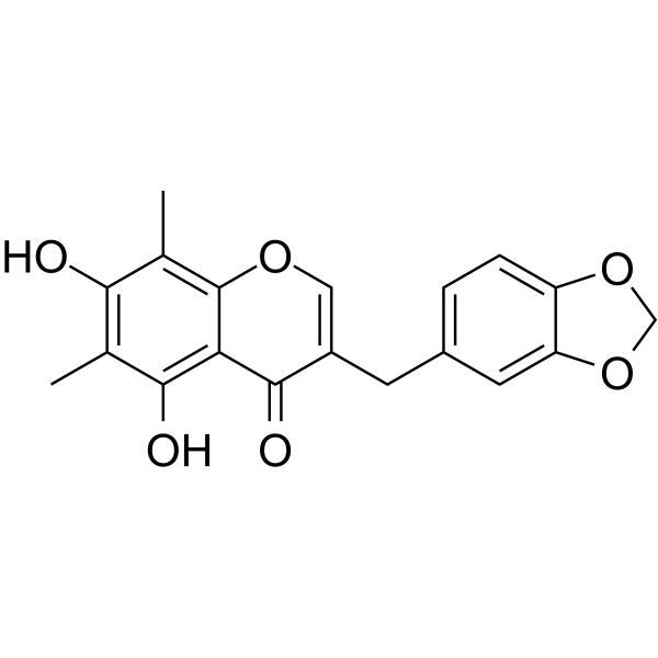
Methylophiopogonone A is a homoisoflavonoid compound found in the tuberous roots of Ophiopogon japonicus. Methylophiopogonone A inhibits lipopolysaccharide-induced NO production in mouse microglia with an IC50 of 19.2 μM. Methylophiopogonone A blocks the proliferation of LX-2 hepatic stellate cells and downregulates TGF-β1-induced expressions of type I collagen and α-SMA. Methylophiopogonone A blocks RELA/EGFR-mediated hepatic stellate cell activation, inhibits the MAPK12 pathway to reduce extracellular matrix accumulation, and simultaneously suppresses inflammatory signal transduction. Methylophiopogonone A can be used in studies related to chronic hepatitis B-associated hepatic fibrosis and neuroinflammation.
Para uso exclusivo en investigación. No vendemos a pacientes.
- Pureza : 99.94%
- No. CAS: 74805-90-6
- Fòrmula: C19H16O6
- Peso molecular:340.33
-
Almacenamiento:
4°C, protect from light
* In solvent : -80°C, 6 months; -20°C, 1 month (protect from light)

To place orders, for customer services and technical support, please contact: MedChemExpress USA
Tel: 609-228-6898 E-mail: [email protected] [email protected]


-
 Actividad biológica
-
Chemical Information
-
Solvente y solubilidad
- Protocolo
- Pureza y Documentación
- Referencias
-
 Help & FAQs




Actividad biológica
Descripciòn
IC50 & Target
[1]|
Collagen I |
In Vitro
Methylophiopogonone A (compound 10) (24 h) potently inhibits lipopolysaccharide-induced nitric oxide production in mouse microglial BV-2 cells, with an IC50 value of 19.2 μM[1].
Methylophiopogonone A (3.13-100 μM; 24-72 h) inhibits the proliferation of immortalized human hepatic stellate LX-2 cells in a time- and dose-dependent manner[2].
Methylophiopogonone A (3.13-25 μM; 24 h) dose-dependently inhibits the expression of type Ⅰ collagen and α-smooth muscle actin (α-SMA) in immortalized human hepatic stellate LX-2 cells induced by TGF-β1[2].
MedChemExpress (MCE) has not independently confirmed the accuracy of these methods. They are for reference only. Further protocols information, click here.
-
Cell Line:immortalized human hepatic stellate LX-2 cells
-
Concentration:3.13, 6.25, 12.5, 25, 50, 100 μM
-
Incubation Time:24 h; 48 h; 72 h
-
Result:Inhibited LX-2 cell viability in a time- and dose-dependent manner.
-
Cell Line:immortalized human hepatic stellate LX-2 cells
-
Concentration:3.13, 6.25, 12.5, 25 μM (in the presence of 5 ng/mL TGF-β1)
-
Incubation Time:24 h
-
Result:Inhibited TGF-β1-induced expression of collagen I and α-SMA.
Reduced protein levels of collagen I in a dose-dependent manner.
Reduced protein levels of α-SMA in a dose-dependent manner.


Chemical Information
-
No. CAS 74805-90-6
-
Appearance Solid
-
Peso molecular 340.33
-
Fòrmula C19H16O6
-
Color Off-white to light yellow
-
SMILES
O=C1C(CC2=CC=C(OCO3)C3=C2)=COC4=C(C)C(O)=C(C)C(O)=C14
-
Structure Classification
-
Initial Source
-
Envío
Room temperature in continental US; may vary elsewhere.
-
Almacenamiento
4°C, protect from light
* In solvent : -80°C, 6 months; -20°C, 1 month (protect from light)


Solvente y solubilidad
In Vitro:
DMSO : 100 mg/mL (293.83 mM; Need ultrasonic; Hygroscopic DMSO has a significant impact on the solubility of product, please use newly opened DMSO)
Please refer to the solubility information to select the appropriate solvent. Once prepared, please aliquot and store the solution to prevent product inactivation from repeated freeze-thaw cycles.
Storage method and period of stock solution: -80°C, 6 months; -20°C, 1 month (protect from light). When stored at -80°C, please use it within 6 months. When stored at -20°C, please use it within 1 month.
Please refer to the solubility information to select the appropriate solvent. Once prepared, please aliquot and store the solution to prevent product inactivation from repeated freeze-thaw cycles.
Storage method and period of stock solution: -80°C, 6 months; -20°C, 1 month (protect from light). When stored at -80°C, please use it within 6 months. When stored at -20°C, please use it within 1 month.
Concentration (start) × Volume (start) = Concentration (final) × Volume (final)
In Vivo:
For the following dissolution methods, please prepare the working solution directly:
It is recommended to prepare fresh solutions and use them promptly within a short period of time.
The percentages shown for the solvents indicate their volumetric ratio in the final prepared solution. If precipitation or phase separation occurs during preparation, heat and/or sonication can be used to aid dissolution.
Add each solvent one by one: 0.5% CMC-Na/saline water
Solubility: 12.5 mg/mL (36.73 mM); Suspended solution; Need ultrasonic

Protocolo
-
Fibrosis/Collagen Morphometry
Fibrosis and collagen morphometry is based on the quantitative visualization of fibrillar collagen deposition in tissue sections using histochemical stains such as Sirius Red (Picrosirius Red) or Masson's trichrome, followed by image-based or polarization-enhanced analysis to estimate collagen proportional area as a surrogate of extracellular matrix accumulation during fibrotic remodeling. Sirius Red combined with polarized light microscopy enhances detection of collagen fibers due to birefringence properties, enabling more specific visualization of collagen type I and III fibrils compared to conventional bright-field histology, while whole-section or region-restricted digital morphometry reduces field-selection bias in fibrosis assessment. Alternative quantitative approaches include second harmonic generation (SHG) and two-photon excited fluorescence microscopy, which enable label-free detection of fibrillar collagen and have been validated against histological staining and biochemica
-
Collagen: Sirius Red Staining
Sirius Red or picrosirius red staining is a histochemical method for visualizing collagen-rich extracellular matrix in tissue sections, and collagen fibers are detected as red-stained structures under bright-field microscopy with enhanced birefringence under polarized light. Picrosirius red is useful for assessing total collagen organization, distribution, and fibrosis burden, but polarized color should not be interpreted as a definitive collagen type I versus type III readout because color is affected by fiber orientation, thickness, and packing.
-
Cell Cytotoxicity Assay
Cytotoxicity assays are usually based on the assessment of cell membrane damage, which can also be indirectly detected by measuring cell viability. Detection methods include MTT assay, CKK-8 assay, LDH assay and ATP assay, etc.
-
ECM-Embedded Organoid (Matrigel/Dome) Culture
ECM-embedded organoid dome culture embeds epithelial stem cells, crypts, organoid fragments, or tumor-derived epithelial cells in a basement-membrane-like hydrogel such as Matrigel, allowing 3D growth, self-organization, lumen formation, budding or cystic morphogenesis, and lineage maintenance under defined niche-factor-containing medium. The primary readouts are organoid establishment efficiency, growth, morphology, passaging capacity, lineage-marker expression, and, when fluorescently labeled lines are used, microscopy- or flow-cytometry-based quantification of population behavior in 3D culture.
-
Connective Tissue: Masson's Trichrome/Collagen Trichrome Staining
Masson’s Trichrome (collagen/trichrome staining) is a histological technique that differentially stains tissue compartments using sequential acidic dyes to distinguish collagen from muscle and cytoplasmic components based on dye affinity and tissue permeability differences, enabling visualization of fibrosis and connective tissue architecture in histological sections. The classical formulation typically uses Weigert's iron hematoxylin for nuclear staining, Biebrich scarlet-acid fuchsin for cytoplasm and muscle, and aniline blue (or light green variants) for collagen, producing a characteristic blue/green collagen signal contrasted against red cytoplasm and dark nuclei. The staining principle relies on selective displacement of smaller dye molecules by larger anionic dyes in collagen-rich regions under controlled acidified conditions, which enhances collagen-specific dye retention. This property makes the method widely used for fibrosis assessment in organs such as heart, liver, lung, a
-
Matrigel/ECM Transwell Invasion Assay
The Matrigel/ECM Transwell invasion assay measures the ability of cells to move toward a chemoattractant while crossing an extracellular-matrix barrier placed on a porous membrane; therefore, the readout reflects both chemotactic motility and matrix invasion rather than migration alone. Matrigel is a basement-membrane-rich matrix derived from Engelbreth-Holm-Swarm mouse sarcoma and has been used as a reconstituted basement membrane barrier in chemoinvasion assays. The assay readout is generated by quantifying cells that reach the underside of the insert membrane or lower compartment after incubation, commonly by staining and counting invaded cells or by fluorescence-based quantification.
-
Research Protocol for Neurological Diseases
PINK1/Parkin-mediated mitophagy pathway is a mitochondrial quality-control signaling axis in which mitochondrial depolarization stabilizes PINK1 on damaged mitochondria, activates Parkin recruitment and E3 ubiquitin ligase activity, promotes ubiquitination of outer mitochondrial membrane proteins, recruits selective autophagy adaptors, and drives lysosomal degradation of damaged mitochondria. In neurological disease research, this pathway is experimentally important because neurons, especially dopaminergic neurons, are highly dependent on mitochondrial integrity, and defective mitochondrial turnover can lead to mitochondrial dysfunction, oxidative stress, impaired neuronal survival, α-synuclein accumulation, and neuroinflammatory damage-associated signals. The genetic disease link is strongest in Parkinson’s disease because mutations in PRKN/parkin cause autosomal recessive juvenile parkinsonism, mutations in PINK1 cause hereditary early-onset Parkinson’s disease, and Drosophila studie
-
Cell Viability Determination by MTT Colorimetric Assay
The following protocol uses the MTT colorimetric assay as a classic literature-established method for assessing cell viability/metabolic activity in cultured mammalian cells. MTT[3-(4,5-dimethylthiazol-2-yl)-2,5-diphenyltetrazolium bromide] is reduced by metabolically active cells to a colored formazan product; the amount of formazan is quantified spectrophotometrically and provides an indirect measure of metabolically active viable cells. Importantly, MTT reduction reflects cellular oxidoreductase/metabolic activity rather than an absolute direct count of living cells, so changes in cellular metabolism can alter the signal independently of cell number.
-
Matrigel Transwell/Boyden Chamber Invasion Assay
Matrigel Transwell/Boyden chamber invasion assay measures the ability of cells to degrade or traverse an extracellular matrix-coated porous membrane and move from an upper chamber toward a chemoattractant in a lower chamber. Invasion is distinguished from migration by coating the membrane with Matrigel or basement membrane matrix; uncoated inserts measure migration, while coated inserts require cells to cross an ECM barrier before reaching the underside of the membrane.


Pureza y Documentación
-
Ficha de datos (275 KB)
-
SDS (252 KB)
- English - EN (252 KB)
- Français - FR (252 KB)
- Deutsch - DE (252 KB)
- Norwegian - NO (252 KB)
- Español - ES (252 KB)
- Swedish - SV (252 KB)
- Italian - IT (252 KB)
- Korean - KR (252 KB)
- Portuguese - PT (252 KB)
-
Instrucciones de manejo (2659 KB)
Referencias
[1]. Li N, et al. Anti-inflammatory homoisoflavonoids from the tuberous roots of Ophiopogon japonicus. Fitoterapia. 2012 Sep;83(6):1042-5. [Content Brief]
[2]. Li G, et al. Active Ingredients and Action Mechanisms of Yi Guan Jian Decoction in Chronic Hepatitis B Patients with Liver Fibrosis. Evidence-based complementary and alternative medicine : eCAM. 2019;2019:2408126. [Content Brief]


Complete Stock Solution Preparation Table
Please refer to the solubility information to select the appropriate solvent. Once prepared, please aliquot and store the solution to prevent product inactivation from repeated freeze-thaw cycles.
Storage method and period of stock solution: -80°C, 6 months; -20°C, 1 month (protect from light). When stored at -80°C, please use it within 6 months. When stored at -20°C, please use it within 1 month.
| Optional Solvent | Concentration Solvent Mass | 1 mg | 5 mg | 10 mg | 25 mg |
|---|---|---|---|---|---|
| DMSO | 1 mM | 2.9383 mL | 14.6916 mL | 29.3832 mL | 73.4581 mL |
| 5 mM | 0.5877 mL | 2.9383 mL | 5.8766 mL | 14.6916 mL | |
| 10 mM | 0.2938 mL | 1.4692 mL | 2.9383 mL | 7.3458 mL | |
| 15 mM | 0.1959 mL | 0.9794 mL | 1.9589 mL | 4.8972 mL | |
| 20 mM | 0.1469 mL | 0.7346 mL | 1.4692 mL | 3.6729 mL | |
| 25 mM | 0.1175 mL | 0.5877 mL | 1.1753 mL | 2.9383 mL | |
| 30 mM | 0.0979 mL | 0.4897 mL | 0.9794 mL | 2.4486 mL | |
| 40 mM | 0.0735 mL | 0.3673 mL | 0.7346 mL | 1.8365 mL | |
| 50 mM | 0.0588 mL | 0.2938 mL | 0.5877 mL | 1.4692 mL | |
| 60 mM | 0.0490 mL | 0.2449 mL | 0.4897 mL | 1.2243 mL | |
| 80 mM | 0.0367 mL | 0.1836 mL | 0.3673 mL | 0.9182 mL | |
| 100 mM | 0.0294 mL | 0.1469 mL | 0.2938 mL | 0.7346 mL |


Methylophiopogonone A Related Classifications
HY-N2441 Related Classifications
 Powered by Bioz