



ML115
Based on 8 publication(s) in Google Scholar
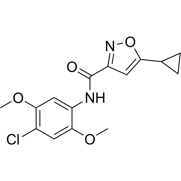
ML115, a molecular probe of the signal transducer, is a selective STAT3 agonist, with an EC50 of 2 nM. ML115 increases the expression of BCL3, a known STAT3-dependent oncogene. ML115 is inactive against the related STAT1, STAT5 and NF-κB anti-targets. ML115 counteracts the effects of Ginsenoside Rc (HY-N0042) on cell viability and inflammatory responses in LPS (HY-D1056)-stimulated H9c2 and RAW264.7 cells, while altering oxidative stress markers. ML115 can be used for the study of breast and prostate cancers.
For research use only. We do not sell to patients.
- Purity : 99.98%
- CAS No.: 912798-42-6
- Formula: C15H15ClN2O4
- Molecular Weight:322.74
-
Storage:
4°C, protect from light
* In solvent : -80°C, 6 months; -20°C, 1 month (protect from light)

To place orders, for customer services and technical support, please contact: MedChemExpress USA
Tel: 609-228-6898 E-mail: [email protected] [email protected]


-
 Biological Activity
-
Chemical Information
-
Solvent & Solubility
- Protocols
- Purity & Documentation
- References
-
 Help & FAQs


-
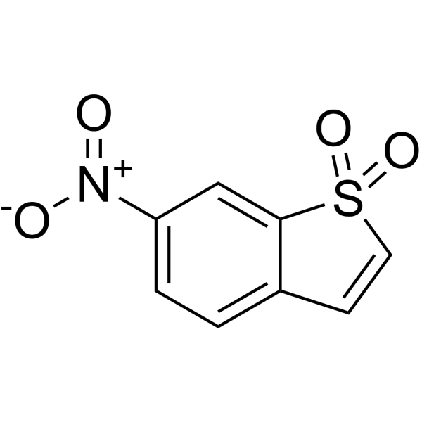
HY-13818
StatticSTAT3 Inhibitor -
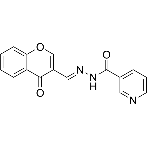
HY-101853
STAT5-IN-1STAT5 Inhibitor -
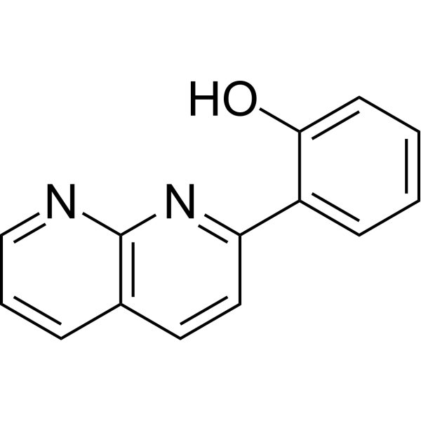
HY-W013523
2-NPSTAT1 Enhancer -
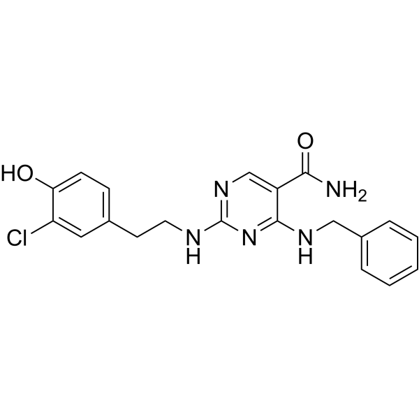
HY-100614
AS1517499STAT6 Inhibitor -
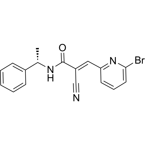
HY-15312
WP1066JAK2/STAT3 Inhibitor -
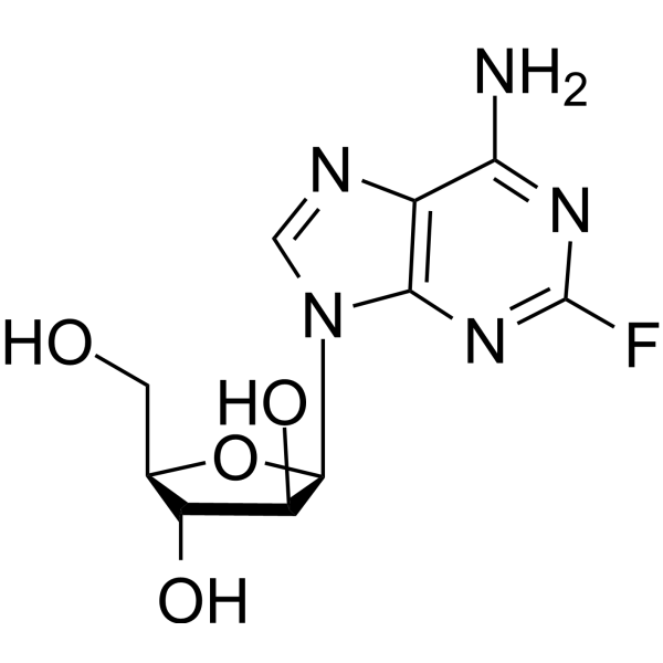
HY-B0069
FludarabineDNA Synthesis Inhibitor -
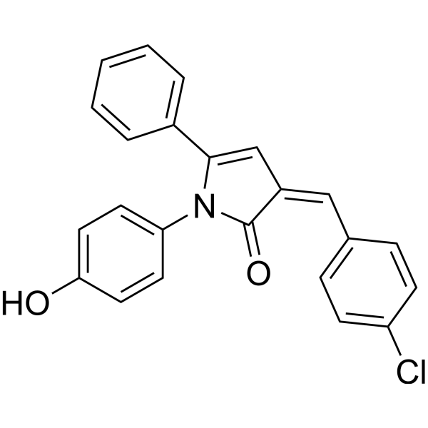
HY-103128
inS3-54A18STAT3 Inhibitor -
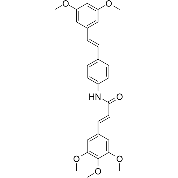
HY-100753
STAT3-IN-1STAT3 Inhibitor -
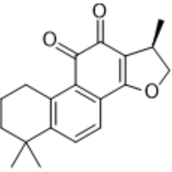
HY-N0174
CryptotanshinoneAutophagy Inducer -
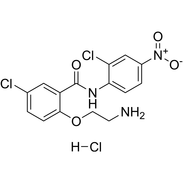
HY-100602
HJC0152STAT3 Inhibitor -
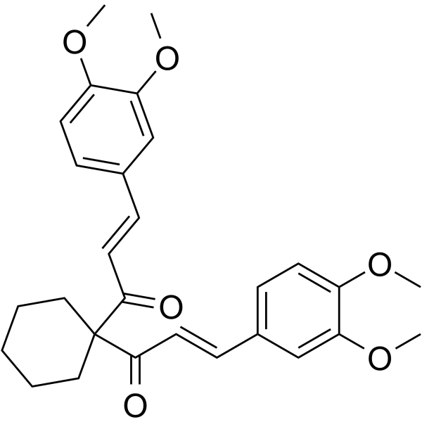
HY-100544
FLLL32JAK2/STAT3 Inhibitor -
HY-17386
RosiglitazonePPARγ Agonist
Publications Citing Use of MedChemExpress (MCE) ML115
More- Imeta. 2026 Jun 14.
- Cell Host Microbe. 2026 Feb 11;34(2):245-262.e8. [Abstract]
- Eur J Pharmacol. 2025 Jun 15:997:177606. [Abstract]
- Biochim Biophys Acta Mol Basis Dis. 2025 Aug;1871(6):167893. [Abstract]
- Basic Clin Pharmacol Toxicol. 2025 Dec;137(6):e70135. [Abstract]
- Discov Oncol. 2026 Jun 3. [Abstract]
- J Mol Histol. 2025 Apr 28;56(3):148. [Abstract]
- bioRxiv. 2025 January 31.


Biological Activity
Description
IC50 & Target
[1]|
Stat-3 2 nM (EC50) |
In Vitro
ML115 (10 µM, 48 h) reverses the Ginsenoside Rc (HY-N0042)-induced improvement in viability of LPS (HY-D1056)-stimulated H9c2 cells, increases MDA levels, and decreases SOD and GSH-Px activities[1].
ML115 (10 µM, 6 h) reverses the Ginsenoside Rc-induced downregulation of IL-1β, TNF-α, iNOS and COX2, and upregulation of Arg1 and Ym1 in LPS-treated RAW264.7 cells, and also reverses the Ginsenoside Rc-induced reduction in IL-1β and TNF-α production[1].
ML115 exhibits no cytotoxicity in HT-1080 and NIH-3T3 cells[2].
MedChemExpress (MCE) has not independently confirmed the accuracy of these methods. They are for reference only. Further protocols information, click here.
-
Cell Line:LPS-treated RAW264.7 cells
-
Concentration:10 µM
-
Incubation Time:6 h
-
Result:Reversed the Ginsenoside Rc-induced downregulation of IL-1β, TNF-α, iNOS and COX2, and upregulation of Arg1 and Ym1 in LPS-treated macrophages.


Chemical Information
-
CAS No. 912798-42-6
-
Appearance Solid
-
Molecular Weight 322.74
-
Formula C15H15ClN2O4
-
Color White to off-white
-
SMILES
O=C(C1=NOC(C2CC2)=C1)NC3=CC(OC)=C(Cl)C=C3OC
-
Shipping
Room temperature in continental US; may vary elsewhere.
-
Storage
4°C, protect from light
* In solvent : -80°C, 6 months; -20°C, 1 month (protect from light)


Publications (8)
-
Journal Impact Factor
-
Most Recent
-
-
Cell Host Microbe
Enterococcus faecalis-derived lactic acid suppresses macrophage activation to facilitate persistent and polymicrobial wound infections. [Abstract]2026 Feb 11;34(2):245-262.e8. PMID: 41605216 -
Eur J Pharmacol
Carnosol inhibits influenza A virus by disrupting the viral envelope and interfering with Jak2/STAT3 signaling pathway. [Abstract]2025 Jun 15:997:177606. PMID: 40216182 -
Biochim Biophys Acta Mol Basis Dis
HNF-1α promotes urethral fibrosis by up-regulating STAT3 transcriptional activity in mice hypospadias. [Abstract]2025 Aug;1871(6):167893. PMID: 40348066 -
Basic Clin Pharmacol Toxicol
Bergaptol Alleviates Oxidative Stress and Inflammation in Intracerebral Haemorrhage Mice via JAK2/STAT3 and NF-κB Pathways, Improving Neurological Function. [Abstract]2025 Dec;137(6):e70135. PMID: 41139697 -
Discov Oncol
PSMC2 drives tumor progression in nasopharyngeal carcinoma by inhibiting ferroptosis through STAT3. [Abstract]2026 Jun 3. PMID: 42234270 -
J Mol Histol
STAT3/FoxO3a/Sirt1 pathway inhibition by ginsenoside Rc ameliorates cardiomyocyte damage in septic cardiomyopathy by altering macrophage polarization. [Abstract]2025 Apr 28;56(3):148. PMID: 40293549 -


Solvent & Solubility
In Vitro:
DMSO : 20 mg/mL (61.97 mM; ultrasonic and warming and heat to 60°C; Hygroscopic DMSO has a significant impact on the solubility of product, please use newly opened DMSO)
Please refer to the solubility information to select the appropriate solvent. Once prepared, please aliquot and store the solution to prevent product inactivation from repeated freeze-thaw cycles.
Storage method and period of stock solution: -80°C, 6 months; -20°C, 1 month (protect from light). When stored at -80°C, please use it within 6 months. When stored at -20°C, please use it within 1 month.
Please refer to the solubility information to select the appropriate solvent. Once prepared, please aliquot and store the solution to prevent product inactivation from repeated freeze-thaw cycles.
Storage method and period of stock solution: -80°C, 6 months; -20°C, 1 month (protect from light). When stored at -80°C, please use it within 6 months. When stored at -20°C, please use it within 1 month.
Concentration (start) × Volume (start) = Concentration (final) × Volume (final)
In Vivo:
Select the appropriate dissolution method based on your experimental animal and administration route.
- For the following dissolution methods, please ensure to first prepare a clear stock solution using an In Vitro approach and then sequentially add co-solvents:
- To ensure reliable experimental results, the clarified stock solution can be appropriately stored based on storage conditions. As for the working solution for In Vivo experiments, it is recommended to prepare freshly and use it on the same day.
- The percentages shown for the solvents indicate their volumetric ratio in the final prepared solution. If precipitation or phase separation occurs during preparation, heat and/or sonication can be used to aid dissolution.
In Vivo Dissolution Calculator
Please enter the basic information of animal experiments:
-
-
-
-
Recommended: Prepare an additional quantity of animals to account for potential losses during experiments.
Please enter your animal formula composition:
-
%DMSO +
Recommended: Keep the proportion of DMSO in working solution below 2% if your animal is weak.
-
%+
-
+%Tween-80 + +
-
%Saline +
The co-solvents required include: DMSO, . All of co-solvents are available by MedChemExpress (MCE). , Tween 80. All of co-solvents are available by MedChemExpress (MCE).
Working solution concentration: 0.22 mg/mL
Method for preparing stock solution: mg drug dissolved in μL DMSO. Stock solution concentration: mg/mL. * In solvent : -80°C, 6 months; -20°C, 1 month (protect from light)
1. Take μL DMSO stock solution;
2. Add μL .
μL , mix evenly;
3. Then add μL Tween 80, mix evenly;
4. Then add μL
Please ensure that the stock solution in the first step is dissolved to a clear state, and add co-solvents in sequence. You can use ultrasonic heating (ultrasonic cleaner, recommended frequency 20-40 kHz), vortexing, etc. to assist dissolution.

Protocols
-
RNA extraction experimental
By lysing cells, releasing RNA, and removing impurities such as proteins and DNA, high-purity RNA products are finally obtained. The commonly used traditional method is the guanidine isothiocyanate/phenol/chloroform method (Trizol), which is suitable for a variety of animal materials including animal tissues, microorganisms, cultured cells, etc., and most plant materials.
-
Cell Cytotoxicity Assay
Cytotoxicity assays are usually based on the assessment of cell membrane damage, which can also be indirectly detected by measuring cell viability. Detection methods include MTT assay, CKK-8 assay, LDH assay and ATP assay, etc.
-
Mammalian live/dead viability and cytotoxicity staining
Live/dead viability and cytotoxicity staining assays are based on the simultaneous detection of intracellular esterase activity in metabolically active (viable) cells and membrane integrity loss in non-viable cells. In commonly used dual-staining approaches, membrane-permeant fluorogenic substrates are converted by intracellular esterases into fluorescent products in live cells, while impermeant DNA-binding dyes selectively enter cells with compromised plasma membranes and label nucleic acids in dead or dying cells, enabling discrimination between viable and non-viable populations by fluorescence microscopy or flow cytometry.
-
ROS/oxidative-stress fluorescent staining
ROS/oxidative-stress fluorescent staining uses cell-permeant fluorogenic probes that become fluorescent after oxidation inside cells or tissues; commonly used examples include DCFH-DA/DCFDA for broad cellular oxidant detection, DHE for superoxide-related signal detection, MitoSOX for mitochondrial superoxide-related signal detection, and CellROX probes for oxidative-stress-associated fluorescence readouts. The assay detects probe oxidation rather than a single ROS species unless the probe and analysis method have been chemically validated for that species. DCFH-DA enters cells, is deacetylated by intracellular esterases to DCFH, and produces fluorescent DCF after oxidation, so the readout is used as an operational measure of total cellular oxidative stress rather than a species-specific ROS measurement. DHE and MitoSOX can report superoxide-related oxidation, but red fluorescence alone can include non-specific ethidium-like oxidation products; HPLC or optimized spectral approaches are
-
CCK-8/WST-8 Cell Proliferation Assay
The CCK-8/WST-8 assay is based on the reduction of the water-soluble tetrazolium salt WST-8 to a water-soluble formazan product by cellular dehydrogenases in metabolically active cells, where the generated formazan amount is proportional to the number of living cells and is quantified by measuring absorbance in the visible range, providing a colorimetric readout for cell viability and proliferation assessment. This class of tetrazolium-based assays improves upon earlier MTT-based systems by producing a water-soluble formazan, eliminating the need for organic solubilization steps and enabling direct spectrophotometric measurement in culture medium.
-
Research Protocol for Inflammation-related Diseases
The NLRP3 inflammasome is a cytosolic innate immune signaling platform that integrates priming signals and danger-signal activation to promote caspase-1 activation, maturation of IL-1β and IL-18, and gasdermin D-mediated pyroptotic cell death. The core experimental logic is to determine whether inflammatory disease phenotypes are driven by increased NLRP3 expression, ASC-containing inflammasome assembly, caspase-1 cleavage, GSDMD cleavage, and extracellular release of IL-1β/IL-18 rather than by nonspecific cell injury alone. The pathway is strongly linked to inflammation-related disease phenotypes because monosodium urate crystals activate NALP3/NLRP3 inflammasome signaling in gout-like crystal inflammation, cholesterol crystals activate NLRP3 inflammasomes in atherogenesis models, and DSS-induced intestinal inflammation has been reported to involve NLRP3 inflammasome activity. However, experimental colitis studies also show context-dependent protective effects of NLRP3 inflammasome co
-
MTT Cell Proliferation Assay
The MTT assay is a colorimetric endpoint assay for estimating viable cell number, cell growth, cytotoxicity, or cell activation in cultured mammalian cells. Living cells reduce the yellow tetrazolium salt MTT into purple/blue formazan, while dead cells do not generate the same signal; the resulting color can be quantified with a multiwell spectrophotometer. MTT reduction is commonly interpreted as a readout of metabolic activity that often correlates with viable cell number, but it should not be treated as a direct cell-counting method unless the assay is optimized for the cell type and experimental condition. Studies show that MTT reduction can involve mitochondrial and non-mitochondrial reducing systems, and formazan may accumulate in intracellular lipid droplets rather than simply marking mitochondria.
-
Cell Viability Determination by MTT Colorimetric Assay
The following protocol uses the MTT colorimetric assay as a classic literature-established method for assessing cell viability/metabolic activity in cultured mammalian cells. MTT[3-(4,5-dimethylthiazol-2-yl)-2,5-diphenyltetrazolium bromide] is reduced by metabolically active cells to a colored formazan product; the amount of formazan is quantified spectrophotometrically and provides an indirect measure of metabolically active viable cells. Importantly, MTT reduction reflects cellular oxidoreductase/metabolic activity rather than an absolute direct count of living cells, so changes in cellular metabolism can alter the signal independently of cell number.


Purity & Documentation
-
Data Sheet (278 KB)
-
SDS (394 KB)
- English - EN (394 KB)
- Français - FR (394 KB)
- Deutsch - DE (394 KB)
- Norwegian - NO (394 KB)
- Español - ES (394 KB)
- Swedish - SV (394 KB)
- Italian - IT (394 KB)
- Korean - KR (394 KB)
- Portuguese - PT (394 KB)
-
Handling Instructions (2659 KB)
References
[1]. Jinzhong Wang MS, et al. STAT3/FoxO3a/Sirt1 pathway inhibition by ginsenoside Rc ameliorates cardiomyocyte damage in septic cardiomyopathy by altering macrophage polarization. J Mol Histol. 2025 Apr 28;56(3):148. [Content Brief]
[2]. Madoux F, et al. Modulators of STAT Transcription Factors for the Targeted Therapy of Cancer (STAT3 Activators). 2009 Aug 27 [updated 2010 Dec 16]. In: Probe Reports from the NIH Molecular Libraries Program [Internet]. Bethesda (MD): National Center for Biotechnology Information (US); 2010–. [Content Brief]


Complete Stock Solution Preparation Table
Please refer to the solubility information to select the appropriate solvent. Once prepared, please aliquot and store the solution to prevent product inactivation from repeated freeze-thaw cycles.
Storage method and period of stock solution: -80°C, 6 months; -20°C, 1 month (protect from light). When stored at -80°C, please use it within 6 months. When stored at -20°C, please use it within 1 month.
| Optional Solvent | Concentration Solvent Mass | 1 mg | 5 mg | 10 mg | 25 mg |
|---|---|---|---|---|---|
| DMSO | 1 mM | 3.0985 mL | 15.4923 mL | 30.9847 mL | 77.4617 mL |
| 5 mM | 0.6197 mL | 3.0985 mL | 6.1969 mL | 15.4923 mL | |
| 10 mM | 0.3098 mL | 1.5492 mL | 3.0985 mL | 7.7462 mL | |
| 15 mM | 0.2066 mL | 1.0328 mL | 2.0656 mL | 5.1641 mL | |
| 20 mM | 0.1549 mL | 0.7746 mL | 1.5492 mL | 3.8731 mL | |
| 25 mM | 0.1239 mL | 0.6197 mL | 1.2394 mL | 3.0985 mL | |
| 30 mM | 0.1033 mL | 0.5164 mL | 1.0328 mL | 2.5821 mL | |
| 40 mM | 0.0775 mL | 0.3873 mL | 0.7746 mL | 1.9365 mL | |
| 50 mM | 0.0620 mL | 0.3098 mL | 0.6197 mL | 1.5492 mL | |
| 60 mM | 0.0516 mL | 0.2582 mL | 0.5164 mL | 1.2910 mL |


ML115 Related Classifications
HY-111152 Related Classifications
 Powered by Bioz