



Myrcene,75% (stabilized with BHT)

Based on 1 Customer Validation
Myrcene (β-Myrcene) is a type of aromatic compound that inhibits TNFα and NF-κB activity. Myrcene has anti-invasive action, inhibits cell cycle, and leads to cancer cell apoptosis. Myrcene has strong blood protection effect, anti-inflammation, and anti-inflammatory activity.
For research use only. We do not sell to patients.
- Purity : 75.0%
- CAS No.: 123-35-3
- Formula: C10H16
- Molecular Weight:136.23
-
Storage:
-20°C, stored under nitrogen
* In solvent : -80°C, 6 months; -20°C, 1 month (stored under nitrogen)

To place orders, for customer services and technical support, please contact: MedChemExpress USA
Tel: 609-228-6898 E-mail: [email protected] [email protected]


-
 Biological Activity
-
Chemical Information
-
Solvent & Solubility
- Protocols
- Purity & Documentation
- References
-
 Help & FAQs


-
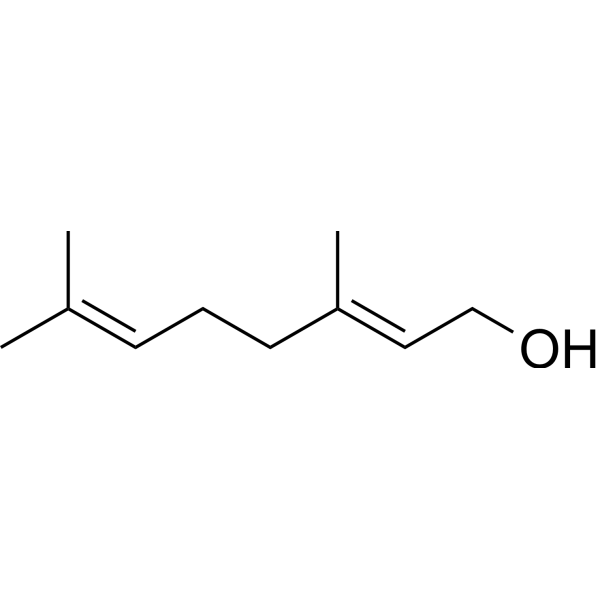
HY-N6952
GeraniolAntibacterial Agent -
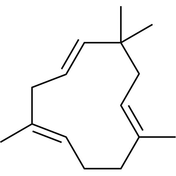
HY-N6968
α-HumuleneCOX Inhibitor -
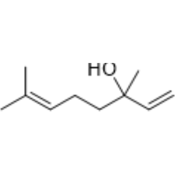
HY-N0368
Linalool,98% (stabilized with MEHQ)Antibiotic -
HY-121615
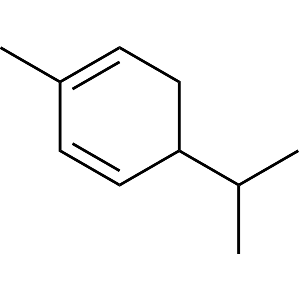
α-PhellandreneInsecticide -
HY-N1415
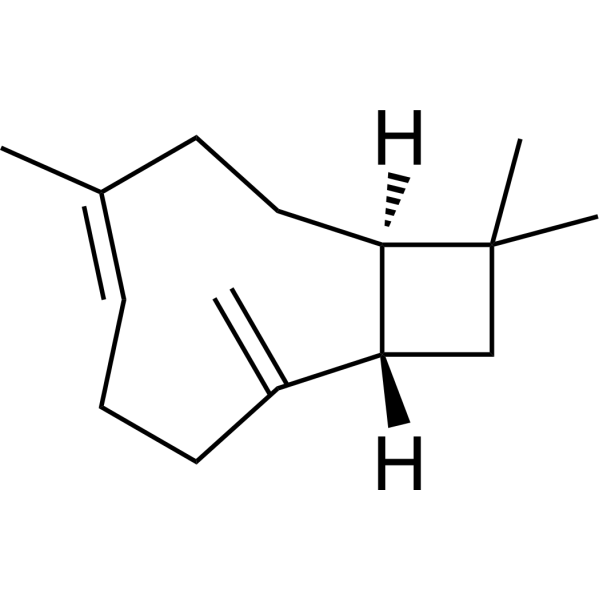
β-CaryophylleneCannabinoid Receptor Agonist -
HY-N7364
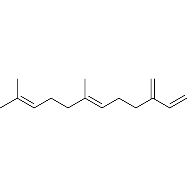
(E)-β-FarneseneAphid Alarm Pheromone -
HY-Z0478
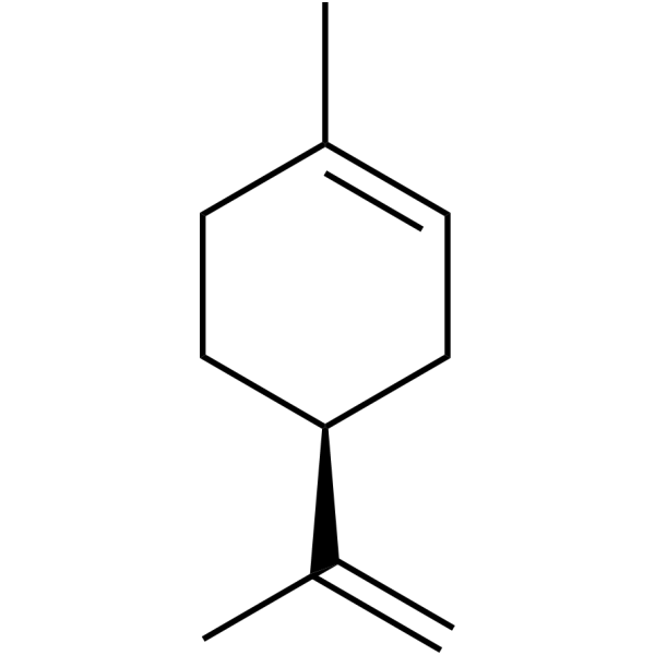
(-)-LimoneneCaMKII Inhibitor -
HY-W020183
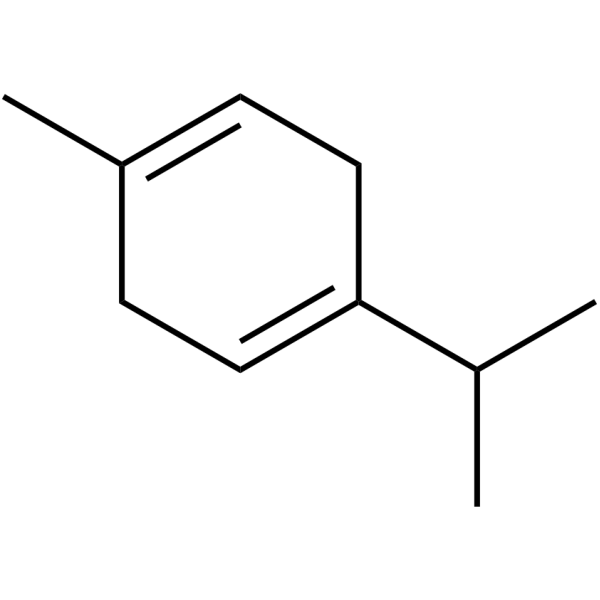
γ-TerpineneAntioxidant Compound


Biological Activity
Description
|
NF-κB |
In Vitro
Myrcene (50 μM, 100 μM; 24 h, 48 h) inhibits 10 ng/mL TNFα-induced NF-κB activity, through causing the inactivation of IKK in MDA-MB-231 cells[2].
Myrcene (0.25-1.0 μg/mL; 24 h, 48 h) inhibits clonal sphere formation in A549 cells, and arrests cell cycle in the G0/G1 phases to cause apoptosis[3].
MedChemExpress (MCE) has not independently confirmed the accuracy of these methods. They are for reference only.
In Vivo
MedChemExpress (MCE) has not independently confirmed the accuracy of these methods. They are for reference only.
- Detection of Apoptosis
- TUNEL staining for apoptotic DNA fragmentation
- Flow cytometric DNA-content cell-cycle staining
- Annexin V plus membrane-impermeant dye apoptosis staining
- BrdU Incorporation Assay
- Cotton Pellet Granuloma
- Carrageenan-Induced Paw Edema
- Apoptosis Solutions
- Protocol for Cell Cycle
- Research Protocol for Inflammation-related Diseases


Chemical Information
-
CAS No. 123-35-3
-
Appearance Liquid (Density: 0.791 g/cm3)
-
Molecular Weight 136.23
-
Formula C10H16
-
Color Colorless to light yellow
-
SMILES
C=CC(CC/C=C(C)/C)=C
-
Synonyms
β-Myrcene
-
Structure Classification
-
Shipping
Room temperature in continental US; may vary elsewhere.
-
Storage
-20°C, stored under nitrogen
* In solvent : -80°C, 6 months; -20°C, 1 month (stored under nitrogen)


Solvent & Solubility
In Vitro:
Ethanol : ≥ 100 mg/mL (734.05 mM)
DMSO : 100 mg/mL (734.05 mM; Need ultrasonic; Hygroscopic DMSO has a significant impact on the solubility of product, please use newly opened DMSO)
* "≥" means soluble, but saturation unknown.
Please refer to the solubility information to select the appropriate solvent. Once prepared, please aliquot and store the solution to prevent product inactivation from repeated freeze-thaw cycles.
Storage method and period of stock solution: -80°C, 6 months; -20°C, 1 month (stored under nitrogen). When stored at -80°C, please use it within 6 months. When stored at -20°C, please use it within 1 month.
Please refer to the solubility information to select the appropriate solvent. Once prepared, please aliquot and store the solution to prevent product inactivation from repeated freeze-thaw cycles.
Storage method and period of stock solution: -80°C, 6 months; -20°C, 1 month (stored under nitrogen). When stored at -80°C, please use it within 6 months. When stored at -20°C, please use it within 1 month.
Concentration (start) × Volume (start) = Concentration (final) × Volume (final)
In Vivo:
Select the appropriate dissolution method based on your experimental animal and administration route.
- For the following dissolution methods, please ensure to first prepare a clear stock solution using an In Vitro approach and then sequentially add co-solvents:
- To ensure reliable experimental results, the clarified stock solution can be appropriately stored based on storage conditions. As for the working solution for In Vivo experiments, it is recommended to prepare freshly and use it on the same day.
- The percentages shown for the solvents indicate their volumetric ratio in the final prepared solution. If precipitation or phase separation occurs during preparation, heat and/or sonication can be used to aid dissolution.
Add each solvent one by one: 10% DMSO 40% PEG300 5% Tween-80 45% Saline
Solubility: ≥ 2.5 mg/mL (18.35 mM); Clear solution
This protocol yields a clear solution of ≥ 2.5 mg/mL (saturation unknown).
Taking 1 mL working solution as an example, add 100 μL DMSO stock solution (25.0 mg/mL) to 400 μL PEG300, and mix evenly; then add 50 μL Tween-80 and mix evenly; then add 450 μL Saline to adjust the volume to 1 mL.
Preparation of Saline: Dissolve 0.9 g sodium chloride in ddH₂O and dilute to 100 mL to obtain a clear Saline solution.
Add each solvent one by one: 10% DMSO 90% (20% SBE-β-CD in Saline)
Solubility: ≥ 2.5 mg/mL (18.35 mM); Clear solution
This protocol yields a clear solution of ≥ 2.5 mg/mL (saturation unknown).
Taking 1 mL working solution as an example, add 100 μL DMSO stock solution (25.0 mg/mL) to 900 μL 20% SBE-β-CD in Saline, and mix evenly.
Preparation of 20% SBE-β-CD in Saline (4°C, storage for one week): 2 g SBE-β-CD powder is dissolved in 10 mL Saline, completely dissolve until clear.
In Vivo Dissolution Calculator
Please enter the basic information of animal experiments:
-
-
-
-
Recommended: Prepare an additional quantity of animals to account for potential losses during experiments.
Please enter your animal formula composition:
-
%DMSO +
Recommended: Keep the proportion of DMSO in working solution below 2% if your animal is weak.
-
%+
-
+%Tween-80 + +
-
%Saline +
The co-solvents required include: DMSO, . All of co-solvents are available by MedChemExpress (MCE). , Tween 80. All of co-solvents are available by MedChemExpress (MCE).
Working solution concentration: 0.22 mg/mL
Method for preparing stock solution: mg drug dissolved in μL DMSO. Stock solution concentration: mg/mL. * In solvent : -80°C, 6 months; -20°C, 1 month (stored under nitrogen)
1. Take μL DMSO stock solution;
2. Add μL .
μL , mix evenly;
3. Then add μL Tween 80, mix evenly;
4. Then add μL
Please ensure that the stock solution in the first step is dissolved to a clear state, and add co-solvents in sequence. You can use ultrasonic heating (ultrasonic cleaner, recommended frequency 20-40 kHz), vortexing, etc. to assist dissolution.


Purity & Documentation
-
Data Sheet (271 KB)
-
SDS (913 KB)
- English - EN (913 KB)
- Français - FR (913 KB)
- Deutsch - DE (913 KB)
- Norwegian - NO (913 KB)
- Español - ES (913 KB)
- Swedish - SV (913 KB)
- Italian - IT (913 KB)
- Korean - KR (913 KB)
- Portuguese - PT (913 KB)
-
Handling Instructions (2659 KB)
References
[1]. Eunson Hwang, et al. Myrcene, an Aromatic Volatile Compound, Ameliorates Human Skin Extrinsic Aging via Regulation of MMPs Production. Am J Chin Med. 2017;45(5):1113-1124. [Content Brief]
[4]. Islam AUS, et al. Myrcene Attenuates Renal Inflammation and Oxidative Stress in the Adrenalectomized Rat Model. Molecules. 2020 Sep 30;25(19):4492. [Content Brief]
[5]. Kumar R, et al. Ameliorative effect of myrcene in mouse model of Alzheimer's disease. Eur J Pharmacol. 2021 Nov 15;911:174529. [Content Brief]


Complete Stock Solution Preparation Table
Please refer to the solubility information to select the appropriate solvent. Once prepared, please aliquot and store the solution to prevent product inactivation from repeated freeze-thaw cycles.
Storage method and period of stock solution: -80°C, 6 months; -20°C, 1 month (stored under nitrogen). When stored at -80°C, please use it within 6 months. When stored at -20°C, please use it within 1 month.
| Optional Solvent | Concentration Solvent Mass | 1 mg | 5 mg | 10 mg | 25 mg |
|---|---|---|---|---|---|
| Ethanol / DMSO | 1 mM | 7.3405 mL | 36.7026 mL | 73.4053 mL | 183.5132 mL |
| 5 mM | 1.4681 mL | 7.3405 mL | 14.6811 mL | 36.7026 mL | |
| 10 mM | 0.7341 mL | 3.6703 mL | 7.3405 mL | 18.3513 mL | |
| 15 mM | 0.4894 mL | 2.4468 mL | 4.8937 mL | 12.2342 mL | |
| 20 mM | 0.3670 mL | 1.8351 mL | 3.6703 mL | 9.1757 mL | |
| 25 mM | 0.2936 mL | 1.4681 mL | 2.9362 mL | 7.3405 mL | |
| 30 mM | 0.2447 mL | 1.2234 mL | 2.4468 mL | 6.1171 mL | |
| 40 mM | 0.1835 mL | 0.9176 mL | 1.8351 mL | 4.5878 mL | |
| 50 mM | 0.1468 mL | 0.7341 mL | 1.4681 mL | 3.6703 mL | |
| 60 mM | 0.1223 mL | 0.6117 mL | 1.2234 mL | 3.0586 mL | |
| 80 mM | 0.0918 mL | 0.4588 mL | 0.9176 mL | 2.2939 mL | |
| 100 mM | 0.0734 mL | 0.3670 mL | 0.7341 mL | 1.8351 mL |


Myrcene,75% (stabilized with BHT) Related Classifications
HY-N0803 Related Classifications
 Powered by Bioz