



N-Feruloylserotonin

Based on 1 Customer Validation
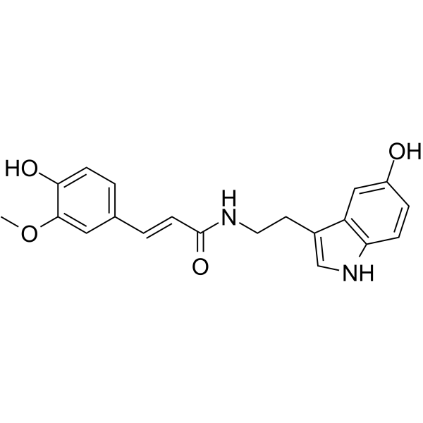
N-Feruloylserotonin ((E/Z)-Moschamine) is a serotonin hydroxycinnamic acid amide. It can be isolated from a variety of plants, particularly the seeds of safflower (Carthamus tinctorius L.). N-Feruloylserotonin inhibits KCl- and 5-HT-induced elevation of intracellular [Ca2+]i. It suppresses PDGF-BB-induced phosphorylation of PDGFRβ and ERK1/2. N-Feruloylserotonin exerts anti-inflammatory effects on aortic endothelial cells. It inhibits atherosclerotic plaque formation in apolipoprotein E-deficient mice.
For research use only. We do not sell to patients.
- Purity : 99.93%
- CAS No.: 68573-23-9
- Formula: C20H20N2O4
- Molecular Weight:352.38
-
Storage:
-20°C, protect from light
* In solvent : -80°C, 6 months; -20°C, 1 month (protect from light)

To place orders, for customer services and technical support, please contact: MedChemExpress USA
Tel: 609-228-6898 E-mail: [email protected] [email protected]


-
 Biological Activity
-
Chemical Information
-
Solvent & Solubility
- Protocols
- Purity & Documentation
- References
-
 Help & FAQs


All Calcium Channel Isoforms
More

Biological Activity
Description
IC50 & Target
[1]|
ERK1 |
ERK2 |
PDGFRβ |
Cellular Effect
|
Cell Line

|
Type | Value | Description | References |
|---|---|---|---|---|
| B16 | IC50 |
216 μM
Compound: 6
|
Inhibition of catecholase activity of tyrosinase in mouse B16 cells assessed as dopachrome formation
Inhibition of catecholase activity of tyrosinase in mouse B16 cells assessed as dopachrome formation
|
[PMID: 19524439] |
| B16 | IC50 |
23 μM
Compound: 6
|
Antimelanogenic activity in mouse B16 cells assessed as inhibition of intracellular melanin accumulation after 2 days
Antimelanogenic activity in mouse B16 cells assessed as inhibition of intracellular melanin accumulation after 2 days
|
[PMID: 19524439] |
| Caco-2 | IC50 |
81 μM
Compound: moschamine
|
Anticancer activity against human Caco2 cells
Anticancer activity against human Caco2 cells
|
[PMID: 25456382] |
| RAW264.7 | IC50 |
123.14 μM
Compound: 2; MC
|
Antiinflammatory activity in mouse RAW264.7 cells assessed as inhibition of LPS-induced nitric oxide production measured after 24 hrs by Griess assay
Antiinflammatory activity in mouse RAW264.7 cells assessed as inhibition of LPS-induced nitric oxide production measured after 24 hrs by Griess assay
|
[PMID: 29102229] |
| RAW264.7 | IC50 |
14.74 μM
Compound: 2; MC
|
Antiinflammatory activity in mouse RAW264.7 cells assessed as inhibition of LPS-induced PGE2 production pretreated for 1 hr followed by LPS addition measured after 24 hrs
Antiinflammatory activity in mouse RAW264.7 cells assessed as inhibition of LPS-induced PGE2 production pretreated for 1 hr followed by LPS addition measured after 24 hrs
|
[PMID: 29102229] |
In Vitro
N-feruloylserotonin (1-100 μM) concentration-dependently inhibits the KCl- and 5-HT-induced elevation of [Ca2+]i in A7r5 cells, exerting a significant effect at concentrations ≥66 μM, but does not affect SOC-mediated Ca2+ influx at 66 μM[1].
N-feruloylserotonin (1-100 μM) inhibits 10% FBS- and PDGF-BB-induced proliferation of A7r5 cells in a concentration-dependent manner[1].
N-feruloylserotonin (1-66 μM) significantly inhibits PDGF-BB-induced migration of A7r5 cells in a concentration-dependent manner[1].
N-feruloylserotonin (10-66 μM; 5-10 minutes) inhibits PDGF-BB-induced phosphorylation of PDGFRβ and ERK1/2 in A7r5 cells in a concentration-dependent manner[1].
MedChemExpress (MCE) has not independently confirmed the accuracy of these methods. They are for reference only. Further protocols information, click here.
-
Cell Line:A7r5 rat aortic vascular smooth muscle cells
-
Concentration:1-100 μM
-
Incubation Time:10% FBS or 10 ng/ml PDGF-BB
for 24 h. -
Result:Inhibited 10% FBS-stimulated A7r5 proliferation in a concentration-dependent manner, causing 60% inhibition at 100 μM.
Inhibited PDGF-BB-stimulated proliferation in a concentration-dependent manner, with significant inhibition observed at 100 μM.
-
Cell Line:A7r5 rat aortic vascular smooth muscle cells
-
Concentration:10-66 μM
-
Incubation Time:5 or 10 minutes (with PDGF-BB stimulation)
-
Result:Attenuated PDGF-BB-induced PDGFRβ tyrosine phosphorylation in a concentration-dependent manner at 5 minutes post-stimulation, but not at 10 minutes.
Significantly inhibited PDGF-BB-induced ERK1/2 phosphorylation in a concentration-dependent manner from 5 to 10 minutes post-stimulation.
In Vivo
MedChemExpress (MCE) has not independently confirmed the accuracy of these methods. They are for reference only.


Chemical Information
-
CAS No. 68573-23-9
-
Appearance Solid
-
Molecular Weight 352.38
-
Formula C20H20N2O4
-
Color White to off-white
-
SMILES
O=C(/C=C/C1=CC=C(C(OC)=C1)O)NCCC2=CNC3=C2C=C(C=C3)O
-
Synonyms
(E/Z)-Moschamine
-
Structure Classification
-
Shipping
Room temperature in continental US; may vary elsewhere.
-
Storage
-20°C, protect from light
* In solvent : -80°C, 6 months; -20°C, 1 month (protect from light)


Solvent & Solubility
In Vitro:
DMSO : 100 mg/mL (283.78 mM; Need ultrasonic; Hygroscopic DMSO has a significant impact on the solubility of product, please use newly opened DMSO)
Please refer to the solubility information to select the appropriate solvent. Once prepared, please aliquot and store the solution to prevent product inactivation from repeated freeze-thaw cycles.
Storage method and period of stock solution: -80°C, 6 months; -20°C, 1 month (protect from light). When stored at -80°C, please use it within 6 months. When stored at -20°C, please use it within 1 month.
Please refer to the solubility information to select the appropriate solvent. Once prepared, please aliquot and store the solution to prevent product inactivation from repeated freeze-thaw cycles.
Storage method and period of stock solution: -80°C, 6 months; -20°C, 1 month (protect from light). When stored at -80°C, please use it within 6 months. When stored at -20°C, please use it within 1 month.
Concentration (start) × Volume (start) = Concentration (final) × Volume (final)

Protocols
-
Kinase activity and phosphorylation assays
Kinase activity assays measure the ability of kinases to transfer phosphate groups from ATP to specific substrates, while phosphorylation assays detect the presence and levels of phosphorylated proteins. Common methods include radiolabeled ATP incorporation (e. g. ,), ADP release detection via bioluminescence (e. g. ,[3]), enzyme-linked immunosorbent assays (ELISA) for phospho-specific epitopes (e. g. ,[6]), and microtiter-based formats for high-throughput screening (e. g. ,[8]). The ADP-Glo assay quantifies kinase activity by measuring ADP produced during phosphorylation using a luciferase-based system. Radiometric assays involve autoradiography or scintillation counting after incorporation of 32P-labeled ATP into substrate proteins. ELISA-based approaches rely on phospho-specific antibodies to detect activated kinases in cell lysates or purified samples.
-
Western Blot
Western blotting (WB) is a commonly used experimental method in molecular biology, biochemistry, and immunogenetics for identifying and quantifying target proteins. It combines gel electrophoresis with immunoassay, enabling researchers to analyze protein expression, post-translational modifications, and molecular weight.
-
Endothelial Tube Formation Assay
Endothelial tube formation assay evaluates the ability of endothelial cells to attach, migrate, align, and organize into capillary-like networks when cultured on gelled basement membrane extract or Matrigel; the readout is the morphology and quantity of tube-like networks, which reflects an in vitro endothelial morphogenesis step related to angiogenesis. Basement membrane extract/Matrigel provides laminin-rich extracellular matrix cues that support endothelial differentiation into capillary-like structures, but it can contain biologically active growth factors, so growth-factor-reduced matrix is preferred when testing defined angiogenic stimulators or inhibitors.
-
Ca2+ Staining Technique
Ca2+ staining is an experimental technique that utilizes specific fluorescent probes (such as Fluo-4 AM, Fura-2, etc.) to qualitatively or quantitatively detect dynamic changes in intracellular Ca2+ concentrations; this is achieved by monitoring the changes in fluorescent signals generated when these probes bind to free intracellular calcium ions. The underlying principle relies primarily on the presence of chelating groups within the probe's molecular structure that possess high affinity for calcium ions.
-
Protocol for Kinase activity and phosphorylation assays
Kinase activity assays measure transfer of phosphate from ATP to a protein or peptide substrate, generating phosphorylated substrate, ADP, or incorporated radiolabeled phosphate as the readout; phosphorylation assays measure site-specific phosphorylation in cells or tissues as a proxy for kinase-pathway activation, inhibition, or substrate regulation. Phosphorylation can be detected by phospho-specific Western blot, immunoprecipitation kinase assay, phospho-immunofluorescence, phospho-flow cytometry, luminescent ADP detection, radiolabeled ATP incorporation, or reporter-based pathway assays, and these readouts can be applied to cancer cells, primary neurons, mouse tumors, organoids, inflammatory macrophages, ferroptosis studies, and mitophagy studies when the kinase target is biologically relevant.
-
Research Protocol for Inflammation-related Diseases
The NLRP3 inflammasome is a cytosolic innate immune signaling platform that integrates priming signals and danger-signal activation to promote caspase-1 activation, maturation of IL-1β and IL-18, and gasdermin D-mediated pyroptotic cell death. The core experimental logic is to determine whether inflammatory disease phenotypes are driven by increased NLRP3 expression, ASC-containing inflammasome assembly, caspase-1 cleavage, GSDMD cleavage, and extracellular release of IL-1β/IL-18 rather than by nonspecific cell injury alone. The pathway is strongly linked to inflammation-related disease phenotypes because monosodium urate crystals activate NALP3/NLRP3 inflammasome signaling in gout-like crystal inflammation, cholesterol crystals activate NLRP3 inflammasomes in atherogenesis models, and DSS-induced intestinal inflammation has been reported to involve NLRP3 inflammasome activity. However, experimental colitis studies also show context-dependent protective effects of NLRP3 inflammasome co


Purity & Documentation
-
Data Sheet (273 KB)
-
SDS (394 KB)
- English - EN (394 KB)
- Français - FR (394 KB)
- Deutsch - DE (394 KB)
- Norwegian - NO (394 KB)
- Español - ES (394 KB)
- Swedish - SV (394 KB)
- Italian - IT (394 KB)
- Korean - KR (394 KB)
- Portuguese - PT (394 KB)
-
Handling Instructions (2659 KB)
References
[1]. Takimoto T, et al. Effect of N-(p-coumaroyl)serotonin and N-feruloylserotonin, major anti-atherogenic polyphenols in safflower seed, on vasodilation, proliferation and migration of vascular smooth muscle cells. Mol Nutr Food Res. 2011;55(10):1561-1571. [Content Brief]
[2]. Katsuda S, et al. Safflower seed polyphenols (N-(p-coumaroyl)serotonin and N-feruloylserotonin) ameliorate atherosclerosis and distensibility of the aortic wall in Kurosawa and Kusanagi-hypercholesterolemic (KHC) rabbits. Hypertens Res. 2009;32(11):944-949. [Content Brief]


Complete Stock Solution Preparation Table
Please refer to the solubility information to select the appropriate solvent. Once prepared, please aliquot and store the solution to prevent product inactivation from repeated freeze-thaw cycles.
Storage method and period of stock solution: -80°C, 6 months; -20°C, 1 month (protect from light). When stored at -80°C, please use it within 6 months. When stored at -20°C, please use it within 1 month.
| Optional Solvent | Concentration Solvent Mass | 1 mg | 5 mg | 10 mg | 25 mg |
|---|---|---|---|---|---|
| DMSO | 1 mM | 2.8378 mL | 14.1892 mL | 28.3785 mL | 70.9461 mL |
| 5 mM | 0.5676 mL | 2.8378 mL | 5.6757 mL | 14.1892 mL | |
| 10 mM | 0.2838 mL | 1.4189 mL | 2.8378 mL | 7.0946 mL | |
| 15 mM | 0.1892 mL | 0.9459 mL | 1.8919 mL | 4.7297 mL | |
| 20 mM | 0.1419 mL | 0.7095 mL | 1.4189 mL | 3.5473 mL | |
| 25 mM | 0.1135 mL | 0.5676 mL | 1.1351 mL | 2.8378 mL | |
| 30 mM | 0.0946 mL | 0.4730 mL | 0.9459 mL | 2.3649 mL | |
| 40 mM | 0.0709 mL | 0.3547 mL | 0.7095 mL | 1.7737 mL | |
| 50 mM | 0.0568 mL | 0.2838 mL | 0.5676 mL | 1.4189 mL | |
| 60 mM | 0.0473 mL | 0.2365 mL | 0.4730 mL | 1.1824 mL | |
| 80 mM | 0.0355 mL | 0.1774 mL | 0.3547 mL | 0.8868 mL | |
| 100 mM | 0.0284 mL | 0.1419 mL | 0.2838 mL | 0.7095 mL |


N-Feruloylserotonin Related Classifications
HY-118824A Related Classifications
 Powered by Bioz