



nor-NOHA
Based on 7 publication(s) in Google Scholar
nor-NOHA is a selective and reversible arginase inhibitor. nor-NOHA induces apoptosis in ARG2-expressing cells under hypoxia. nor-NOHA has anti-leukemic activity. nor-NOHA can used in study of endothelial dysfunction, immunosuppression and metabolism.
For research use only. We do not sell to patients.
- CAS No.: 189302-40-7
- Formula: C5H12N4O3
- Molecular Weight:176.17
-
Storage:
Please store the product under the recommended conditions in the Certificate of Analysis.


To place orders, for customer services and technical support, please contact: MedChemExpress USA
Tel: 609-228-6898 E-mail: [email protected] [email protected]


-
 Biological Activity
-
Chemical Information
-
Solvent & Solubility
- Protocols
- Purity & Documentation
- References
-
 Help & FAQs


Publications Citing Use of MedChemExpress (MCE) nor-NOHA
More- Cell. 2025 Dec 11;188(25):7291-7308.e23. [Abstract]
- Free Radic Biol Med. 2026 Apr:247:516-527. [Abstract]
- Chin Med. 2026 May 28;21(1):151. [Abstract]
- Front Cell Dev Biol. 2021 Dec 23;9:741911. [Abstract]
- FASEB J. 2025 Sep 30;39(18):e71057. [Abstract]
- Clin Immunol. 2024 Nov:268:110355. [Abstract]
- Res Sq. 2026 May 8:rs.3.rs-9404453. [Abstract]
 Customer Validation & Images

-
Flow Cytometry
-
Cell Proliferation/Viability Assay
-
RT-PCR
-
Bio/Physico-chemical Assay


Biological Activity
Description
In Vitro
nor-NOHA (0.1-1 mM, 72 h) induces apoptosis in a dose-dependent manner under hypoxia (1.5% O2) in K562 cells[1]. nor-NOHA (1 mM, 72 h) attenuates hypoxia-mediated resistance towards imatinib in K562 or KCL22 cells[1].
MedChemExpress (MCE) has not independently confirmed the accuracy of these methods. They are for reference only.
In Vivo
MedChemExpress (MCE) has not independently confirmed the accuracy of these methods. They are for reference only.
Clinical Trial
| NCT Number | Sponsor | Condition | Start Date |
Phase

|
|---|---|---|---|---|
| NCT01329991 | Plexxikon| | 2011-05 | PHASE1 |


Chemical Information
-
CAS No. 189302-40-7
-
Molecular Weight 176.17
-
Formula C5H12N4O3
-
SMILES
O=C(O)[C@@H](N)CCNC(NO)=N
-
Synonyms
Nω-Hydroxy-nor-L-arginine
-
Shipping
Room temperature in continental US; may vary elsewhere.
-
Storage
Please store the product under the recommended conditions in the Certificate of Analysis.


Publications (7)
-
Journal Impact Factor
-
Most Recent
-
Cell
2025 Dec 11;188(25):7291-7308.e23. PMID: 41161316
nor-NOHA purchased from MedChemExpress. Usage Cited in: Cell. 2025 Dec 11;188(25):7291-7308.e23. [Abstract]
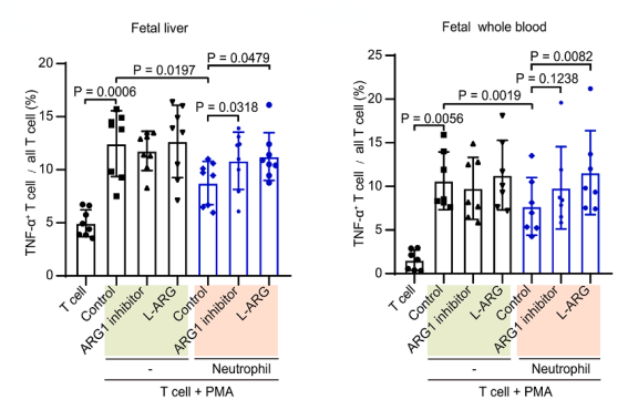
Bar plots showing proportions of TNF-α+ T cells in fetal liver whole blood following PMA simulation. T cells were cultured alone (light green) or co-cultured with matched fetal neutrophils (orange) in the presence or absence of ARG1 inhibitor (nonor-NOHA monoacetate; overnight) or L-arginine supplementation, as indicated on the x axis.
-
Free Radic Biol Med
Endothelial Arg2 regulates HIMM-induced mitochondrial hyperfission via affecting arginine metabolism. [Abstract]2026 Apr:247:516-527. PMID: 41720362 -
Chin Med
Licoisoflavone A potentiates oxaliplatin efficacy in colorectal cancer by dual targeting arginine metabolism of tumor microenvironment and cancer cells. [Abstract]2026 May 28;21(1):151. PMID: 42210399 -
Front Cell Dev Biol
mTOR Signaling Regulates the Development and Therapeutic Efficacy of PMN-MDSCs in Acute GVHD. [Abstract]2021 Dec 23;9:741911. PMID: 35004668
nor-NOHA purchased from MedChemExpress. Usage Cited in: Front Cell Dev Biol. 2021 Dec 23;9:741911. [Abstract]
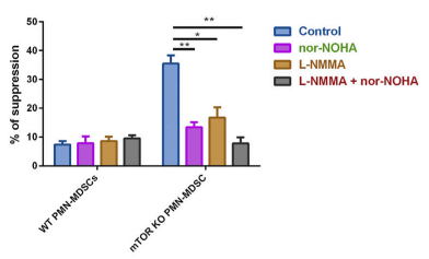
nor-NOHA monoacetate (nor-NOHA) efficiently reduced immunosuppressive activity. Arg1 inhibitor (nor-NOHA) and/or iNOS inhibitor [L-NG-Monomethylarginine, Acetate Salt (L-NMMA)] were added in PMN-MDSCs’ suppressive function assays.
-
FASEB J
Spatial Metabolism of Primary Limited Cutaneous Amyloidosis Based on Mass Spectrometry Imaging. [Abstract]2025 Sep 30;39(18):e71057. PMID: 40955599
nor-NOHA purchased from MedChemExpress. Usage Cited in: FASEB J. 2025 Sep 30;39(18):e71057. [Abstract]
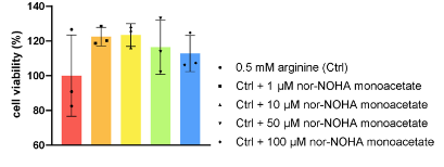
nonor-NOHA monoacetate (1, 10, 50, 100 μM) increased HaCaT cell viability.
nor-NOHA purchased from MedChemExpress. Usage Cited in: FASEB J. 2025 Sep 30;39(18):e71057. [Abstract]
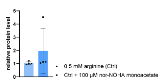
nonor-NOHA monoacetate (100 μM) increased K5 expression in HaCaT cells.
-
Clin Immunol
MAPK signaling pathway induced LOX-1+ polymorphonuclear myeloid-derived suppressor cells in biliary atresia. [Abstract]2024 Nov:268:110355. PMID: 39237078 -
Res Sq
Protective coinfection: influenza reprograms myeloid cells to limit CD8 T cell-mediated malaria pathology. [Abstract]2026 May 8:rs.3.rs-9404453. PMID: 42147182


Solvent & Solubility
In Vitro:
H2O : 250 mg/mL (1419.08 mM; Need ultrasonic)
* Note: If you choose water as the stock solution, please dilute it to the working solution, then filter and sterilize it with a 0.22 μm filter before use.
* Note: If you choose water as the stock solution, please dilute it to the working solution, then filter and sterilize it with a 0.22 μm filter before use.
Concentration (start) × Volume (start) = Concentration (final) × Volume (final)


Purity & Documentation
References
[1]. Ng KP, et al. The arginase inhibitor Nω-hydroxy-nor-arginine (nor-NOHA) induces apoptosis in leukemic cells specifically under hypoxic conditions but CRISPR/Cas9 excludes arginase 2 (ARG2) as the functional target. PLoS One. 2018 Oct 11;13(10):e0205254. [Content Brief]
[2]. Jung C, et al. Arginase inhibition mediates cardioprotection during ischaemia-reperfusion. Cardiovasc Res. 2010 Jan 1;85(1):147-54. [Content Brief]


Complete Stock Solution Preparation Table
| Optional Solvent | Concentration Solvent Mass | 1 mg | 5 mg | 10 mg | 25 mg |
|---|---|---|---|---|---|
| H2O | 1 mM | 5.6763 mL | 28.3817 mL | 56.7634 mL | 141.9084 mL |
| 5 mM | 1.1353 mL | 5.6763 mL | 11.3527 mL | 28.3817 mL | |
| 10 mM | 0.5676 mL | 2.8382 mL | 5.6763 mL | 14.1908 mL | |
| 15 mM | 0.3784 mL | 1.8921 mL | 3.7842 mL | 9.4606 mL | |
| 20 mM | 0.2838 mL | 1.4191 mL | 2.8382 mL | 7.0954 mL | |
| 25 mM | 0.2271 mL | 1.1353 mL | 2.2705 mL | 5.6763 mL | |
| 30 mM | 0.1892 mL | 0.9461 mL | 1.8921 mL | 4.7303 mL | |
| 40 mM | 0.1419 mL | 0.7095 mL | 1.4191 mL | 3.5477 mL | |
| 50 mM | 0.1135 mL | 0.5676 mL | 1.1353 mL | 2.8382 mL | |
| 60 mM | 0.0946 mL | 0.4730 mL | 0.9461 mL | 2.3651 mL | |
| 80 mM | 0.0710 mL | 0.3548 mL | 0.7095 mL | 1.7739 mL | |
| 100 mM | 0.0568 mL | 0.2838 mL | 0.5676 mL | 1.4191 mL |
* Note: If you choose water as the stock solution, please dilute it to the working solution, then filter and sterilize it with a 0.22 μm filter before use.


nor-NOHA Related Classifications
HY-112885 Related Classifications
 Powered by Bioz