



ON 108600

Based on 1 Customer Validation
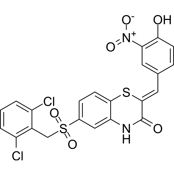
ON 108600 (108600) is a CK2 (CK2α1: IC50 = 50 nM; CK2α2 = 5 nM), TNIK (IC50 = 5 nM) and DYRK (DYRK1A: IC50 = 16 nM; DYRK1B = 7 nM; DYRK2: = 28 nM). ON 108600 suppresses the growth of CD44high/CD24low breast cancer stem cell (BCSC) populations, induces G2/M arrest and apoptosis in triple negative breast cancer (TNBC) cells, and inhibits tubulin polymerization. ON 108600 can be used for the study of chemotherapy-resistant and metastatic TNBC.
Nur für Forschungszwecke. Wir verkaufen nicht an Patienten.
- Reinheit : 98.65%
- CAS. Nr.: 1585246-23-6
- Formel: C22H14Cl2N2O6S2
- Molecular Weight:537.39
-
Speicherung:Powder -20°C, 3 years , 4°C, 2 years ; In solvent -80°C, 6 months , -20°C, 1 month

To place orders, for customer services and technical support, please contact: MedChemExpress USA
Tel: 609-228-6898 E-mail: [email protected] [email protected]


-
 Biologische Aktivität
-
Chemical Information
-
Lösungsmittel & Löslichkeit
- Protokoll
- Reinheit & Dokumentation
- Verweise
-
 Help & FAQs


-
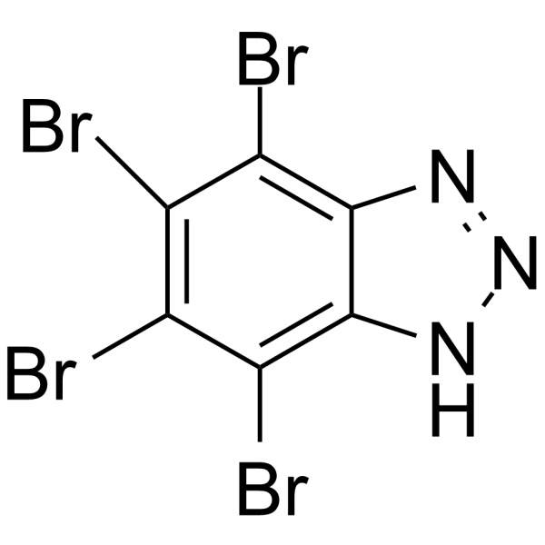
HY-14394
TBBCK2 Inhibitor -
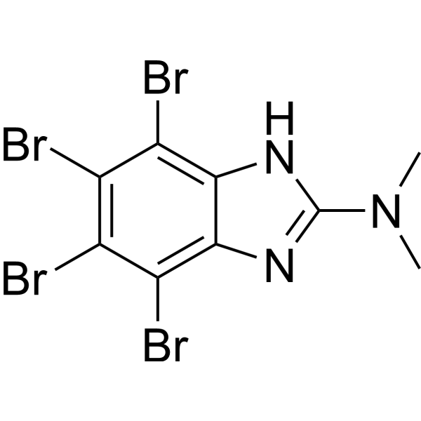
HY-15535
DMATCasein Kinase Inhibitor -
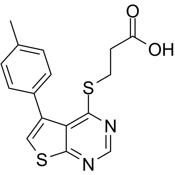
HY-15479
TTP 22Casein Kinase Inhibitor -
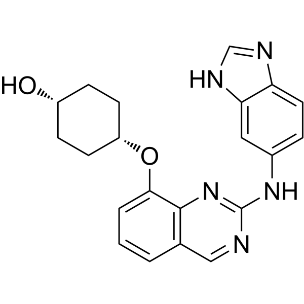
HY-100830
NCB-0846TRAF2- and NCK-interacting Inhibitor -
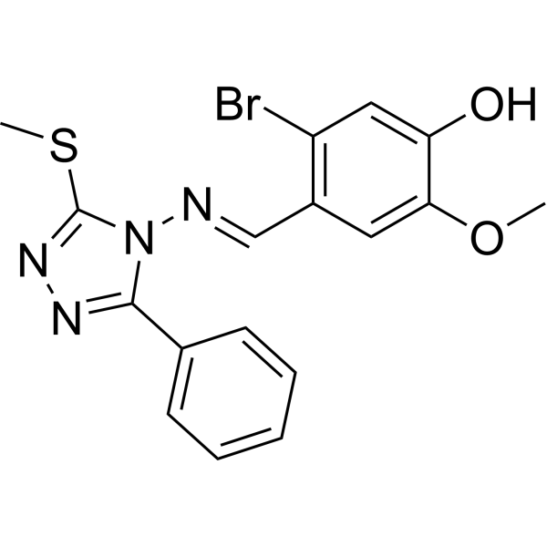
HY-115519
(E/Z)-GO289CK2 Inhibitor -
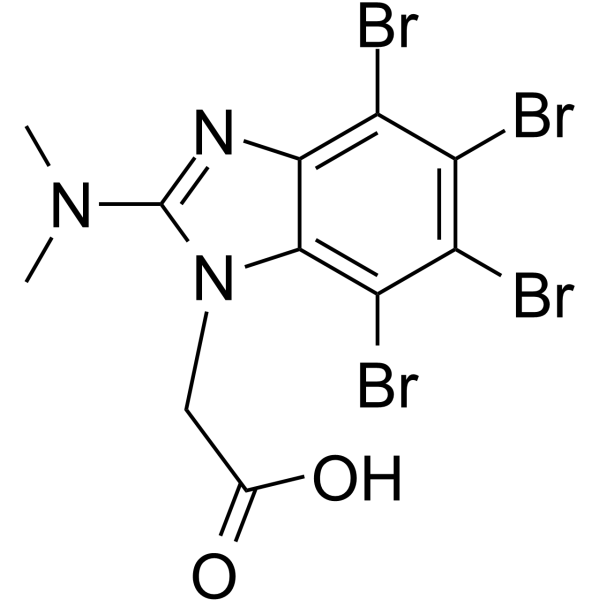
HY-135906
CK2/ERK8-IN-1CK2/ERK8 Inhibitor -
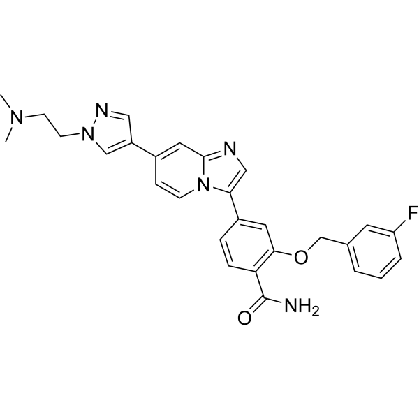
HY-101029
MBM-55Nek2 Inhibitor -
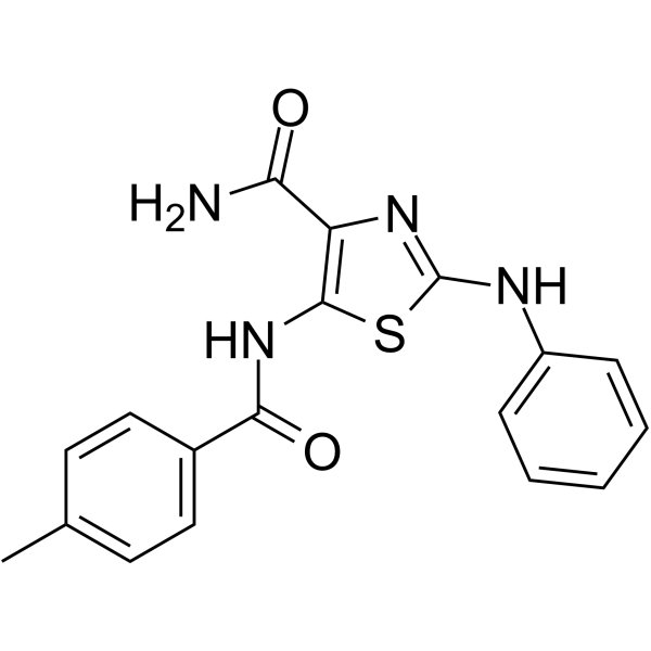
HY-124745
KY-05009TNIK Inhibitor -
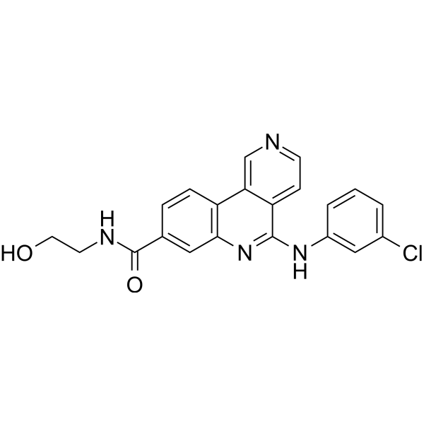
HY-132175
CK2 inhibitor 2CK2 Inhibitor -
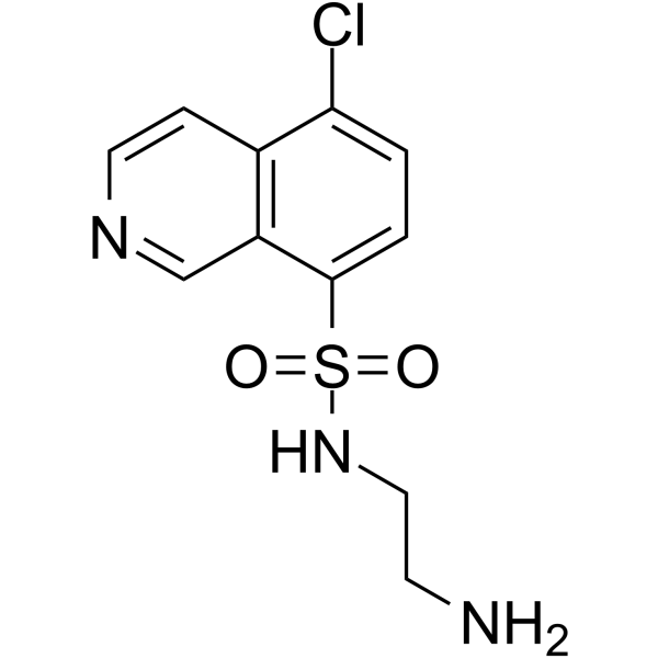
HY-133028
CKI-7 free baseCK1 Inhibitor -
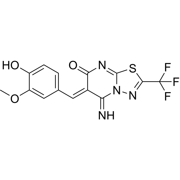
HY-114653
SRPIN803CK2/SRPK1 Dual Inhibitor -
HY-123954
BTX-A51CK1α Inhibitor


Biologische Aktivität
Beschreibung
IC50 & Target
[1]|
DYRK2 0.028 μM (IC50) |
DYRK1A 0.016 μM (IC50) |
DYRK1B 0.007 μM (IC50) |
CK2α2 0.005 μM (IC50) |
CK2α1 0.05 μM (IC50) |
Cellular Effect
|
Cell Line

|
Type | Value | Description | References |
|---|---|---|---|---|
| HT-22 | IC50 |
1 μM
Compound: 29; 108600
|
Cytotoxicity against mouse HT-22 cells assessed as reduction in cell viability incubated for 48 hrs by MTS assay
Cytotoxicity against mouse HT-22 cells assessed as reduction in cell viability incubated for 48 hrs by MTS assay
|
36876904 |
| MCF-10A | GI50 |
2.5 μM
|
Shows little or no toxicity against MCF-10A.
Shows little or no toxicity against MCF-10A.
|
34344863 |
| MDA-MB-231 | GI50 |
0.12 μM
|
Inhibits the viability of MDA-MB-231.
Inhibits the viability of MDA-MB-231.
|
34344863 |
| MDA-MB-157 | GI50 |
0.18 μM
|
Inhibits the viability of MDA-MB-157.
Inhibits the viability of MDA-MB-157.
|
34344863 |
| MDA-MB-436 | GI50 |
0.2 μM
|
Inhibits the viability of MDA-MB-436.
Inhibits the viability of MDA-MB-436.
|
34344863 |
| MDA-MB-468 | GI50 |
0.2 μM
|
Inhibits the viability of MDA-MB-468.
Inhibits the viability of MDA-MB-468.
|
34344863 |
| MDA-MB-453 | GI50 |
0.75 μM
|
Inhibits the viability of MDA-MB-453.
Inhibits the viability of MDA-MB-453.
|
34344863 |
| BT-20 | GI50 |
0.25 μM
|
Inhibits the viability of BT-20.
Inhibits the viability of BT-20.
|
34344863 |
| Hs-578T | GI50 |
0.15 μM
|
Inhibits the viability of Hs-587T.
Inhibits the viability of Hs-587T.
|
34344863 |
| HCC1806 | GI50 |
0.15 μM
|
Inhibits the viability of HCC1806.
Inhibits the viability of HCC1806.
|
34344863 |
| MCF7 | GI50 |
0.5 μM
|
Inhibits the viability of MCF7.
Inhibits the viability of MCF7.
|
34344863 |
| BT-474 | GI50 |
0.19 μM
|
Inhibits the viability of BT-474.
Inhibits the viability of BT-474.
|
34344863 |
| PBMC | GI50 |
> 10 μM
|
Inhibits the viability of PBMC.
Inhibits the viability of PBMC.
|
34344863 |
In Vitro
ON 108600 (108600) (24-72 h) inhibits the viability of TNBC cell lines MDA-MB-231, MDA-MB-468, Hs578T and BT-20, with GI50 values of 0.1-0.2 μM, and shows little or no toxicity against normal cells including MCF-10A, NBSC-1, NBSC-2, PBMC, HFL and hMSC-TERT (GI50 > 2.5 μM)[1].
ON 108600 (0.1-3 μM; 24 h) inhibits phosphorylation of CK2α substrate AKT1 (Ser129), DYRK1A substrates CYCLIN D1 (Thr286) and p27 (Ser10), and TNIK substrate AXIN2 in MDA-MB-231 and Hs578T cells[1].
ON 108600 (10-1000 nM; 72 h) suppresses colony and mammosphere formation of CD44high/CD24low BCSC populations purified from MDA-MB-231 and Hs578T cells[1].
ON 108600 exhibits inhibition against recombinant CK2α1, CK2α2, TNIK, DYRK1A, DYRK1B and DYRK2 kinases[1].
ON 108600 (72 h) induces G2/M arrest and apoptosis in MDA-MB-231 cells and CTG1883 TNBC organoids[1].
ON 108600 (1 μM; 12 h) induces abnormal mitotic spindle formation with multipolar spindles and multiple centrosomes in HeLa cells[1].
ON 108600 (72 h) inhibits growth and colony formation of Paclitaxel (HY-B0015)-resistant MDA-MB-231 and BT-20 cells (MDA-MB-231-TR and BT-20-TR) with the same efficiency as parental paclitaxel-sensitive cells[1].
ON 108600 (2.5 and 5 μM; 48 h) inhibits cell viability, induces G0/G1 arrest and apoptosis in metastatic canine mammary gland tumor (CMGT) cells[2].
MedChemExpress (MCE) has not independently confirmed the accuracy of these methods. They are for reference only.
-
Cell Line:MDA-MB-231 (CD44high/CD24low), Hs578T (CD44high/CD24low)
-
Concentration:10, 100, 250, 500, 1000 nM
-
Incubation Time:72 h
-
Result:Suppressed colony formation in a dose-dependent manner.
Suppressed mammosphere formation in a dose-dependent manner.
-
Cell Line:MDA-MB-231, Hs578T
-
Concentration:0.1, 0.5, 1.0, 1.5, 3.0 μM
-
Incubation Time:24 h
-
Result:Inhibited phosphorylation of AKT1 (Ser129), CYCLIN D1 (Thr286) and p27 (Ser10).
Reduced AXIN2 expression in a dose-dependent manner.
-
Cell Line:MDA-MB-231, CTG1883 organoids
-
Concentration:0.125, 0.25, 0.5 μM
-
Incubation Time:24, 48, 72 h
-
Result:Induced G2/M arrest and increased sub-G1 fraction.
-
Cell Line:MDA-MB-231, CTG1883 organoids
-
Concentration:0.25, 0.5, 1.0, 1.5, 3.0 μM
-
Incubation Time:24,48 h
-
Result:Induced PARP and Caspase 3 cleavage in a dose-dependent manner.
-
Cell Line:HeLa
-
Concentration:1 μM
-
Incubation Time:12 h
-
Result:Induced abnormal mitotic spindle formation with multipolar spindles and multiple centrosomes.
-
Cell Line:Canine mammary gland tumor (CMGT) cells
-
Concentration:1, 2, 2.5, 3, 4, 5 μM
-
Incubation Time:24, 48 h
-
Result:Inhibited cell viability in a dose- and time-dependent manner, with approximately 50% reduction at 2.5-5 μM for 48 h.
-
Cell Line:Canine mammary gland tumor (CMGT) cells
-
Concentration:2.5, 5 μM
-
Incubation Time:48 h
-
Result:Increased late apoptotic cells from 2.65% to 29.65% and 48.50%, respectively.
-
Cell Line:Canine mammary gland tumor (CMGT) cells
-
Concentration:2.5, 5 μM
-
Incubation Time:48 h
-
Result:Induced G0/G1 arrest and increased G0/G1 population from 61.6% to 65.2% and 73.7%, respectively.
-
Cell Line:Canine mammary gland tumor (CMGT) cells
-
Concentration:2.5, 5 μM
-
Incubation Time:48 h
-
Result:Induced nuclear damage.
In Vivo
ON 108600 (50 or 100 mg/kg; i.p.; every other day for 21 days) inhibits tumor growth in MDA-MB-231 xenograft model in nude mice, without observable toxicity[1].
ON 108600 (100 mg/kg; i.p.; every other day for 21 days) suppresses tumor growth in TM00098 (Paclitaxel (HY-B0015)-sensitive TNBC PDX) model in NSG mice, without adverse effects on body weight[1].
ON 108600 (100 mg/kg; i.p.; every other day for 12-24 days) in combination with Paclitaxel synergistically suppresses tumor growth in chemotherapy-resistant TNBC PDX models (CTG1883 and CTG2397)[1].
ON 108600 (100 mg/kg; i.p.; every other day for 14 days) in combination with Paclitaxel (HY-B0015) suppresses growth of pre-established lung metastases in the MDA-MB-231/LM2-4/mCherry metastatic model in NSG mice[1].
MedChemExpress (MCE) has not independently confirmed the accuracy of these methods. They are for reference only.
-
Animal Model:CTG1883 PDX tumor-bearing female NSG mice (8-12 weeks old)[1]
-
Dosage:100 mg/kg
-
Administration:i.p.; every other day for 5 days
-
Result:Inhibited phosphorylation of pAKT1 Ser129 and pCYCLIN D1 Thr286.
Reduced C-MYC expression in tumors.
-
Animal Model:MDA-MB-231 xenograft-bearing female athymic nude mice (8-12 weeks old)[1]
-
Dosage:50, 100 mg/kg
-
Administration:i.p.; every other day for 21 days
-
Result:Inhibited tumor growth in a dose-dependent manner, with no observable toxicity or body weight loss.
-
Animal Model:TM00098 PDX tumor-bearing female NSG mice (8-12 weeks old)[1]
-
Dosage:100 mg/kg
-
Administration:i.p.; every other day for 21 days
-
Result:Suppressed tumor growth without adverse effects on body weight.
-
Animal Model:CTG1883 and CTG2397 PDX tumor-bearing female NSG mice (8-12 weeks old)[1]
-
Dosage:100 mg/kg
-
Administration:i.p.; every other day for 12-24 days
-
Result:Suppressed tumor growth.
-
Animal Model:MDA-MB-231/LM2-4/mCherry metastatic model (lung metastases) in female NSG mice (7 weeks old)[1]
-
Dosage:100 mg/kg
-
Administration:i.p.; every other day for 14 days
-
Result:Suppressed growth of pre-established lung metastases.


Chemical Information
-
CAS. Nr. 1585246-23-6
-
Appearance Solid
-
Molecular Weight 537.39
-
Formel C22H14Cl2N2O6S2
-
Color Brown to reddish brown
-
SMILES
ClC(C=CC=C1Cl)=C1CS(=O)(C2=CC=C(S/C(C(N3)=O)=C\C4=CC([N+]([O-])=O)=C(C=C4)O)C3=C2)=O
-
Synonyms
108600
-
Versand
Room temperature in continental US; may vary elsewhere.
-
Speicherung
Powder -20°C 3 years 4°C 2 years In solvent -80°C 6 months -20°C 1 month


Lösungsmittel & Löslichkeit
In Vitro:
DMSO : 100 mg/mL (186.08 mM; Need ultrasonic; Hygroscopic DMSO has a significant impact on the solubility of product, please use newly opened DMSO)
Please refer to the solubility information to select the appropriate solvent. Once prepared, please aliquot and store the solution to prevent product inactivation from repeated freeze-thaw cycles.
Storage method and period of stock solution: -80°C, 6 months; -20°C, 1 month. When stored at -80°C, please use it within 6 months. When stored at -20°C, please use it within 1 month.
Please refer to the solubility information to select the appropriate solvent. Once prepared, please aliquot and store the solution to prevent product inactivation from repeated freeze-thaw cycles.
Storage method and period of stock solution: -80°C, 6 months; -20°C, 1 month. When stored at -80°C, please use it within 6 months. When stored at -20°C, please use it within 1 month.
Konzentration (Stammlösung) × Volumen (Stammlösung) = Konzentration (Ziellösung) × Volumen (Ziellösung)


Reinheit & Dokumentation
-
Data Sheet (293 KB)
-
SDS (251 KB)
- English - EN (251 KB)
- Français - FR (251 KB)
- Deutsch - DE (251 KB)
- Norwegian - NO (251 KB)
- Español - ES (251 KB)
- Swedish - SV (251 KB)
- Italian - IT (251 KB)
- Korean - KR (251 KB)
- Portuguese - PT (251 KB)
-
Handling Instructions (2659 KB)
Verweise


Complete Stock Solution Preparation Table
Please refer to the solubility information to select the appropriate solvent. Once prepared, please aliquot and store the solution to prevent product inactivation from repeated freeze-thaw cycles.
Storage method and period of stock solution: -80°C, 6 months; -20°C, 1 month. When stored at -80°C, please use it within 6 months. When stored at -20°C, please use it within 1 month.
| Optional Solvent | Concentration Solvent Mass | 1 mg | 5 mg | 10 mg | 25 mg |
|---|---|---|---|---|---|
| DMSO | 1 mM | 1.8608 mL | 9.3042 mL | 18.6085 mL | 46.5211 mL |
| 5 mM | 0.3722 mL | 1.8608 mL | 3.7217 mL | 9.3042 mL | |
| 10 mM | 0.1861 mL | 0.9304 mL | 1.8608 mL | 4.6521 mL | |
| 15 mM | 0.1241 mL | 0.6203 mL | 1.2406 mL | 3.1014 mL | |
| 20 mM | 0.0930 mL | 0.4652 mL | 0.9304 mL | 2.3261 mL | |
| 25 mM | 0.0744 mL | 0.3722 mL | 0.7443 mL | 1.8608 mL | |
| 30 mM | 0.0620 mL | 0.3101 mL | 0.6203 mL | 1.5507 mL | |
| 40 mM | 0.0465 mL | 0.2326 mL | 0.4652 mL | 1.1630 mL | |
| 50 mM | 0.0372 mL | 0.1861 mL | 0.3722 mL | 0.9304 mL | |
| 60 mM | 0.0310 mL | 0.1551 mL | 0.3101 mL | 0.7754 mL | |
| 80 mM | 0.0233 mL | 0.1163 mL | 0.2326 mL | 0.5815 mL | |
| 100 mM | 0.0186 mL | 0.0930 mL | 0.1861 mL | 0.4652 mL |


ON 108600 Related Classifications
HY-156816 Related Classifications
 Powered by Bioz
Keywords
- ON 108600
- 1585246-23-6
- 108600
- ON108600
- ON-108600
- Casein Kinase
- DYRK
- Apoptosis
- Microtubule/Tubulin
- CK2α1
- CK2α2
- TNIK
- DYRK1A
- DYRK1B
- DYRK2
- multi-kinase inhibitor
- triple negative breast cancer (TNBC)
- breast cancer stem cell (BCSC)
- CD44high/CD24low
- G2/M arrest
- apoptosis
- tubulin polymerization
- chemotherapy-resistant
- metastasis
- MDA-MB-231
- Hs578T
- BT-20
- PDX model
- paclitaxel-resistant
- Inhibitor
- inhibitor
- inhibit