



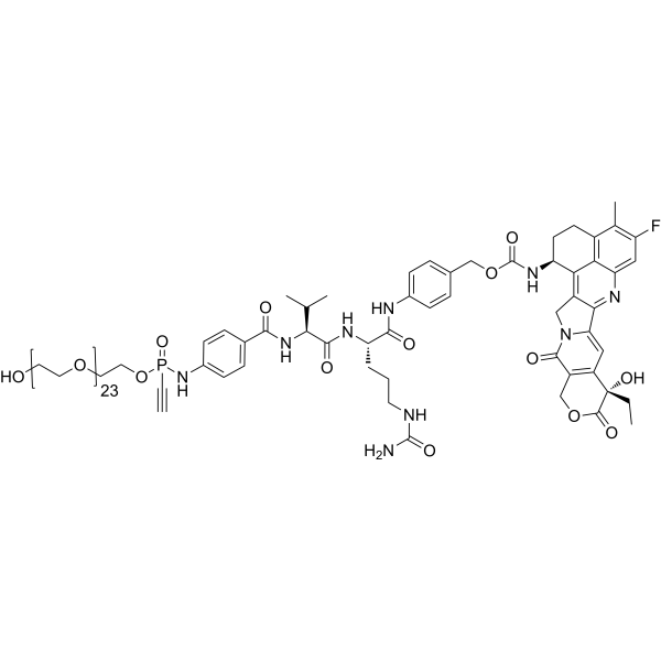
P5(PEG24)-VC-PAB-Exatecan

Based on 1 Customer Validation
P5 (PEG24)-VC-PAB-Exatecan is a TOP1 inhibitor payload with antibody-conjugation-dependent activity. Conjugation of P5 (PEG24)-VC-PAB-Exatecan with Trastuzumab (HY-P9907) generates a DAR8 antibody-drug conjugate (ADCs) with antibody-like pharmacokinetic properties. P5 (PEG24)-VC-PAB-Exatecan induces S-phase and G2-M-phase cell cycle arrest, DNA damage and apoptosis in target-positive tumor cells, and releases damage-associated molecular patterns (DAMP) related to immunogenic cell death (ICD). The ADCs prepared from it exert bystander killing effects on non-target tumor cells. ADCs based on P5 (PEG24)-VC-PAB-Exatecan exhibit linker stability in vitro and in vivo, show in vivo efficacy, and can be used in research related to HER2-positive cancers.
For research use only. We do not sell to patients.
- Purity : 98.59%
- CAS No.: 2928571-43-9
- Formula: C100H151FN9O36P
- Molecular Weight:2105.28
-
Storage:
-20°C, stored under nitrogen
* In solvent : -80°C, 6 months; -20°C, 1 month (stored under nitrogen)

To place orders, for customer services and technical support, please contact: MedChemExpress USA
Tel: 609-228-6898 E-mail: [email protected] [email protected]


-
 Biological Activity
-
Chemical Information
-
Solvent & Solubility
- Protocols
- Purity & Documentation
- References
-
 Help & FAQs




Biological Activity
Description
IC50 & Target
|
Top1 |
In Vitro
Under optimized conditions, P5 (PEG24)-VC-PAB-Exatecan (LP5) (16 h; 25°C) is conjugated with Trastuzumab (HY-P9907) to produce a DAR8 ADC with a monomer yield of up to 99% and minimal aggregates[1].
P5 (PEG24)-VC-PAB-Exatecan (0.05-3 mg/mL; 7 d) potently inhibits the viability of HER2-positive SKBR-3 cells (EC50=12.5 ng/mL, maximum cell death rate 96%) and HCC-78 cells (EC50=97.6 ng/mL, maximum cell death rate 88%), but shows no activity against HER2-negative MDA-MB-468 cells[1].
Trastuzumab-LP5 DAR8 (0.05-3 mg/mL; 5 d) potently induces bystander killing of HER2-negative MDA-MB-468 cells during co-culture with HER2-positive SKBR-3 cells, and exhibits superior activity to Enhertu[1].
DAR8 ADC (0.5 mg/mL; 72 h) induces robust DNA damage in HER2-positive SKBR-3 cells, as evidenced by increased levels of phosphorylated H2AX, activated caspase 3, and cleaved PARP following treatment at 0.5 mg/mL for 72 h[1].
DAR8 ADC (3 mg/mL; 48 h) induces immunogenic cell death in HER2-positive SKBR-3 cells, which is characterized by increased cell surface calreticulin expression, ATP release, and HMGB1 release after treatment at 3 mg/mL for 48 h[1].
MedChemExpress (MCE) has not independently confirmed the accuracy of these methods. They are for reference only. Further protocols information, click here.
-
Cell Line:HER2-positive SKBR-3 cells, HER2-positive HCC-78 cells, HER2-negative MDA-MB-468 cells
-
Concentration:0.05-3 mg/mL DAR8 ADC
-
Incubation Time:7 days
-
Result:Inhibited SKBR-3 cell viability with an EC50 of 12.5 ng/mL and achieved a maximum of 96% dead cells.
Inhibited HCC-78 cell viability with an EC50 of 97.6 ng/mL and achieved a maximum of 88% dead cells.
Showed no cytotoxic effect on HER2-negative MDA-MB-468 cells.
-
Cell Line:HER2-positive SKBR-3 cells, HER2-negative MDA-MB-468 cells (co-cultured; MDA-MB-468 cultured alone)
-
Concentration:0.05-3 mg/mL DAR8 ADC
-
Incubation Time:5 days
-
Result:Induced bystander killing of HER2-negative MDA-MB-468 cells in co-culture with HER2-positive SKBR-3 cells, showing a tendency toward lower IC50 values and higher maximum cell killing compared with Enhertu.
Showed no effect on MDA-MB-468 cells cultured alone.
In Vivo
When conjugated with IgG1 to form a DAR8 antibody-drug conjugate (ADC), P5 (PEG24)-VC-PAB-Exatecan (10 mg/kg; intravenous injection; single administration) exhibits antibody-like in vivo pharmacokinetic stability, and fully maintains a drug-to-antibody ratio of 8 during 21 days of circulation in rats[1].
P5 (PEG24)-VC-PAB-Exatecan (20 mg/kg; intravenous injection; single administration), as the conjugated payload in Trastuzumab-LP5 DAR8, exhibits complete in vivo stability in mice, with the DAR maintained at the level of 8 throughout the 7-day circulation period[1].
MedChemExpress (MCE) has not independently confirmed the accuracy of these methods. They are for reference only.
-
Animal Model:CB17-Scid (female, subcutaneous xenograft of NCI-N87 HER2-positive cells)[1]
-
Dosage:0.25 mg/kg; 0.5 mg/kg; 1 mg/kg; 2 mg/kg
-
Administration:i.v.; single dose
-
Result:Achieved complete tumor regression in 10/10 mice at 2 mg/kg.
Achieved complete tumor regression in 8/10 mice at 1 mg/kg.
Achieved complete tumor regression in 8/10 mice at 0.5 mg/kg.
Achieved complete tumor regression in 0/10 mice at 0.25 mg/kg.
Demonstrated superior tumor growth inhibition over all tested dose levels compared to Enhertu, including greater efficacy at 0.5 mg/kg than Enhertu at 1 mg/kg.


Chemical Information
-
CAS No. 2928571-43-9
-
Appearance Oil
-
Molecular Weight 2105.28
-
Formula C100H151FN9O36P
-
Color White to light yellow
-
SMILES
O=C(N[C@@H](C(C)C)C(N[C@@H](CCCNC(N)=O)C(NC(C=C1)=CC=C1COC(N[C@H]2CCC3=C4C2=C5C(C(N6C5)=CC([C@](CC)(O)C(OC7)=O)=C7C6=O)=NC4=CC(F)=C3C)=O)=O)=O)C8=CC=C(NP(C#C)(OCCOCCOCCOCCOCCOCCOCCOCCOCCOCCOCCOCCOCCOCCOCCOCCOCCOCCOCCOCCOCCOCCOCCOCCO)=O)C=C8
-
Shipping
Room temperature in continental US; may vary elsewhere.
-
Storage
-20°C, stored under nitrogen
* In solvent : -80°C, 6 months; -20°C, 1 month (stored under nitrogen)


Solvent & Solubility
In Vitro:
DMSO : 100 mg/mL (47.50 mM; Need ultrasonic; Hygroscopic DMSO has a significant impact on the solubility of product, please use newly opened DMSO)
Please refer to the solubility information to select the appropriate solvent. Once prepared, please aliquot and store the solution to prevent product inactivation from repeated freeze-thaw cycles.
Storage method and period of stock solution: -80°C, 6 months; -20°C, 1 month (stored under nitrogen). When stored at -80°C, please use it within 6 months. When stored at -20°C, please use it within 1 month.
Please refer to the solubility information to select the appropriate solvent. Once prepared, please aliquot and store the solution to prevent product inactivation from repeated freeze-thaw cycles.
Storage method and period of stock solution: -80°C, 6 months; -20°C, 1 month (stored under nitrogen). When stored at -80°C, please use it within 6 months. When stored at -20°C, please use it within 1 month.
Concentration (start) × Volume (start) = Concentration (final) × Volume (final)

Protocols
-
Apoptosis
Apoptosis, also called programmed cell death, is generally characterized by distinct morphological characteristics.
-
TUNEL staining for apoptotic DNA fragmentation
TUNEL staining detects DNA strand breaks by using terminal deoxynucleotidyl transferase to add labeled nucleotides to exposed 3′-OH DNA termini, generating either microscopic staining in fixed cells or tissue sections, or fluorescence/cytometric signal in cell suspensions. TUNEL positivity reflects DNA fragmentation but should not be interpreted alone as definitive apoptosis, because TUNEL can also label necrotic, autolytic, mechanically damaged, or DNA-repair-associated DNA breaks.
-
Flow cytometric DNA-content cell-cycle staining
Flow cytometric DNA-content cell-cycle staining measures the fluorescence intensity of DNA-bound fluorochromes in single cells or nuclei to estimate DNA content distributions, allowing assignment of populations to G0/G1, S, and G2/M phases by DNA histogram deconvolution. Propidium iodide (PI) intercalates into DNA, and PI fluorescence is proportional to cellular DNA content when staining is performed under conditions that make DNA accessible and minimize non-DNA signal. Cells with G2/M DNA content are expected to show approximately twice the fluorescence intensity of G0/G1 cells, while S-phase cells occupy intermediate fluorescence values. PI-based DNA-content analysis can also detect cells with fractional DNA content, often reported as sub-G1, when DNA fragmentation and extraction during staining reduce retained DNA signal in apoptotic cells. DAPI is an alternative DNA fluorochrome for univariate DNA-content analysis, while bivariate approaches combining DNA content with proliferation
-
Cell Cytotoxicity Assay
Cytotoxicity assays are usually based on the assessment of cell membrane damage, which can also be indirectly detected by measuring cell viability. Detection methods include MTT assay, CKK-8 assay, LDH assay and ATP assay, etc.
-
Mammalian live/dead viability and cytotoxicity staining
Live/dead viability and cytotoxicity staining assays are based on the simultaneous detection of intracellular esterase activity in metabolically active (viable) cells and membrane integrity loss in non-viable cells. In commonly used dual-staining approaches, membrane-permeant fluorogenic substrates are converted by intracellular esterases into fluorescent products in live cells, while impermeant DNA-binding dyes selectively enter cells with compromised plasma membranes and label nucleic acids in dead or dying cells, enabling discrimination between viable and non-viable populations by fluorescence microscopy or flow cytometry.
-
Annexin V plus membrane-impermeant dye apoptosis staining
Annexin V-based apoptosis assays rely on the detection of phosphatidylserine (PS) externalization from the inner leaflet of the plasma membrane to the outer leaflet, an early biochemical hallmark of apoptosis. Fluorescently labeled Annexin V binds PS in a calcium-dependent manner, enabling identification of early apoptotic cells by flow cytometry or fluorescence microscopy. When combined with a membrane-impermeant DNA-binding dye (e. g. , propidium iodide), this approach allows discrimination between viable (Annexin V−/dye−), early apoptotic (Annexin V+/dye−), and late apoptotic or necrotic (Annexin V+/dye+) cell populations by assessing membrane integrity and PS exposure.
-
BrdU Incorporation Assay
Bromodeoxyuridine (BrdU) incorporation assay is based on the principle that BrdU, a thymidine analog, is incorporated into newly synthesized DNA during the S phase of the cell cycle, thereby serving as a marker of DNA replication and cellular proliferation. Incorporated BrdU can be detected using anti-BrdU antibodies following DNA denaturation, enabling visualization or quantification of proliferating cells through immunochemical detection methods such as immunofluorescence or immunohistochemistry.
-
Apoptosis Solutions
Apoptosis is a regulated, generally non-lytic cell-death pathway that removes unwanted, damaged, infected, or abnormal cells through coordinated morphological changes, caspase activation, DNA fragmentation, and membrane remodeling. The intrinsic apoptosis pathway is controlled mainly by mitochondrial outer membrane permeabilization, BCL-2 family proteins, cytochrome c release, apoptosome formation, caspase-9 activation, and downstream executioner caspase-3/7 activation. The extrinsic apoptosis pathway is initiated by death receptors such as Fas, TNFR, and TRAIL receptors, which recruit adaptor proteins and activate caspase-8 before engaging executioner caspases or mitochondrial amplification through BID cleavage. Apoptosis is linked to many phenotypes, including cancer cell killing, tissue homeostasis, immune regulation, neurodegeneration, infection response, and treatment-induced cytotoxicity; unresolved questions include how apoptosis interacts with necroptosis, pyroptosis, ferroptos
-
Protocol for Cell Cycle
Cell-cycle analysis by flow cytometry measures DNA content in single cells to estimate the fraction of cells in G0/G1, S, and G2/M phases. Propidium iodide intercalates into DNA, and after RNA removal with RNase, fluorescence intensity reflects cellular DNA content: 2N cells are assigned to G0/G1, cells between 2N and 4N to S phase, and 4N cells to G2/M. DNA-content analysis alone cannot reliably separate G0 from G1 or G2 from M. Ki-67 can distinguish quiescent G0 cells from cycling cells, EdU or BrdU incorporation marks active DNA synthesis in S phase, and phospho-histone H3 staining identifies mitotic cells within the 4N population.
-
Genotoxicity/Mutagenicity Study
The bacterial reverse mutation assay detects point mutations that restore amino-acid prototrophy in auxotrophic Salmonella typhimurium or Escherichia coli tester strains; after exposure to a test article, mutagenic activity is read out as an increased number of revertant colonies on minimal agar compared with the vehicle control. The assay uses tester strains with different mutation targets so that base-substitution and frameshift mutagens can be detected, and testing is performed with and without exogenous mammalian metabolic activation because some chemicals require biotransformation to become mutagenic.


Purity & Documentation
-
Data Sheet (285 KB)
-
SDS (252 KB)
- English - EN (252 KB)
- Français - FR (252 KB)
- Deutsch - DE (252 KB)
- Norwegian - NO (252 KB)
- Español - ES (252 KB)
- Swedish - SV (252 KB)
- Italian - IT (252 KB)
- Korean - KR (252 KB)
- Portuguese - PT (252 KB)
-
Handling Instructions (2659 KB)
References


Complete Stock Solution Preparation Table
Please refer to the solubility information to select the appropriate solvent. Once prepared, please aliquot and store the solution to prevent product inactivation from repeated freeze-thaw cycles.
Storage method and period of stock solution: -80°C, 6 months; -20°C, 1 month (stored under nitrogen). When stored at -80°C, please use it within 6 months. When stored at -20°C, please use it within 1 month.
| Optional Solvent | Concentration Solvent Mass | 1 mg | 5 mg | 10 mg | 25 mg |
|---|---|---|---|---|---|
| DMSO | 1 mM | 0.4750 mL | 2.3750 mL | 4.7500 mL | 11.8749 mL |
| 5 mM | 0.0950 mL | 0.4750 mL | 0.9500 mL | 2.3750 mL | |
| 10 mM | 0.0475 mL | 0.2375 mL | 0.4750 mL | 1.1875 mL | |
| 15 mM | 0.0317 mL | 0.1583 mL | 0.3167 mL | 0.7917 mL | |
| 20 mM | 0.0237 mL | 0.1187 mL | 0.2375 mL | 0.5937 mL | |
| 25 mM | 0.0190 mL | 0.0950 mL | 0.1900 mL | 0.4750 mL | |
| 30 mM | 0.0158 mL | 0.0792 mL | 0.1583 mL | 0.3958 mL | |
| 40 mM | 0.0119 mL | 0.0594 mL | 0.1187 mL | 0.2969 mL |


P5(PEG24)-VC-PAB-Exatecan Related Classifications
HY-163099 Related Classifications
 Powered by Bioz