



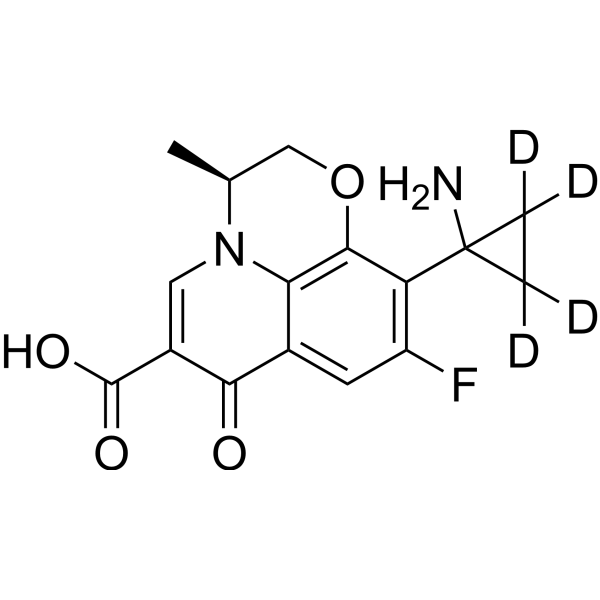
Pazufloxacin-d4
Pazufloxacin-d4 is deuterium labeled Pazufloxacin (HY-B0724B). Pazufloxacin is an orally active fluoroquinolone antimicrobial agent. Pazufloxacin inhibits DNA gyrase with IC50 values of 0.88 μg/mL (E. coli) and 1.9 μg/mL (P. aeruginosa). Pazufloxacin exhibits broad-spectrum antimicrobial activity, with MIC90 values ranging from 0.025 to 100 μg/mL against Gram-positive and Gram-negative bacteria, non-fermenting bacteria, Legionella spp., and anaerobic bacteria. Pazufloxacin is indicated for research on systemic infections, lung infections, urinary tract infections, and Legionella pneumonia.
연구목적의 판매만을 진행합니다. 환자를 대상으로 한 판매는 하지 않습니다.
- CAS No.: 1346602-96-7
- 화학식: C16H11D4FN2O4
- 분자량:322.32
-
보관:
Please store the product under the recommended conditions in the Certificate of Analysis.

To place orders, for customer services and technical support, please contact: MedChemExpress USA
Tel: 609-228-6898 E-mail: [email protected] [email protected]


-
 Biological Activity
-
Chemical Information
- Protocol
- 순도&문서
- References
-
 Help & FAQs


All DNA/RNA Synthesis Isoforms
More

Biological Activity
제품 설명
In Vitro
Stable heavy isotopes of hydrogen, carbon, and other elements have been incorporated into drug molecules, largely as tracers for quantitation during the drug development process. Deuteration has gained attention because of its potential to affect the pharmacokinetic and metabolic profiles of drugs[1].
MedChemExpress (MCE) has not independently confirmed the accuracy of these methods. They are for reference only. Further protocols information, click here.
신청
1. This compound can be used as a tracer
2. This compound can be used as an internal standard for quantitative analysis by NMR, GC-MS, or LC-MS.


Chemical Information
-
CAS No. 1346602-96-7
-
Unlabeled CAS 127045-41-4
-
분자량 322.32
-
화학식 C16H11D4FN2O4
-
SMILES
O=C1C2=CC(F)=C(C3(C([2H])([2H])C3([2H])[2H])N)C4=C2N(C=C1C(O)=O)[C@@H](C)CO4
-
Synonyms
T3761-d4
-
선적
Room temperature in continental US; may vary elsewhere.
-
보관
Please store the product under the recommended conditions in the Certificate of Analysis.

Protocol
-
Research Protocol for Infectious Diseases
Infectious-disease experiments test how pathogens interact with host barriers, innate immune receptors, inflammatory signaling, pathogen replication, and tissue injury; pattern-recognition receptors such as TLRs, RIG-I-like receptors, NOD-like receptors, and inflammasomes detect microbial molecules and activate NF-κB, interferon, and cytokine responses. The central hypothesis is that infection severity reflects the balance between pathogen burden and host response: protective inflammation restricts pathogen growth, whereas excessive or mislocalized inflammation contributes to tissue damage and disease phenotype. Unresolved questions include which host pathways are protective versus pathogenic, why some infection models fail to translate to human disease, and which combined readouts best predict clinically relevant infection outcomes.
-
Anaerobic Bacterial Culture
Anaerobic bacterial culture detects viable bacteria that can grow under oxygen-depleted conditions; the readout is visible colony formation or broth turbidity after incubation in a chamber, jar, pouch, bag, or roll-tube system that maintains anaerobiosis. Oxygen control is central to the method because recovery depends on limiting oxygen exposure during collection, transport, inoculation, and incubation.
-
Cell Cytotoxicity Assay
Cytotoxicity assays are usually based on the assessment of cell membrane damage, which can also be indirectly detected by measuring cell viability. Detection methods include MTT assay, CKK-8 assay, LDH assay and ATP assay, etc.
-
Gram Staining of Tissue Sections
Gram staining of tissue sections is a histochemical technique used to differentiate Gram-positive and Gram-negative bacteria within histological specimens based on differences in bacterial cell wall structure and dye retention, adapted from classical bacteriological Gram staining into tissue-compatible “histological Gram stain” variants. In tissue applications, modifications of the Brown-Hopps and Brown-Brenn methods are commonly used to improve differentiation of microorganisms embedded within host connective tissue and to reduce overstaining or loss of Gram-negative signal, which are known limitations of earlier approaches. The principle relies on crystal violet-iodine complex retention in Gram-positive organisms and subsequent decolorization and counterstaining steps that allow contrast visualization of Gram-negative organisms against tissue background.


순도&문서
References
[1]. Russak EM, et al. Impact of Deuterium Substitution on the Pharmacokinetics of Pharmaceuticals. Ann Pharmacother. 2019;53(2):211-216. [Content Brief]
[2]. Fukuoka, Y., et al., In vitro and in vivo antibacterial activities of T-3761, a new quinolone derivative. Antimicrob Agents Chemother, 1993. 37(3): p. 384-92. [Content Brief]
[3]. Muratani, T., M. Inoue, and S. Mitsuhashi, In vitro activity of T-3761, a new fluoroquinolone. Antimicrob Agents Chemother, 1992. 36(10): p. 2293-303. [Content Brief]
[4]. Takei M, et al. Target preference of 15 quinolones against Staphylococcus aureus, based on antibacterial activities and target inhibition. Antimicrob Agents Chemother. 2001 Dec;45(12):3544-7. [Content Brief]
[5]. Sousa J, et al. First liquid chromatography method for the simultaneous determination of levofloxacin, Pazufloxacin hydrochloride, gatifloxacin, moxifloxacin and trovafloxacin in human plasma. J Chromatogr B Analyt Technol Biomed Life Sci. 2013 Jul 1;930:104-11. [Content Brief]
[6]. Morinaga Y, et al. In vivo efficacy of sivelestat in combination with Pazufloxacin hydrochloride against Legionella pneumonia. Exp Lung Res. 2010 Oct;36(8):484-90. [Content Brief]

Calculators
Concentration (start) × Volume (start) = Concentration (final) × Volume (final)


Pazufloxacin-d4 Related Classifications
HY-B0724BS Related Classifications
 Powered by Bioz