



Prostaglandin J2

Based on 1 Customer Validation
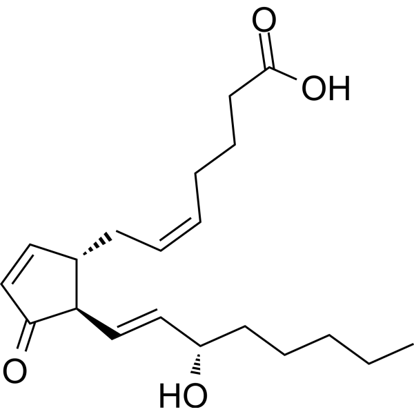
Prostaglandin J2 (PGJ2), an endogenous metabolite of Prostaglandin D2 (PGD2; HY-101988), is a potent PGD2 receptor (DP) agonist with Kis of 0.9 nM and 6.6 nM for hDP and hCRTH2, respectively. Prostaglandin J2 stimulates intracellular cyclic AMP production with an EC50 value of 1.2 nM. Prostaglandin J2 induces oxidative stress and neuronal apoptosis. Prostaglandin J2 induces the accumulation/aggregation of ubiquitinated (Ub) proteins. Prostaglandin J2 is highly neurotoxic and potentially contributes to many neurodegenerative conditions, including Alzheimer's (AD) and Parkinson's diseases (PD).
For research use only. We do not sell to patients.
- Purity : 98.1%
- CAS No.: 60203-57-8
- Formula: C20H30O4
- Molecular Weight:334.45
-
Storage:
Solution, -20°C, 2 years

To place orders, for customer services and technical support, please contact: MedChemExpress USA
Tel: 609-228-6898 E-mail: [email protected] [email protected]


-
 Biological Activity
-
Chemical Information
- Protocols
- Purity & Documentation
- References
-
 Help & FAQs


-
HY-101952
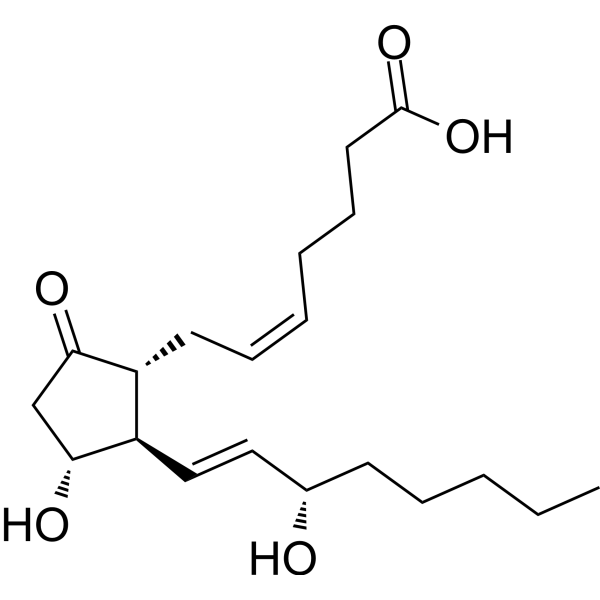
Prostaglandin E2Prostaglandin Receptor Agonist -
HY-109590
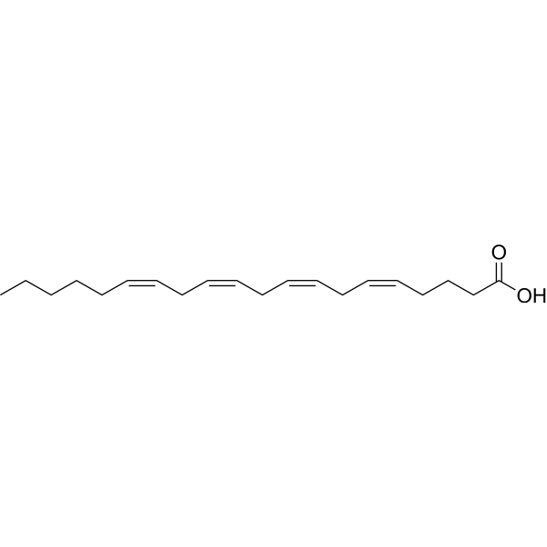
Arachidonic acidPolyunsaturated Fatty Acid -
HY-B0131
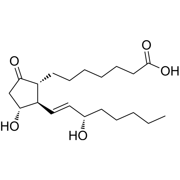
Prostaglandin E1Vasodilator -
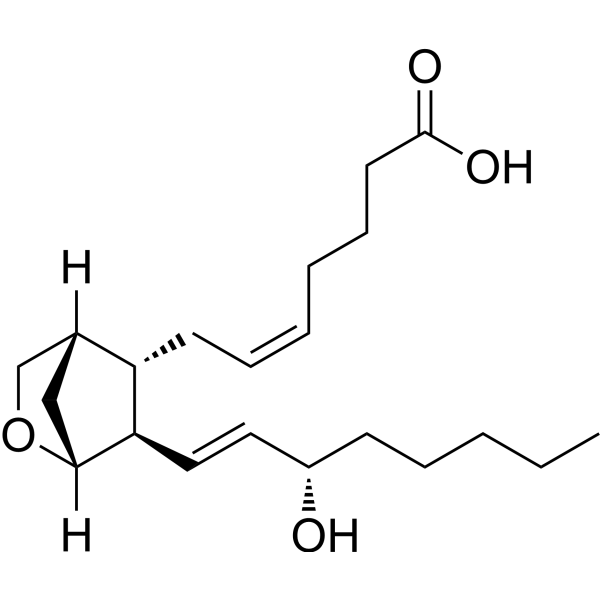
HY-108566
U-46619TXA2 Agonist -
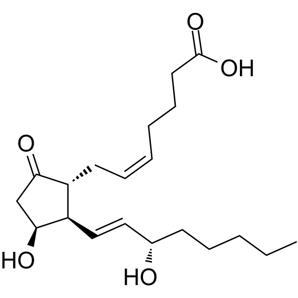
HY-130223
11β-Prostaglandin E2Prostanoid Derivative -
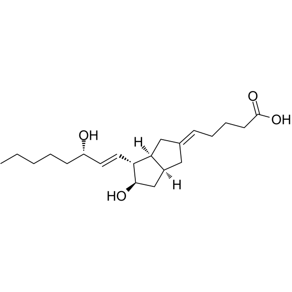
HY-112322
CarbacyclinProstaglandin Receptor Agonist -
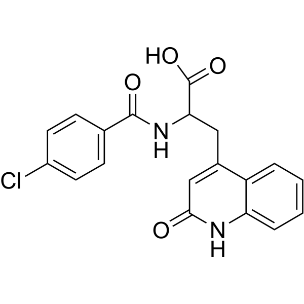
HY-B0360
RebamipideGastroprotective Agent -
HY-113205
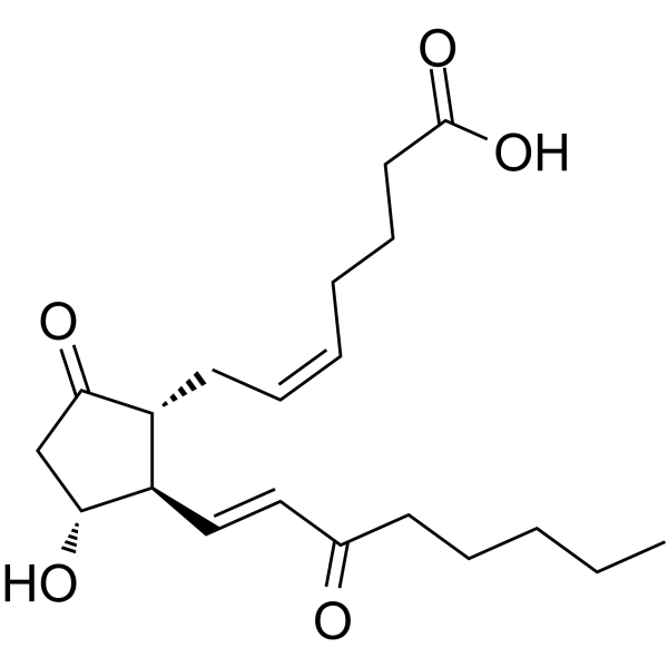
15-keto-Prostaglandin E2PGE2 Metabolite -

HY-B0360R
Rebamipide (Standard)Gastroprotective Agent -
HY-118282
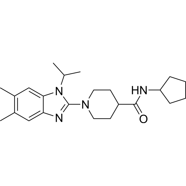
mPGES1-IN-7mPGES-1 Inhibitor
All Endogenous Metabolite Isoforms
More

Biological Activity
Description
|
hDP 0.9 nM (Ki) |
hCRTH2 6.6 nM (Ki) |
hEP1 15.678 μM (Ki) |
hEP2 989 nM (Ki) |
hEP3 319 nM (Ki) |
hEP4 1065 nM (Ki) |
hFP 553 nM (Ki) |
hIP >25 μM (Ki) |
hTP 6426 nM (Ki) |
Human Endogenous Metabolite |
In Vivo
MedChemExpress (MCE) has not independently confirmed the accuracy of these methods. They are for reference only.
-
Animal Model:Sixteen-week-old Sprague Dawley male rats[4]
-
Dosage:33.4 μg/injection
-
Administration:Unilateral (right side) injections to the SNpc; once per week for 2 or 4 weeks
-
Result:Induced progressive dopaminergic neuronal loss in the rat substantia nigra pars compacta (SNpc).
Developed parkinsonian-like motor deficits in a progressive manner.


Chemical Information
-
CAS No. 60203-57-8
-
Appearance Liquid
-
Molecular Weight 334.45
-
Formula C20H30O4
-
Color Colorless to light yellow
-
SMILES
CCCCC[C@H](O)/C=C/[C@@H]1[C@H](C=CC1=O)C/C=C\CCCC(O)=O
-
Synonyms
PGJ2
-
Structure Classification
-
Initial Source
-
Shipping
Room temperature in continental US; may vary elsewhere.
-
Storage
Solution, -20°C, 2 years

Protocols
-
Neurotoxicity Study
This protocol assesses in vitro neurotoxicity by combining neuronal viability, mitochondrial/metabolic activity, neurite outgrowth, and optional neuronal network function readouts. Calcein-AM or resazurin/PrestoBlue readouts estimate viable or metabolically active cells; βIII-tubulin immunofluorescence detects neuronal morphology and neurite networks; TMRE detects mitochondrial membrane potential; and MEA recordings detect functional changes in neuronal network activity.
-
Apoptosis
Apoptosis, also called programmed cell death, is generally characterized by distinct morphological characteristics.
-
TUNEL staining for apoptotic DNA fragmentation
TUNEL staining detects DNA strand breaks by using terminal deoxynucleotidyl transferase to add labeled nucleotides to exposed 3′-OH DNA termini, generating either microscopic staining in fixed cells or tissue sections, or fluorescence/cytometric signal in cell suspensions. TUNEL positivity reflects DNA fragmentation but should not be interpreted alone as definitive apoptosis, because TUNEL can also label necrotic, autolytic, mechanically damaged, or DNA-repair-associated DNA breaks.
-
Annexin V plus membrane-impermeant dye apoptosis staining
Annexin V-based apoptosis assays rely on the detection of phosphatidylserine (PS) externalization from the inner leaflet of the plasma membrane to the outer leaflet, an early biochemical hallmark of apoptosis. Fluorescently labeled Annexin V binds PS in a calcium-dependent manner, enabling identification of early apoptotic cells by flow cytometry or fluorescence microscopy. When combined with a membrane-impermeant DNA-binding dye (e. g. , propidium iodide), this approach allows discrimination between viable (Annexin V−/dye−), early apoptotic (Annexin V+/dye−), and late apoptotic or necrotic (Annexin V+/dye+) cell populations by assessing membrane integrity and PS exposure.
-
ROS/oxidative-stress fluorescent staining
ROS/oxidative-stress fluorescent staining uses cell-permeant fluorogenic probes that become fluorescent after oxidation inside cells or tissues; commonly used examples include DCFH-DA/DCFDA for broad cellular oxidant detection, DHE for superoxide-related signal detection, MitoSOX for mitochondrial superoxide-related signal detection, and CellROX probes for oxidative-stress-associated fluorescence readouts. The assay detects probe oxidation rather than a single ROS species unless the probe and analysis method have been chemically validated for that species. DCFH-DA enters cells, is deacetylated by intracellular esterases to DCFH, and produces fluorescent DCF after oxidation, so the readout is used as an operational measure of total cellular oxidative stress rather than a species-specific ROS measurement. DHE and MitoSOX can report superoxide-related oxidation, but red fluorescence alone can include non-specific ethidium-like oxidation products; HPLC or optimized spectral approaches are
-
Apoptosis Solutions
Apoptosis is a regulated, generally non-lytic cell-death pathway that removes unwanted, damaged, infected, or abnormal cells through coordinated morphological changes, caspase activation, DNA fragmentation, and membrane remodeling. The intrinsic apoptosis pathway is controlled mainly by mitochondrial outer membrane permeabilization, BCL-2 family proteins, cytochrome c release, apoptosome formation, caspase-9 activation, and downstream executioner caspase-3/7 activation. The extrinsic apoptosis pathway is initiated by death receptors such as Fas, TNFR, and TRAIL receptors, which recruit adaptor proteins and activate caspase-8 before engaging executioner caspases or mitochondrial amplification through BID cleavage. Apoptosis is linked to many phenotypes, including cancer cell killing, tissue homeostasis, immune regulation, neurodegeneration, infection response, and treatment-induced cytotoxicity; unresolved questions include how apoptosis interacts with necroptosis, pyroptosis, ferroptos


Purity & Documentation
-
Data Sheet (276 KB)
-
SDS (394 KB)
- English - EN (394 KB)
- Français - FR (394 KB)
- Deutsch - DE (394 KB)
- Norwegian - NO (394 KB)
- Español - ES (394 KB)
- Swedish - SV (394 KB)
- Italian - IT (394 KB)
- Korean - KR (394 KB)
- Portuguese - PT (394 KB)
-
Handling Instructions (2659 KB)
References
[1]. D H Wright, et al. Characterization of the recombinant human prostanoid DP receptor and identification of L-644,698, a novel selective DP agonist. Br J Pharmacol. 1998 Apr;123(7):1317-24. [Content Brief]
[2]. Nicole Sawyer, et al. Molecular pharmacology of the human prostaglandin D2 receptor, CRTH2. Br J Pharmacol. 2002 Dec;137(8):1163-72. [Content Brief]
[3]. Maria E Figueiredo-Pereira, et al. Prostaglandin J2: a potential target for halting inflammation-induced neurodegeneration. Ann N Y Acad Sci. 2016 Jan;1363(1):125-37. [Content Brief]
[4]. Chuhyon Corwin, et al. Prostaglandin D2/J2 signaling pathway in a rat model of neuroinflammation displaying progressive parkinsonian-like pathology: potential novel therapeutic targets. J Neuroinflammation. 2018 Sep 20;15(1):272. [Content Brief]

Calculators
Concentration (start) × Volume (start) = Concentration (final) × Volume (final)


Prostaglandin J2 Related Classifications
HY-113366 Related Classifications
 Powered by Bioz
Keywords
- Prostaglandin J2
- 60203-57-8
- PGJ2
- Prostaglandin J 2
- Prostaglandin J-2
- PGJ 2
- PGJ-2
- Prostaglandin Receptor
- Endogenous Metabolite
- endogenous metabolite
- prostaglandin D2
- PGD2
- DP
- Hdp
- hCRTH2
- cyclic AMP
- oxidative stress
- neuronal apoptosis
- ubiquitinated
- neurodegenerative
- Alzheimer's
- AD
- Parkinson's diseases
- PD
- Inhibitor
- inhibitor
- inhibit