



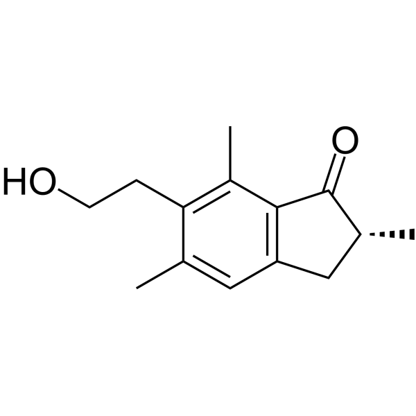
Pterosin B

Based on 1 Customer Validation
Pterosin B is an orally active indanone. Pterosin B can be obtained from Pteridium aquilinum. Pterosin B is a Sik3 signaling inhibitor. Pterosin B inhibits Klf5 expression and reduces β-amyloid deposition. Pterosin B prevents chondrocyte hypertrophy and osteoarthritis in mice. Pterosin B inhibits cardiomyocyte hypertrophy, improves cognitive impairment, and lowers blood glucose. Pterosin B can be used in research on arthritis, Alzheimer's disease, pathological cardiac hypertrophy and diabetes.
For research use only. We do not sell to patients.
- Purity : 99.98%
- CAS No.: 34175-96-7
- Formula: C14H18O2
- Molecular Weight:218.30
-
Storage:Powder -20°C, 3 years , 4°C, 2 years ; In solvent -80°C, 2 years , -20°C, 1 year

To place orders, for customer services and technical support, please contact: MedChemExpress USA
Tel: 609-228-6898 E-mail: [email protected] [email protected]


-
 Biological Activity
-
Chemical Information
-
Solvent & Solubility
- Protocols
- Purity & Documentation
- References
-
 Help & FAQs


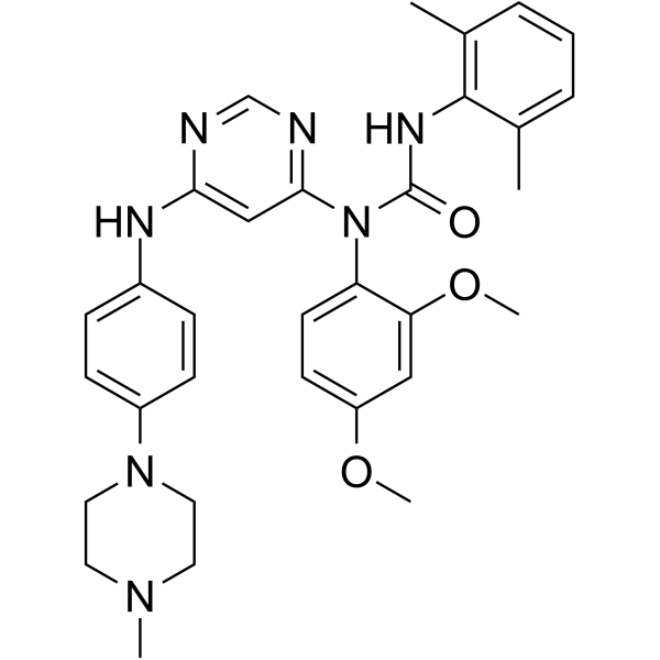
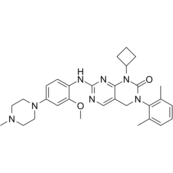

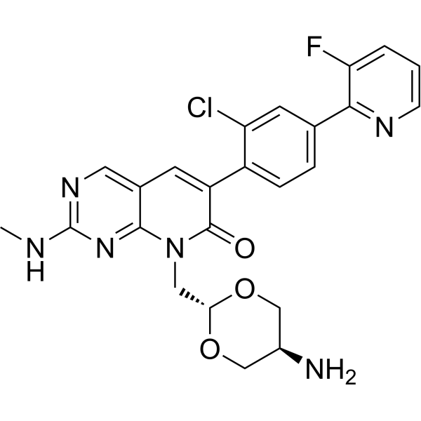


Biological Activity
Description
IC50 & Target
[2]|
QSK/SIK3 |
Cellular Effect
|
Cell Line

|
Type | Value | Description | References |
|---|---|---|---|---|
| HCT-116 | IC50 |
50.1 μM
Compound: 7
|
Cytotoxicity against human HCT116 cells assessed as reduction in cell viability after 48 hrs by MTT assay
Cytotoxicity against human HCT116 cells assessed as reduction in cell viability after 48 hrs by MTT assay
|
[PMID: 28532669] |
In Vitro
Pterosin B (300 μM; 5 days) reduces the amounts of Sik3, phosphorylated Hdac4, and phosphorylated Crtc1 in primary chondrocytes, similar to the effect of 4-OH tamoxifen[2].
Pterosin B (10-50 μM; 48 h) inhibits Angiotensin II-induced cardiomyocyte hypertrophy in H9c2 cells, reducing hypertrophy-related gene expression, cell size, and protein synthesis[3].
Pterosin B (1, 5 μM) promotes the phenotypic shift of lipopolysaccharide-induced BV-2 cells from M1 to M2, inhibits Klf5 expression, and attenuates Lipopolysaccharide-induced microglial glycolysis[4].
Pterosin B (300 μM; 36 h) inhibits the SIK3 signaling pathway, suppressing the cytoplasmic localization of HDAC5 and CRTC2 in HEK293 cells[5].
MedChemExpress (MCE) has not independently confirmed the accuracy of these methods. They are for reference only. Further protocols information, click here.
In Vivo
Pterosin B (5-20 mg/kg; p.o.; 8 weeks) ameliorates cognitive deficits, reduces β-amyloid deposition, and inhibits excessive microglia activation in APP/PS1 mice[4].
Pterosin B (0.1% in diet; p.o.; 1 month) lowers blood glucose levels and enhances insulin responses in db/db mice[5].
MedChemExpress (MCE) has not independently confirmed the accuracy of these methods. They are for reference only.
-
Animal Model:C57BL/6NCrSlc male mice (male, 8-week-old or 13-week-old) with destabilized medial meniscus (DMM) surgery-induced osteoarthritis model[2]
-
Dosage:900 μM
-
Administration:Intra-articular injection, three times a week, for 8 weeks (in 8-week-old mice) or 12 weeks (in 13-week-old mice)
-
Result:Led to better OARSI scores in 8-week-old mice, indicating a protective effect against osteoarthritis development.
Reduced the Col10 expression activated by DMM in the non-calcified zone of articular cartilage.
Resulted in better OARSI scores and reduced Col10 expression in noncalcified areas of articular cartilage in 13-week-old mice.
-
Animal Model:Male APP/PS1 mice (male, weight: 40 g, 6-month-old) with Alzheimer's disease model[4]
-
Dosage:5 mg/kg/day and 20 mg/kg/day (dissolved in 0.5% sodium carboxymethyl)
-
Administration:Oral administration, for 8 weeks
-
Result:Reduced escape latency and increased the number of platform crossings and target-zone duration.
Improved learning and memory.
Rescued the decline in spontaneous alternation rate.
Revealed a significant decrease in the Aβ1-42-positive area.
Improved the number and arrangement of Nissl bodies in the hippocampal DG region.
Decreased the number of microglia in the cortex and CA1 region and inhibited the elevation of mRNA levels of inflammatory factors such as Tnfa, Il6, and Il1b in the hippocampus and serum.


Chemical Information
-
CAS No. 34175-96-7
-
Appearance Solid
-
Molecular Weight 218.30
-
Formula C14H18O2
-
Color White to light yellow
-
SMILES
O=C1[C@H](C)CC2=C1C(C)=C(CCO)C(C)=C2
-
Structure Classification
-
Initial Source
-
Shipping
Room temperature in continental US; may vary elsewhere.
-
Storage
Powder -20°C 3 years 4°C 2 years In solvent -80°C 2 years -20°C 1 year


Solvent & Solubility
In Vitro:
DMSO : 250 mg/mL (1145.21 mM; Need ultrasonic; Hygroscopic DMSO has a significant impact on the solubility of product, please use newly opened DMSO)
Please refer to the solubility information to select the appropriate solvent. Once prepared, please aliquot and store the solution to prevent product inactivation from repeated freeze-thaw cycles.
Storage method and period of stock solution: -80°C, 2 years; -20°C, 1 year. When stored at -80°C, please use it within 2 years. When stored at -20°C, please use it within 1 year.
Please refer to the solubility information to select the appropriate solvent. Once prepared, please aliquot and store the solution to prevent product inactivation from repeated freeze-thaw cycles.
Storage method and period of stock solution: -80°C, 2 years; -20°C, 1 year. When stored at -80°C, please use it within 2 years. When stored at -20°C, please use it within 1 year.
Concentration (start) × Volume (start) = Concentration (final) × Volume (final)
In Vivo:
Select the appropriate dissolution method based on your experimental animal and administration route.
- For the following dissolution methods, please ensure to first prepare a clear stock solution using an In Vitro approach and then sequentially add co-solvents:
- To ensure reliable experimental results, the clarified stock solution can be appropriately stored based on storage conditions. As for the working solution for In Vivo experiments, it is recommended to prepare freshly and use it on the same day.
- The percentages shown for the solvents indicate their volumetric ratio in the final prepared solution. If precipitation or phase separation occurs during preparation, heat and/or sonication can be used to aid dissolution.
Add each solvent one by one: 10% DMSO 40% PEG300 5% Tween-80 45% Saline
Solubility: ≥ 2.75 mg/mL (12.60 mM); Clear solution
This protocol yields a clear solution of ≥ 2.75 mg/mL (saturation unknown).
Taking 1 mL working solution as an example, add 100 μL DMSO stock solution (27.5 mg/mL) to 400 μL PEG300, and mix evenly; then add 50 μL Tween-80 and mix evenly; then add 450 μL Saline to adjust the volume to 1 mL.
Preparation of Saline: Dissolve 0.9 g sodium chloride in ddH₂O and dilute to 100 mL to obtain a clear Saline solution.
Add each solvent one by one: 10% DMSO 90% (20% SBE-β-CD in Saline)
Solubility: ≥ 2.75 mg/mL (12.60 mM); Clear solution
This protocol yields a clear solution of ≥ 2.75 mg/mL (saturation unknown).
Taking 1 mL working solution as an example, add 100 μL DMSO stock solution (27.5 mg/mL) to 900 μL 20% SBE-β-CD in Saline, and mix evenly.
Preparation of 20% SBE-β-CD in Saline (4°C, storage for one week): 2 g SBE-β-CD powder is dissolved in 10 mL Saline, completely dissolve until clear.
In Vivo Dissolution Calculator
Please enter the basic information of animal experiments:
-
-
-
-
Recommended: Prepare an additional quantity of animals to account for potential losses during experiments.
Please enter your animal formula composition:
-
%DMSO +
Recommended: Keep the proportion of DMSO in working solution below 2% if your animal is weak.
-
%+
-
+%Tween-80 + +
-
%Saline +
The co-solvents required include: DMSO, . All of co-solvents are available by MedChemExpress (MCE). , Tween 80. All of co-solvents are available by MedChemExpress (MCE).
Working solution concentration: 0.22 mg/mL
Method for preparing stock solution: mg drug dissolved in μL DMSO. Stock solution concentration: mg/mL.
1. Take μL DMSO stock solution;
2. Add μL .
μL , mix evenly;
3. Then add μL Tween 80, mix evenly;
4. Then add μL
Please ensure that the stock solution in the first step is dissolved to a clear state, and add co-solvents in sequence. You can use ultrasonic heating (ultrasonic cleaner, recommended frequency 20-40 kHz), vortexing, etc. to assist dissolution.

Protocols
-
RNA extraction experimental
By lysing cells, releasing RNA, and removing impurities such as proteins and DNA, high-purity RNA products are finally obtained. The commonly used traditional method is the guanidine isothiocyanate/phenol/chloroform method (Trizol), which is suitable for a variety of animal materials including animal tissues, microorganisms, cultured cells, etc., and most plant materials.
-
Collagen-Induced Arthritis
Collagen-induced arthritis (CIA) is an autoimmune murine model of rheumatoid arthritis in which immunization with type II collagen (CII) emulsified in an adjuvant induces a T cell- and autoantibody-driven inflammatory arthritis characterized by synovial hyperplasia, immune cell infiltration, and joint destruction. The model typically relies on genetically susceptible mouse strains (e. g. , DBA/1) and reproduces key features of human rheumatoid arthritis, including anti-collagen immune responses and progressive joint inflammation. Disease onset generally occurs within ~3-4 weeks after immunization, depending on antigen/adjuvant combinations and protocol variation. The immunopathology is driven by adaptive immune activation against CII, leading to systemic and local joint inflammation mediated by pro-inflammatory cytokines and effector immune cells, making CIA a standard preclinical platform for evaluating immunomodulatory and anti-arthritic interventions.
-
Amyloid: Congo Red Amyloid Staining
Congo red amyloid staining is a histochemical method used to detect extracellular amyloid deposits in tissue sections based on the affinity of Congo red dye for β-pleated sheet-rich protein aggregates. When bound to amyloid, Congo red produces characteristic apple-green birefringence under polarized light microscopy, which is widely regarded as a diagnostic feature of amyloid deposition in histopathology. The diagnostic principle relies on the combination of dye binding (congophilia) and optical anisotropy under polarized illumination, which distinguishes amyloid from most non-amyloid eosinophilic extracellular deposits in routine histological evaluation. Amyloid identification by Congo red staining remains a cornerstone in diagnostic pathology despite the availability of adjunct methods such as immunohistochemistry and mass spectrometry, particularly because of its ability to localize deposits directly within tissue architecture. The specificity of Congo red-positive deposits is incre
-
Research Protocol for Metabolic Diseases
AMP-activated protein kinase, AMPK, is a conserved cellular energy sensor that responds to reduced cellular energy status and coordinates metabolism by increasing ATP-generating catabolic pathways while suppressing ATP-consuming anabolic processes. In metabolic disease research, the AMPK pathway is experimentally relevant because it regulates hepatic lipid synthesis, fatty acid oxidation, glucose production, skeletal-muscle glucose disposal, mTORC1-linked biosynthesis, autophagy, mitochondrial homeostasis, and whole-body energy balance. The central pathway logic is that energy stress, metformin, exercise-like stimulation, or direct AMPK activators increase AMPKα Thr172 phosphorylation and downstream substrate phosphorylation, including ACC and RAPTOR. Phosphorylation of ACC suppresses lipogenesis and supports fatty acid oxidation, whereas phosphorylation of RAPTOR suppresses mTORC1 signaling and links cellular energy status to growth and protein synthesis control. The pathway is linked
-
Cardiac Morphometry
Cardiac morphometry is based on quantitative histological and stereological assessment of myocardial structure, including cardiomyocyte size, number, and extracellular matrix composition, to evaluate cardiac growth and remodeling under physiological or pathological conditions. Design-based stereology is considered a reference framework for obtaining unbiased estimates of structural parameters such as cardiomyocyte number, volume, and tissue architecture, enabling quantitative comparison across experimental groups. Histological image-based morphometry further enables measurement of cardiomyocyte cross-sectional area and collagen deposition using microscopy combined with image analysis software, allowing assessment of hypertrophy and fibrosis in cardiac remodeling models. These morphometric readouts reflect underlying biological processes such as cardiomyocyte hypertrophy, loss, or structural reorganization during disease progression or experimental stress.
-
Alzheimer’s Disease Modeling
Alzheimer’s Disease (AD) is a neurodegenerative disorder characterized by a progressive decline in cognitive functions and loss of specific types of neurons and synapses. Alzheimer's symptoms can be simulated in mice by injecting drugs (such as Aβ) or genetically modified.


Purity & Documentation
-
Data Sheet (277 KB)
-
SDS (393 KB)
- English - EN (393 KB)
- Français - FR (393 KB)
- Deutsch - DE (393 KB)
- Norwegian - NO (393 KB)
- Español - ES (393 KB)
- Swedish - SV (393 KB)
- Italian - IT (393 KB)
- Korean - KR (393 KB)
- Portuguese - PT (393 KB)
-
Handling Instructions (2659 KB)
References
[2]. Yahara Y, et al. Pterosin B prevents chondrocyte hypertrophy and osteoarthritis in mice by inhibiting Sik3. Nat Commun. 2016 Mar 24;7:10959. [Content Brief]
[4]. Zhang Y, et al. Pterosin B improves cognitive dysfunction by promoting microglia M1/M2 polarization through inhibiting Klf5/Parp14 pathway. Phytomedicine. 2024 Dec;135:156152. [Content Brief]


Complete Stock Solution Preparation Table
Please refer to the solubility information to select the appropriate solvent. Once prepared, please aliquot and store the solution to prevent product inactivation from repeated freeze-thaw cycles.
Storage method and period of stock solution: -80°C, 2 years; -20°C, 1 year. When stored at -80°C, please use it within 2 years. When stored at -20°C, please use it within 1 year.
| Optional Solvent | Concentration Solvent Mass | 1 mg | 5 mg | 10 mg | 25 mg |
|---|---|---|---|---|---|
| DMSO | 1 mM | 4.5809 mL | 22.9043 mL | 45.8085 mL | 114.5213 mL |
| 5 mM | 0.9162 mL | 4.5809 mL | 9.1617 mL | 22.9043 mL | |
| 10 mM | 0.4581 mL | 2.2904 mL | 4.5809 mL | 11.4521 mL | |
| 15 mM | 0.3054 mL | 1.5270 mL | 3.0539 mL | 7.6348 mL | |
| 20 mM | 0.2290 mL | 1.1452 mL | 2.2904 mL | 5.7261 mL | |
| 25 mM | 0.1832 mL | 0.9162 mL | 1.8323 mL | 4.5809 mL | |
| 30 mM | 0.1527 mL | 0.7635 mL | 1.5270 mL | 3.8174 mL | |
| 40 mM | 0.1145 mL | 0.5726 mL | 1.1452 mL | 2.8630 mL | |
| 50 mM | 0.0916 mL | 0.4581 mL | 0.9162 mL | 2.2904 mL | |
| 60 mM | 0.0763 mL | 0.3817 mL | 0.7635 mL | 1.9087 mL | |
| 80 mM | 0.0573 mL | 0.2863 mL | 0.5726 mL | 1.4315 mL | |
| 100 mM | 0.0458 mL | 0.2290 mL | 0.4581 mL | 1.1452 mL |


Pterosin B Related Classifications
HY-N1570 Related Classifications
 Powered by Bioz