



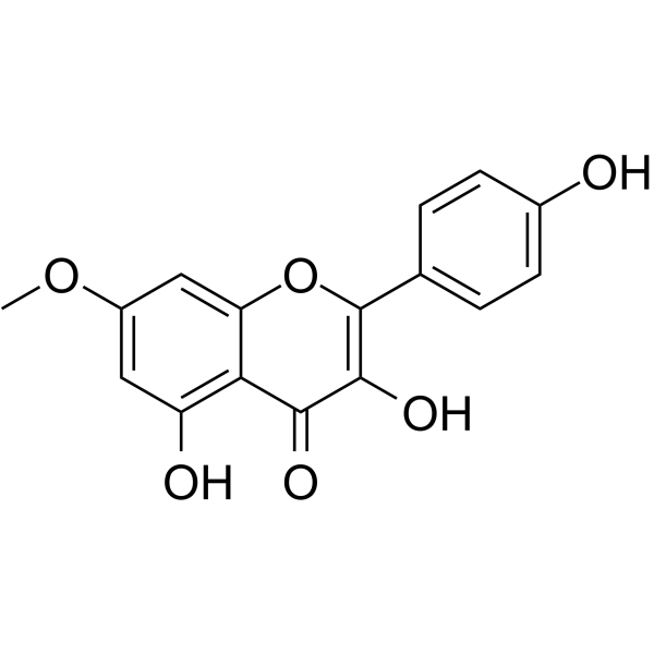
Rhamnocitrin
Based on 4 publication(s) in Google Scholar
Rhamnocitrin is an anti-inflammatory and antioxidant agent that targets STIM-1, NFATc3 and MAPK pathways and can scavenge DPPH (IC50=28.38 mM). Rhamnocitrin selectively inhibits oxidative stress and inflammatory responses in vascular endothelial cells and neurons. Rhamnocitrin up-regulates miR-185 to inhibit STIM-1-mediated store-operated calcium entry (SOCE), thereby blocking NFATc3 nuclear translocation and downstream inflammatory factor expression, while inducing heme oxygenase HO-1 expression and regulating the ERK/p38 MAPK pathway, inhibiting antioxidant and pro-inflammatory cytokines (such as IL-6, IL-8) and adhesion molecules (such as ICAM-1, VCAM-1). Rhamnocitrin can be used in the study of endothelial-related inflammatory diseases (such as sepsis, acute lung injury, atherosclerosis) and neuroprotection (such as oxidative damage of PC12 cells).
For research use only. We do not sell to patients.
- Purity : 99.38%
- CAS No.: 569-92-6
- Formula: C16H12O6
- Molecular Weight:300.26
-
Storage:
4°C, protect from light
* In solvent : -80°C, 6 months; -20°C, 1 month (protect from light)

To place orders, for customer services and technical support, please contact: MedChemExpress USA
Tel: 609-228-6898 E-mail: [email protected] [email protected]


-
 Biological Activity
-
Chemical Information
-
Solvent & Solubility
- Protocols
- Purity & Documentation
- References
-
 Help & FAQs


-
HY-N0160
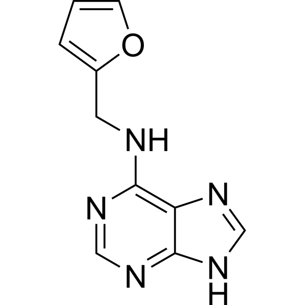
KinetinPlant Hormone -

HY-N0160R
Kinetin (Standard)Plant Hormone -
HY-N4183
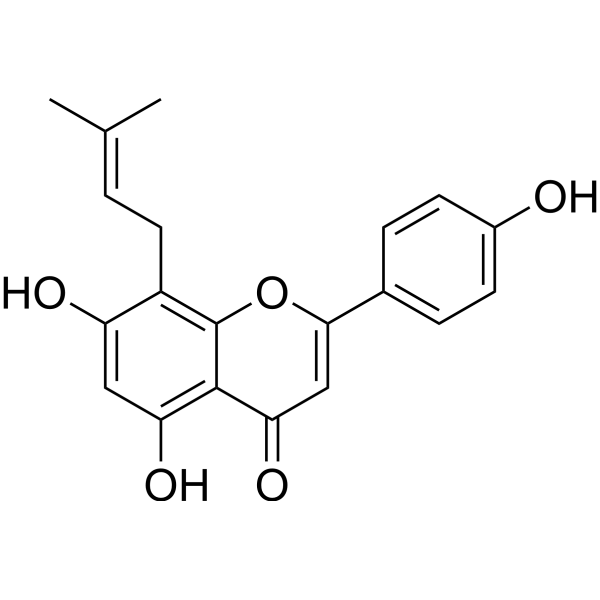
Licoflavone CViral Endonuclease Inhibitor -
HY-N1414
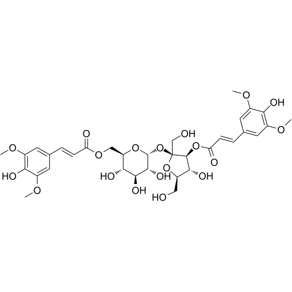
(E)-3',6-DisinapoylsucroseTrkB/BDNF Regulator -

HY-N1353R
Rhamnocitrin (Standard)Anti-Inflammatory Agent -

HY-N1414R
(E)-3',6-Disinapoylsucrose (Standard)Chinese Herbal Ingredient
Publications Citing Use of MedChemExpress (MCE) Rhamnocitrin
More Customer Validation & Images

-
Cell Proliferation/Viability Assay
-
IF
-
Flow Cytometry
-
WB


Biological Activity
Description
In Vitro
Rhamnocitrin (40-80 μM; 48 h) significantly reduces apoptosis in serum deprivation-induced rat adrenal pheochromocytoma PC12 cells, and has no significant effect on STIM-1/Orai-1-mediated SOCE and downstream NFATc3 signaling[2].
Rhamnocitrin (0.1-10 μM; 24 h) inhibits the expression of IL-6, IL-8, MCP-1, ICAM-1, VCAM-1 and PAI-1 in a concentration-dependent manner in LPS-induced human umbilical vein endothelial cells (HUVECs) experiments, and inhibits STIM-1-mediated store-operated calcium entry (SOCE)[3].
MedChemExpress (MCE) has not independently confirmed the accuracy of these methods. They are for reference only. Further protocols information, click here.
-
Cell Line:Human umbilical vein endothelial cells (HUVECs)
-
Concentration:0.1 μM, 1 μM, 10 μM
-
Incubation Time:24 h pretreatment + 6 h LPS stimulation
-
Result:Dose-dependently reduced protein levels of ICAM-1, VCAM-1, and PAI-1 induced by LPS.
Significantly suppressed mRNA expressions of IL-6, IL-8, and MCP-1 at 1 μM and 10 μM.
Attenuated nuclear translocation of NFATc3, a downstream target of SOCE in a concentration-dependent manner.
Cell Viability**: No significant cytotoxicity was observed at concentrations ≤10 μM (CCK-8 assay).
-
Cell Line:Rat pheochromocytoma PC12 cells
-
Concentration:40 μM, 60 μM, 80 μM
-
Incubation Time:48 h
-
Result:Cell Viability: Dose-dependent increased in cell viability, with 60 μM and 80 μM groups significantly higher than the vehicle control.
Cell Apoptosis: Resulted a reduction in sub-G0/G1 phase cells (apoptotic cells) at 60 μM and 80 μM, indicating decreased apoptosis.
WB: Persistently attenuated of p38 MAPK, while increased ERK1/2 activation transiently at 60 μM.
In Vivo
MedChemExpress (MCE) has not independently confirmed the accuracy of these methods. They are for reference only.


Chemical Information
-
CAS No. 569-92-6
-
Appearance Solid
-
Molecular Weight 300.26
-
Formula C16H12O6
-
Color White to yellow
-
SMILES
O=C1C(O)=C(C2=CC=C(O)C=C2)OC3=CC(OC)=CC(O)=C13
-
Structure Classification
-
Initial Source
-
Shipping
Room temperature in continental US; may vary elsewhere.
-
Storage
4°C, protect from light
* In solvent : -80°C, 6 months; -20°C, 1 month (protect from light)


Publications (4)
-
Journal Impact Factor
-
Most Recent
-
Nat Biomed Eng
DeepDrugDiscovery identifies blood-brain barrier permeable autophagy enhancers for Alzheimer's disease. [Abstract]2026 Apr 24. PMID: 42032039 -
-
J Ethnopharmacol
Integrating bioinformatic analysis, network pharmacology, molecular docking and experimental validation to explore the mechanism of Tian Long Cha against influenza virus. [Abstract]2025 Jun 12:349:119964. PMID: 40368255 -
Sci Rep
Potential antiviral activity of rhamnocitrin against influenza virus H3N2 by inhibiting cGAS/STING pathway in vitro. [Abstract]2024 Nov 16;14(1):28287. PMID: 39550441
Rhamnocitrin purchased from MedChemExpress. Usage Cited in: Sci Rep. 2024 Nov 16;14(1):28287. [Abstract]
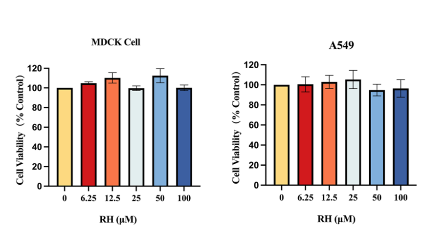
MDCK and A549 cells were incubated with different concentrations of Rhamnocitrin (RH) (0-100 µM). MDCK and A549 cell viability after 48 and 24 h of treatment, respectively, were measured via CCK-8 assay.
Rhamnocitrin purchased from MedChemExpress. Usage Cited in: Sci Rep. 2024 Nov 16;14(1):28287. [Abstract]
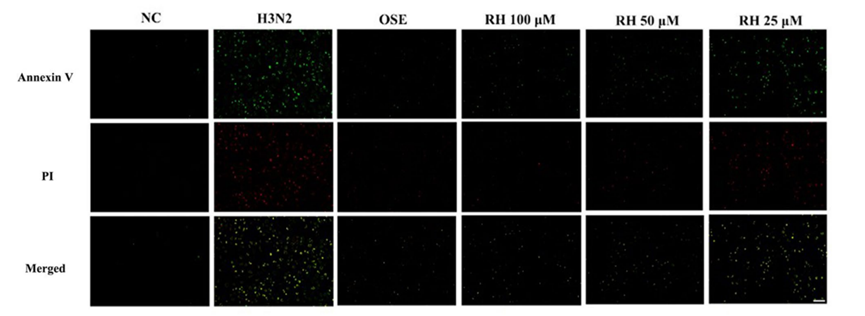
Effects of Rhamnocitrin (RH) (25, 50, 100 μM) on the apoptosis of H3N2-infected MDCK cells.
Rhamnocitrin purchased from MedChemExpress. Usage Cited in: Sci Rep. 2024 Nov 16;14(1):28287. [Abstract]
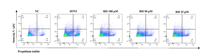
The effect of Rhamnocitrin (RH) (25, 50, 100 μM) on apoptotic A549 cells infected with H3N2 virus for 24 h was detected by flow cytometry analysis
Rhamnocitrin purchased from MedChemExpress. Usage Cited in: Sci Rep. 2024 Nov 16;14(1):28287. [Abstract]
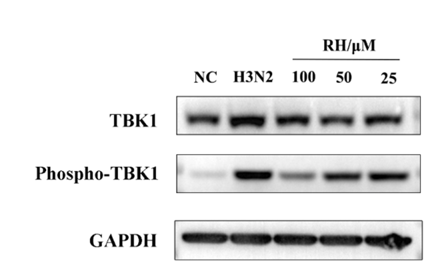
Protein expressions of TBK1, phosphp-TBK1 were detected via western blot analysis treated with Rhamnocitrin (RH) (25, 50, 100 μM).


Solvent & Solubility
In Vitro:
DMSO : 100 mg/mL (333.04 mM; Need ultrasonic; Hygroscopic DMSO has a significant impact on the solubility of product, please use newly opened DMSO)
Please refer to the solubility information to select the appropriate solvent. Once prepared, please aliquot and store the solution to prevent product inactivation from repeated freeze-thaw cycles.
Storage method and period of stock solution: -80°C, 6 months; -20°C, 1 month (protect from light). When stored at -80°C, please use it within 6 months. When stored at -20°C, please use it within 1 month.
Please refer to the solubility information to select the appropriate solvent. Once prepared, please aliquot and store the solution to prevent product inactivation from repeated freeze-thaw cycles.
Storage method and period of stock solution: -80°C, 6 months; -20°C, 1 month (protect from light). When stored at -80°C, please use it within 6 months. When stored at -20°C, please use it within 1 month.
Concentration (start) × Volume (start) = Concentration (final) × Volume (final)
In Vivo:
Select the appropriate dissolution method based on your experimental animal and administration route.
- For the following dissolution methods, please ensure to first prepare a clear stock solution using an In Vitro approach and then sequentially add co-solvents:
- To ensure reliable experimental results, the clarified stock solution can be appropriately stored based on storage conditions. As for the working solution for In Vivo experiments, it is recommended to prepare freshly and use it on the same day.
- The percentages shown for the solvents indicate their volumetric ratio in the final prepared solution. If precipitation or phase separation occurs during preparation, heat and/or sonication can be used to aid dissolution.
Add each solvent one by one: 10% DMSO 40% PEG300 5% Tween-80 45% Saline
Solubility: ≥ 2.08 mg/mL (6.93 mM); Clear solution
This protocol yields a clear solution of ≥ 2.08 mg/mL (saturation unknown).
Taking 1 mL working solution as an example, add 100 μL DMSO stock solution (20.8 mg/mL) to 400 μL PEG300, and mix evenly; then add 50 μL Tween-80 and mix evenly; then add 450 μL Saline to adjust the volume to 1 mL.
Preparation of Saline: Dissolve 0.9 g sodium chloride in ddH₂O and dilute to 100 mL to obtain a clear Saline solution.
In Vivo Dissolution Calculator
Please enter the basic information of animal experiments:
-
-
-
-
Recommended: Prepare an additional quantity of animals to account for potential losses during experiments.
Please enter your animal formula composition:
-
%DMSO +
Recommended: Keep the proportion of DMSO in working solution below 2% if your animal is weak.
-
%+
-
+%Tween-80 + +
-
%Saline +
The co-solvents required include: DMSO, . All of co-solvents are available by MedChemExpress (MCE). , Tween 80. All of co-solvents are available by MedChemExpress (MCE).
Working solution concentration: 0.22 mg/mL
Method for preparing stock solution: mg drug dissolved in μL DMSO. Stock solution concentration: mg/mL. * In solvent : -80°C, 6 months; -20°C, 1 month (protect from light)
1. Take μL DMSO stock solution;
2. Add μL .
μL , mix evenly;
3. Then add μL Tween 80, mix evenly;
4. Then add μL
Please ensure that the stock solution in the first step is dissolved to a clear state, and add co-solvents in sequence. You can use ultrasonic heating (ultrasonic cleaner, recommended frequency 20-40 kHz), vortexing, etc. to assist dissolution.

Protocols
-
Cytoplasmic-Nuclear Fractionated Protein Extraction
Cytoplasmic-nuclear fractionated protein extraction separates soluble cytoplasmic proteins from nuclear-enriched proteins by mild plasma-membrane permeabilization, differential centrifugation, washing of nuclei, and extraction of nuclear proteins for downstream immunoblotting or related molecular analysis. The readout is the relative abundance of a protein in cytoplasmic and nuclear fractions, commonly assessed by western blotting together with compartment markers such as tubulin or pyruvate kinase for cytoplasm and lamin, nucleoporin, hnRNP, H2AX, or Lamin B for nuclear fractions.
-
RNA extraction experimental
By lysing cells, releasing RNA, and removing impurities such as proteins and DNA, high-purity RNA products are finally obtained. The commonly used traditional method is the guanidine isothiocyanate/phenol/chloroform method (Trizol), which is suitable for a variety of animal materials including animal tissues, microorganisms, cultured cells, etc., and most plant materials.
-
Cell Cytotoxicity Assay
Cytotoxicity assays are usually based on the assessment of cell membrane damage, which can also be indirectly detected by measuring cell viability. Detection methods include MTT assay, CKK-8 assay, LDH assay and ATP assay, etc.
-
LPS-Induced Endotoxemia/Systemic Inflammation
Lipopolysaccharide (LPS)-induced endotoxemia is a widely used in vivo model of acute systemic inflammation in which LPS, a Gram-negative bacterial endotoxin, activates innate immune signaling primarily through TLR4, leading to rapid and transient induction of pro-inflammatory cytokines such as TNF-α, IL-6, and IL-1β in circulation and tissues. This cytokine surge is commonly used as a measurable readout of systemic inflammatory activation and immune dysregulation, and is typically assessed within hours after intraperitoneal LPS administration in mouse models of endotoxemia. The model captures key features of systemic inflammatory response syndrome, including cytokine release, immune cell activation, and downstream tissue responses, and has been used to evaluate anti-inflammatory interventions such as cytokine modulation, lipid mediators, and immune cell-targeting therapies.
-
ROS/oxidative-stress fluorescent staining
ROS/oxidative-stress fluorescent staining uses cell-permeant fluorogenic probes that become fluorescent after oxidation inside cells or tissues; commonly used examples include DCFH-DA/DCFDA for broad cellular oxidant detection, DHE for superoxide-related signal detection, MitoSOX for mitochondrial superoxide-related signal detection, and CellROX probes for oxidative-stress-associated fluorescence readouts. The assay detects probe oxidation rather than a single ROS species unless the probe and analysis method have been chemically validated for that species. DCFH-DA enters cells, is deacetylated by intracellular esterases to DCFH, and produces fluorescent DCF after oxidation, so the readout is used as an operational measure of total cellular oxidative stress rather than a species-specific ROS measurement. DHE and MitoSOX can report superoxide-related oxidation, but red fluorescence alone can include non-specific ethidium-like oxidation products; HPLC or optimized spectral approaches are
-
Endothelial Tube Formation Assay
Endothelial tube formation assay evaluates the ability of endothelial cells to attach, migrate, align, and organize into capillary-like networks when cultured on gelled basement membrane extract or Matrigel; the readout is the morphology and quantity of tube-like networks, which reflects an in vitro endothelial morphogenesis step related to angiogenesis. Basement membrane extract/Matrigel provides laminin-rich extracellular matrix cues that support endothelial differentiation into capillary-like structures, but it can contain biologically active growth factors, so growth-factor-reduced matrix is preferred when testing defined angiogenic stimulators or inhibitors.
-
Research Protocol for Cardiovascular Diseases
Cardiovascular disease can be modeled as maladaptive cardiac remodeling, where ischemic injury or pressure overload activates inflammatory signaling, fibroblast activation, extracellular-matrix deposition, cardiomyocyte hypertrophy, vascular remodeling, and progressive ventricular dysfunction. The TGF-β/SMAD axis is a central profibrotic pathway after myocardial injury and pressure overload, while innate immune and cytokine pathways regulate leukocyte recruitment, scar formation, and adverse remodeling. Key unresolved questions include which inflammatory signals are reparative versus harmful, when fibrosis is protective versus maladaptive, and whether pathway inhibition improves function without weakening necessary infarct healing or compensatory remodeling.
-
Research Protocol for Inflammation-related Diseases
The NLRP3 inflammasome is a cytosolic innate immune signaling platform that integrates priming signals and danger-signal activation to promote caspase-1 activation, maturation of IL-1β and IL-18, and gasdermin D-mediated pyroptotic cell death. The core experimental logic is to determine whether inflammatory disease phenotypes are driven by increased NLRP3 expression, ASC-containing inflammasome assembly, caspase-1 cleavage, GSDMD cleavage, and extracellular release of IL-1β/IL-18 rather than by nonspecific cell injury alone. The pathway is strongly linked to inflammation-related disease phenotypes because monosodium urate crystals activate NALP3/NLRP3 inflammasome signaling in gout-like crystal inflammation, cholesterol crystals activate NLRP3 inflammasomes in atherogenesis models, and DSS-induced intestinal inflammation has been reported to involve NLRP3 inflammasome activity. However, experimental colitis studies also show context-dependent protective effects of NLRP3 inflammasome co
-
Inhalation Toxicity Study
Inhalation toxicity studies expose rodents to a controlled aerosol, vapor, gas, or smoke atmosphere and assess respiratory and systemic toxicity using exposure-atmosphere characterization, clinical observations, body and organ weights, bronchoalveolar lavage fluid, histopathology, blood chemistry, hematology, and, when included, molecular endpoints such as transcriptomics, proteomics, lipidomics, or tissue burden analysis. The primary biological readouts are airway irritation, pulmonary inflammation, cytotoxicity, altered surfactant or lipid homeostasis, impaired particle clearance, and tissue remodeling, reflected by BALF cell differentials, BALF protein, LDH, phosphatase activities, cytokines, lung weight, microscopic respiratory-tract lesions, and retained lung burden.


Purity & Documentation
-
Data Sheet (280 KB)
-
SDS (393 KB)
- English - EN (393 KB)
- Français - FR (393 KB)
- Deutsch - DE (393 KB)
- Norwegian - NO (393 KB)
- Español - ES (393 KB)
- Swedish - SV (393 KB)
- Italian - IT (393 KB)
- Korean - KR (393 KB)
- Portuguese - PT (393 KB)
-
Handling Instructions (2659 KB)
References
[1]. Li Y, et al. Simultaneous Determination of Formononetin, Calycosin and Rhamnocitrin from Astragalus Complanatus by UHPLC-MS-MS in Rat Plasma: Application to a Pharmacokinetic Study. J Chromatogr Sci. 2016 Jun 19. [Content Brief]
[2]. Hong JT, et al. Regulation of heme oxygenase-1 expression and MAPK pathways in response to kaempferol and rhamnocitrin in PC12 cells. Toxicol Appl Pharmacol. 2009 May 15;237(1):59-68. [Content Brief]
[3]. Lin T, et al. Rhamnocitrin extracted from Nervilia fordii inhibited vascular endothelial activation via miR-185/STIM-1/SOCE/NFATc3. Phytomedicine. 2020 Dec;79:153350. [Content Brief]


Complete Stock Solution Preparation Table
Please refer to the solubility information to select the appropriate solvent. Once prepared, please aliquot and store the solution to prevent product inactivation from repeated freeze-thaw cycles.
Storage method and period of stock solution: -80°C, 6 months; -20°C, 1 month (protect from light). When stored at -80°C, please use it within 6 months. When stored at -20°C, please use it within 1 month.
| Optional Solvent | Concentration Solvent Mass | 1 mg | 5 mg | 10 mg | 25 mg |
|---|---|---|---|---|---|
| DMSO | 1 mM | 3.3304 mL | 16.6522 mL | 33.3045 mL | 83.2612 mL |
| 5 mM | 0.6661 mL | 3.3304 mL | 6.6609 mL | 16.6522 mL | |
| 10 mM | 0.3330 mL | 1.6652 mL | 3.3304 mL | 8.3261 mL | |
| 15 mM | 0.2220 mL | 1.1101 mL | 2.2203 mL | 5.5507 mL | |
| 20 mM | 0.1665 mL | 0.8326 mL | 1.6652 mL | 4.1631 mL | |
| 25 mM | 0.1332 mL | 0.6661 mL | 1.3322 mL | 3.3304 mL | |
| 30 mM | 0.1110 mL | 0.5551 mL | 1.1101 mL | 2.7754 mL | |
| 40 mM | 0.0833 mL | 0.4163 mL | 0.8326 mL | 2.0815 mL | |
| 50 mM | 0.0666 mL | 0.3330 mL | 0.6661 mL | 1.6652 mL | |
| 60 mM | 0.0555 mL | 0.2775 mL | 0.5551 mL | 1.3877 mL | |
| 80 mM | 0.0416 mL | 0.2082 mL | 0.4163 mL | 1.0408 mL | |
| 100 mM | 0.0333 mL | 0.1665 mL | 0.3330 mL | 0.8326 mL |


Rhamnocitrin Related Classifications
HY-N1353 Related Classifications
 Powered by Bioz