



Rhapontin
Based on 2 publication(s) in Google Scholar
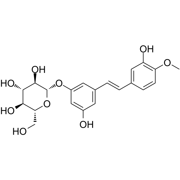
Rhapontin (Rhaponiticin) is an orally aactive SIRT1 agonist and AMPK activator with anti-inflammatory and anti-fibrotic activities. Rhapontin inhibits NLRP3 inflammasome activation by activating SIRT1 and inhibits TGF-β/Smad signaling via the AMPK pathway. Rhapontin reduces intestinal and lung inflammation, inhibits fibroblast differentiation and extracellular matrix deposition, and enhances tight junction protein expression to repair epithelial barriers. Rhapontin can be used in the study of inflammatory bowel diseases (such as ulcerative colitis) and pulmonary fibrosis.
For research use only. We do not sell to patients.
- Purity : 99.73%
- CAS No.: 155-58-8
- Formula: C21H24O9
- Molecular Weight:420.41
-
Storage:
4°C, protect from light
* In solvent : -80°C, 6 months; -20°C, 1 month (protect from light)

To place orders, for customer services and technical support, please contact: MedChemExpress USA
Tel: 609-228-6898 E-mail: [email protected] [email protected]


-
 Biological Activity
-
Chemical Information
-
Solvent & Solubility
- Protocols
- Purity & Documentation
- References
-
 Help & FAQs


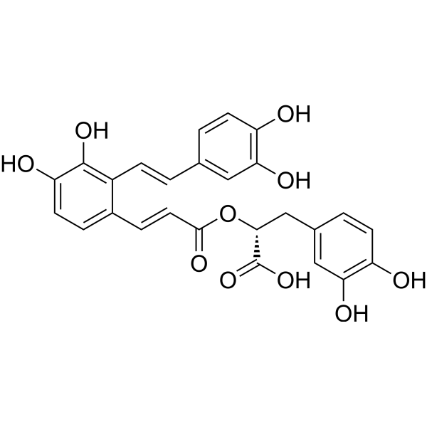
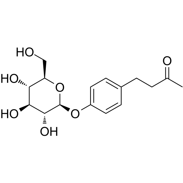
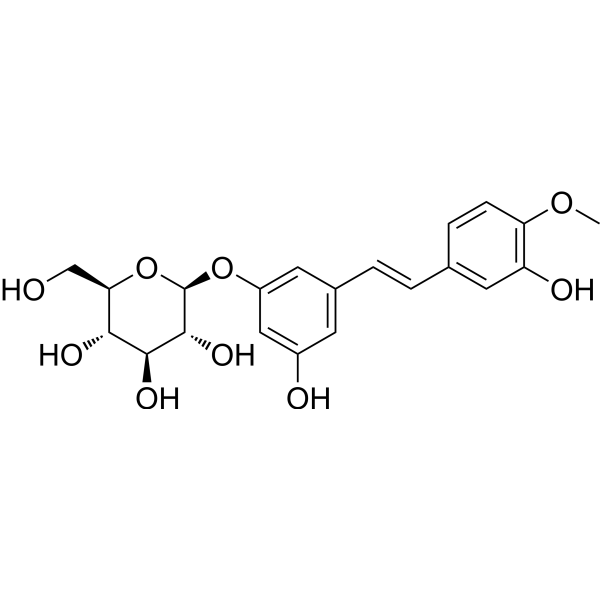

Publications Citing Use of MedChemExpress (MCE) Rhapontin
More

Biological Activity
Description
Cellular Effect
|
Cell Line

|
Type | Value | Description | References |
|---|---|---|---|---|
| HCCLM3 | IC50 |
>100 μM
Compound: 28
|
Antiproliferative activity against human HCCLM3 cells assessed as inhibition of cell growth measured after 48 hrs by MTT assay
Antiproliferative activity against human HCCLM3 cells assessed as inhibition of cell growth measured after 48 hrs by MTT assay
|
[PMID: 34931827] |
| HepG2 | IC50 |
>100 μM
Compound: 28
|
Antiproliferative activity against human HepG2 cells assessed as inhibition of cell growth measured after 48 hrs by MTT assay
Antiproliferative activity against human HepG2 cells assessed as inhibition of cell growth measured after 48 hrs by MTT assay
|
[PMID: 34931827] |
| KB | IC50 |
34 nM
Compound: RHA
|
Antitumor activity against FR-positive human KB cells assessed as decrease in cell viability measured after 48 hrs by MTT proliferation assay
Antitumor activity against FR-positive human KB cells assessed as decrease in cell viability measured after 48 hrs by MTT proliferation assay
|
[PMID: 23177726] |
In Vitro
Rhapontin (0.4-20 μM; 24 h) inhibits NLRP3 inflammasome activation, reduces IL-1β secretion, and reverses the activation of NF-κB and MAPK signaling pathways by upregulating SIRT1 expression in LPS-stimulated THP-1 macrophages[1].
Rhapontin (0.4-10 μM; 24 h) inhibits NLRP3 inflammasome activation, reduces IL-1β secretion, and reverses the activation of NF-κB and MAPK signaling pathways by upregulating SIRT1 expression in LPS-stimulated THP-1 macrophages[1].
Rhapontin (5-10 μM; 24 h) increases the expression of tight junction proteins ZO-1 and occludin, reduces the Bcl-2/Bax apoptotic protein ratio, and restores epithelial barrier function in NCM460 colon epithelial cells stimulated with TNF-α (10 ng/mL; 6 h)[1].
MedChemExpress (MCE) has not independently confirmed the accuracy of these methods. They are for reference only. Further protocols information, click here.
-
Cell Line:Human intestinal colonic epithelial cells NCM460
-
Concentration:0.4 µM, 2 µM, 10 µM
-
Incubation Time:24 h
-
Result:Significantly reduced the protein levels of NLRP3, cleaved caspase-1, and pro-inflammatory cytokines (IL-1β, TNF-α), while upregulating SIRT1 expression.
Inhibited phosphorylation of NF-κB p65 and MAPKs (ERK, JNK, p38) compared to the LPS-stimulated control group.
In Vivo
Rhapontin (25-100 mg/kg; gavage; once a day; 14 days) significantly reduces lung tissue collagen deposition, inhibits TGF-β/Smad signaling pathway, activates AMPK and downregulates Lox2 and HIF-1α expression, and improves lung function[2] in the Bleomycin (HY-108345)-induced pulmonary fibrosis model in mice[1].
MedChemExpress (MCE) has not independently confirmed the accuracy of these methods. They are for reference only.
-
Animal Model:C57BL/6 female mice (18-22 g, 6-8 weeks old), DSS-induced acute colitis model[1]
-
Dosage:100 mg/kg
-
Administration:Gavage, once daily for 8 days (days 1-8 post-DSS induction).
-
Result:Reduced disease activity index (DAI) score by 35% compared to the DSS group, with significant improvements in body weight loss, diarrhea, and hematochezia.
Histological analysis showed decreased inflammatory cell infiltration (lamina propria and submucosa), reduced crypt damage, and restored colon length.
MPO activity, a marker of neutrophil infiltration, was reduced by 42%.
Western blot showed decreased NLRP3, cleaved caspase-1, and TGF-β1 protein levels, while ZO-1 and occludin expressions were increased by 58% and 45%, respectively.
-
Animal Model:C57BL/6 male mice (20-22 g, 6-8 weeks old) with Bleomycin (HY-108345)-induced pulmonary fibrosis model[2]
-
Dosage:25, 50, 100 mg/kg; Prednisone (6.5 mg/kg, positive control).
-
Administration:Gavage, once daily for 14 days (days 7-21 post-bleomycin instillation).
-
Result:At 100 mg/kg, reduced pulmonary index (lung weight/body weight) by 28% compared to the model group, with decreased collagen deposition.
Reduced TGF-β1, α-SMA, and Lox2 protein expressions by 41%, 37%, and 53%, respectively, while AMPK phosphorylation (p-AMPK/AMPK ratio) was increased by 62%.
ELISA showed decreased BALF levels of IL-1β.


Chemical Information
-
CAS No. 155-58-8
-
Appearance Solid
-
Molecular Weight 420.41
-
Formula C21H24O9
-
Color Off-white to yellow
-
SMILES
O[C@H]([C@@H](O)[C@@H]1O)[C@@H](O[C@@H]1CO)OC2=CC(/C=C/C3=CC(O)=C(OC)C=C3)=CC(O)=C2
-
Synonyms
Rhaponiticin
-
Structure Classification
-
Initial Source
-
Shipping
Room temperature in continental US; may vary elsewhere.
-
Storage
4°C, protect from light
* In solvent : -80°C, 6 months; -20°C, 1 month (protect from light)


Publications (2)
-
Journal Impact Factor
-
Most Recent
-
Redox Biol
Gaylussacin, a stilbene glycoside, inhibits chronic obstructive pulmonary disease in mice. [Abstract]2025 Sep:85:103744. PMID: 40614364 -
Biomed Res Int
Anti-influenza A Virus Effects and Mechanisms of Emodin and Its Analogs via Regulating PPAR α/ γ-AMPK-SIRT1 Pathway and Fatty Acid Metabolism. [Abstract]2021 Sep 9;2021:9066938. PMID: 34540999


Solvent & Solubility
In Vitro:
DMSO : 250 mg/mL (594.66 mM; Need ultrasonic; Hygroscopic DMSO has a significant impact on the solubility of product, please use newly opened DMSO)
Please refer to the solubility information to select the appropriate solvent. Once prepared, please aliquot and store the solution to prevent product inactivation from repeated freeze-thaw cycles.
Storage method and period of stock solution: -80°C, 6 months; -20°C, 1 month (protect from light). When stored at -80°C, please use it within 6 months. When stored at -20°C, please use it within 1 month.
Please refer to the solubility information to select the appropriate solvent. Once prepared, please aliquot and store the solution to prevent product inactivation from repeated freeze-thaw cycles.
Storage method and period of stock solution: -80°C, 6 months; -20°C, 1 month (protect from light). When stored at -80°C, please use it within 6 months. When stored at -20°C, please use it within 1 month.
Concentration (start) × Volume (start) = Concentration (final) × Volume (final)
In Vivo:
Select the appropriate dissolution method based on your experimental animal and administration route.
- For the following dissolution methods, please ensure to first prepare a clear stock solution using an In Vitro approach and then sequentially add co-solvents:
- To ensure reliable experimental results, the clarified stock solution can be appropriately stored based on storage conditions. As for the working solution for In Vivo experiments, it is recommended to prepare freshly and use it on the same day.
- The percentages shown for the solvents indicate their volumetric ratio in the final prepared solution. If precipitation or phase separation occurs during preparation, heat and/or sonication can be used to aid dissolution.
Add each solvent one by one: 10% DMSO 40% PEG300 5% Tween-80 45% Saline
Solubility: ≥ 2.08 mg/mL (4.95 mM); Clear solution
This protocol yields a clear solution of ≥ 2.08 mg/mL (saturation unknown).
Taking 1 mL working solution as an example, add 100 μL DMSO stock solution (20.8 mg/mL) to 400 μL PEG300, and mix evenly; then add 50 μL Tween-80 and mix evenly; then add 450 μL Saline to adjust the volume to 1 mL.
Preparation of Saline: Dissolve 0.9 g sodium chloride in ddH₂O and dilute to 100 mL to obtain a clear Saline solution.
Add each solvent one by one: 10% DMSO 90% (20% SBE-β-CD in Saline)
Solubility: ≥ 2.08 mg/mL (4.95 mM); Clear solution
This protocol yields a clear solution of ≥ 2.08 mg/mL (saturation unknown).
Taking 1 mL working solution as an example, add 100 μL DMSO stock solution (20.8 mg/mL) to 900 μL 20% SBE-β-CD in Saline, and mix evenly.
Preparation of 20% SBE-β-CD in Saline (4°C, storage for one week): 2 g SBE-β-CD powder is dissolved in 10 mL Saline, completely dissolve until clear.
In Vivo Dissolution Calculator
Please enter the basic information of animal experiments:
-
-
-
-
Recommended: Prepare an additional quantity of animals to account for potential losses during experiments.
Please enter your animal formula composition:
-
%DMSO +
Recommended: Keep the proportion of DMSO in working solution below 2% if your animal is weak.
-
%+
-
+%Tween-80 + +
-
%Saline +
The co-solvents required include: DMSO, . All of co-solvents are available by MedChemExpress (MCE). , Tween 80. All of co-solvents are available by MedChemExpress (MCE).
Working solution concentration: 0.22 mg/mL
Method for preparing stock solution: mg drug dissolved in μL DMSO. Stock solution concentration: mg/mL. * In solvent : -80°C, 6 months; -20°C, 1 month (protect from light)
1. Take μL DMSO stock solution;
2. Add μL .
μL , mix evenly;
3. Then add μL Tween 80, mix evenly;
4. Then add μL
Please ensure that the stock solution in the first step is dissolved to a clear state, and add co-solvents in sequence. You can use ultrasonic heating (ultrasonic cleaner, recommended frequency 20-40 kHz), vortexing, etc. to assist dissolution.

Protocols
-
Collagen: Sirius Red Staining
Sirius Red or picrosirius red staining is a histochemical method for visualizing collagen-rich extracellular matrix in tissue sections, and collagen fibers are detected as red-stained structures under bright-field microscopy with enhanced birefringence under polarized light. Picrosirius red is useful for assessing total collagen organization, distribution, and fibrosis burden, but polarized color should not be interpreted as a definitive collagen type I versus type III readout because color is affected by fiber orientation, thickness, and packing.
-
RNA extraction experimental
By lysing cells, releasing RNA, and removing impurities such as proteins and DNA, high-purity RNA products are finally obtained. The commonly used traditional method is the guanidine isothiocyanate/phenol/chloroform method (Trizol), which is suitable for a variety of animal materials including animal tissues, microorganisms, cultured cells, etc., and most plant materials.
-
Western Blot
Western blotting (WB) is a commonly used experimental method in molecular biology, biochemistry, and immunogenetics for identifying and quantifying target proteins. It combines gel electrophoresis with immunoassay, enabling researchers to analyze protein expression, post-translational modifications, and molecular weight.
-
ECM-Embedded Organoid (Matrigel/Dome) Culture
ECM-embedded organoid dome culture embeds epithelial stem cells, crypts, organoid fragments, or tumor-derived epithelial cells in a basement-membrane-like hydrogel such as Matrigel, allowing 3D growth, self-organization, lumen formation, budding or cystic morphogenesis, and lineage maintenance under defined niche-factor-containing medium. The primary readouts are organoid establishment efficiency, growth, morphology, passaging capacity, lineage-marker expression, and, when fluorescently labeled lines are used, microscopy- or flow-cytometry-based quantification of population behavior in 3D culture.
-
Connective Tissue: Masson's Trichrome/Collagen Trichrome Staining
Masson’s Trichrome (collagen/trichrome staining) is a histological technique that differentially stains tissue compartments using sequential acidic dyes to distinguish collagen from muscle and cytoplasmic components based on dye affinity and tissue permeability differences, enabling visualization of fibrosis and connective tissue architecture in histological sections. The classical formulation typically uses Weigert's iron hematoxylin for nuclear staining, Biebrich scarlet-acid fuchsin for cytoplasm and muscle, and aniline blue (or light green variants) for collagen, producing a characteristic blue/green collagen signal contrasted against red cytoplasm and dark nuclei. The staining principle relies on selective displacement of smaller dye molecules by larger anionic dyes in collagen-rich regions under controlled acidified conditions, which enhances collagen-specific dye retention. This property makes the method widely used for fibrosis assessment in organs such as heart, liver, lung, a
-
Research Protocol for Inflammation-related Diseases
The NLRP3 inflammasome is a cytosolic innate immune signaling platform that integrates priming signals and danger-signal activation to promote caspase-1 activation, maturation of IL-1β and IL-18, and gasdermin D-mediated pyroptotic cell death. The core experimental logic is to determine whether inflammatory disease phenotypes are driven by increased NLRP3 expression, ASC-containing inflammasome assembly, caspase-1 cleavage, GSDMD cleavage, and extracellular release of IL-1β/IL-18 rather than by nonspecific cell injury alone. The pathway is strongly linked to inflammation-related disease phenotypes because monosodium urate crystals activate NALP3/NLRP3 inflammasome signaling in gout-like crystal inflammation, cholesterol crystals activate NLRP3 inflammasomes in atherogenesis models, and DSS-induced intestinal inflammation has been reported to involve NLRP3 inflammasome activity. However, experimental colitis studies also show context-dependent protective effects of NLRP3 inflammasome co
-
Matrigel Transwell/Boyden Chamber Invasion Assay
Matrigel Transwell/Boyden chamber invasion assay measures the ability of cells to degrade or traverse an extracellular matrix-coated porous membrane and move from an upper chamber toward a chemoattractant in a lower chamber. Invasion is distinguished from migration by coating the membrane with Matrigel or basement membrane matrix; uncoated inserts measure migration, while coated inserts require cells to cross an ECM barrier before reaching the underside of the membrane.
-
Protocol For Protein Expression And Purification
Recombinant protein expression in Escherichia coli followed by purification of a His-tagged soluble protein by immobilized metal affinity chromatography (IMAC), with optional MBP fusion and TEV tag removal when the construct includes these elements. The biological readout is production of the encoded target protein, detected as an inducible band at the expected molecular mass by SDS-PAGE and quantified by total protein assay or chromatographic absorbance; the purification readout is enrichment of the target protein in elution fractions after selective binding of polyhistidine residues to immobilized Ni2+/metal-chelate resin and elution by imidazole-containing buffer. Expression is driven by an inducible bacterial expression system, commonly T7/lac-based, in which IPTG or lactose/auto-induction activates transcription and translation of the cloned gene; lower induction temperature, lower inducer concentration, induction timing, and solubility-enhancing fusion tags can influence the frac
-
Fibrosis/Collagen Morphometry
Fibrosis and collagen morphometry is based on the quantitative visualization of fibrillar collagen deposition in tissue sections using histochemical stains such as Sirius Red (Picrosirius Red) or Masson's trichrome, followed by image-based or polarization-enhanced analysis to estimate collagen proportional area as a surrogate of extracellular matrix accumulation during fibrotic remodeling. Sirius Red combined with polarized light microscopy enhances detection of collagen fibers due to birefringence properties, enabling more specific visualization of collagen type I and III fibrils compared to conventional bright-field histology, while whole-section or region-restricted digital morphometry reduces field-selection bias in fibrosis assessment. Alternative quantitative approaches include second harmonic generation (SHG) and two-photon excited fluorescence microscopy, which enable label-free detection of fibrillar collagen and have been validated against histological staining and biochemica
-
DSS-Induced Colitis
Dextran sulfate sodium (DSS)-induced colitis is generated by administering DSS in mouse drinking water, producing epithelial injury, barrier disruption, weight loss, diarrhea, fecal blood, colon shortening, histologic mucosal damage, and inflammatory mediator changes; the model is mainly used to study acute or chronic intestinal inflammation resembling selected features of ulcerative colitis. DSS injury is interpreted through clinical and tissue readouts rather than a single molecular endpoint: daily body weight, stool consistency, and bleeding are combined into a disease activity index, while colon length, histology, cytokines, myeloperoxidase activity, intestinal permeability, and tight-junction markers provide complementary measures of inflammation and barrier damage.
-
TNBS-Induced Colitis
TNBS-induced colitis is produced by intrarectal delivery of 2,4,6-trinitrobenzene sulfonic acid in ethanol, where ethanol disrupts the mucosal barrier and TNBS haptenates colonic proteins, generating immune-mediated colonic inflammation with weight loss, diarrhea, ulceration, transmural injury, inflammatory-cell infiltration, and cytokine responses. The model is used as an experimental intestinal inflammation model with Crohn’s disease–like features, especially when Th1-type responses, IL-12–dependent inflammation, chronic relapsing inflammation, or fibrosis-related endpoints are studied.
-
Matrigel/ECM Transwell Invasion Assay
The Matrigel/ECM Transwell invasion assay measures the ability of cells to move toward a chemoattractant while crossing an extracellular-matrix barrier placed on a porous membrane; therefore, the readout reflects both chemotactic motility and matrix invasion rather than migration alone. Matrigel is a basement-membrane-rich matrix derived from Engelbreth-Holm-Swarm mouse sarcoma and has been used as a reconstituted basement membrane barrier in chemoinvasion assays. The assay readout is generated by quantifying cells that reach the underside of the insert membrane or lower compartment after incubation, commonly by staining and counting invaded cells or by fluorescence-based quantification.
-
Pyroptosis Solutions
Pyroptosis is a lytic inflammatory cell-death pathway executed by gasdermin pores, most classically through inflammasome-mediated activation of caspase-1, cleavage of gasdermin D, membrane pore formation, LDH release, and secretion of IL-1β and IL-18. The canonical pathway is commonly modeled by priming cells with an inflammatory signal such as LPS to induce pro-IL-1β and inflammasome components, followed by an activation signal such as ATP or nigericin to activate NLRP3, ASC speck formation, caspase-1 cleavage, GSDMD cleavage, cytokine release, and pyroptotic membrane rupture. The non-canonical pathway is triggered when cytosolic LPS activates mouse caspase-11 or human caspase-4/5, leading to GSDMD cleavage and pyroptosis, and this can secondarily activate NLRP3-dependent IL-1β release. Pyroptosis is linked to inflammatory injury, infection, cancer, liver disease, ocular disease, placental inflammation, and other disease phenotypes, but unresolved questions include which gasdermin fam


Purity & Documentation
-
Data Sheet (279 KB)
-
SDS (394 KB)
- English - EN (394 KB)
- Français - FR (394 KB)
- Deutsch - DE (394 KB)
- Norwegian - NO (394 KB)
- Español - ES (394 KB)
- Swedish - SV (394 KB)
- Italian - IT (394 KB)
- Korean - KR (394 KB)
- Portuguese - PT (394 KB)
-
Handling Instructions (2659 KB)
References
[1]. Wei W, et al. Rhapontin ameliorates colonic epithelial dysfunction in experimental colitis through SIRT1 signaling. Int Immunopharmacol. 2017 Jan;42:185-194. [Content Brief]
[2]. Tao L, et al. Protective role of rhapontin in experimental pulmonary fibrosis in vitro and in vivo. Int Immunopharmacol. 2017 Jun;47:38-46. [Content Brief]
[3]. Hibasami H, et al. Induction of apoptosis by rhapontin having stilbene moiety, a component of rhubarb (Rheum officinale Baillon) in human stomach cancer KATO III cells. Oncol Rep. 2007 Aug;18(2):347-51. [Content Brief]


Complete Stock Solution Preparation Table
Please refer to the solubility information to select the appropriate solvent. Once prepared, please aliquot and store the solution to prevent product inactivation from repeated freeze-thaw cycles.
Storage method and period of stock solution: -80°C, 6 months; -20°C, 1 month (protect from light). When stored at -80°C, please use it within 6 months. When stored at -20°C, please use it within 1 month.
| Optional Solvent | Concentration Solvent Mass | 1 mg | 5 mg | 10 mg | 25 mg |
|---|---|---|---|---|---|
| DMSO | 1 mM | 2.3786 mL | 11.8932 mL | 23.7863 mL | 59.4658 mL |
| 5 mM | 0.4757 mL | 2.3786 mL | 4.7573 mL | 11.8932 mL | |
| 10 mM | 0.2379 mL | 1.1893 mL | 2.3786 mL | 5.9466 mL | |
| 15 mM | 0.1586 mL | 0.7929 mL | 1.5858 mL | 3.9644 mL | |
| 20 mM | 0.1189 mL | 0.5947 mL | 1.1893 mL | 2.9733 mL | |
| 25 mM | 0.0951 mL | 0.4757 mL | 0.9515 mL | 2.3786 mL | |
| 30 mM | 0.0793 mL | 0.3964 mL | 0.7929 mL | 1.9822 mL | |
| 40 mM | 0.0595 mL | 0.2973 mL | 0.5947 mL | 1.4866 mL | |
| 50 mM | 0.0476 mL | 0.2379 mL | 0.4757 mL | 1.1893 mL | |
| 60 mM | 0.0396 mL | 0.1982 mL | 0.3964 mL | 0.9911 mL | |
| 80 mM | 0.0297 mL | 0.1487 mL | 0.2973 mL | 0.7433 mL | |
| 100 mM | 0.0238 mL | 0.1189 mL | 0.2379 mL | 0.5947 mL |


Rhapontin Related Classifications
HY-N0671 Related Classifications
 Powered by Bioz