



RyRs activator 3
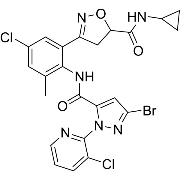
RyRs activator 3 (compound A4) is an effective insecticide against diamondback moths (M. separata) and diamondback moths (P. xylostella). The LC50 value of RyRs activator 3 against diamondback moth is 3.27 mg/L. RyRs activator 3 can bind to ryanodine receptor, increase cytoplasmic Ca2+ concentration, and produce biological toxicity.
Nos produits utilisent uniquement pour la recherche. Nous ne vendons pas aux patients.
- CAS No.: 2850333-35-4
- Formule: C23H19BrCl2N6O3
- Masse moléculaire:578.25
-
Stockage:
Please store the product under the recommended conditions in the Certificate of Analysis.

To place orders, for customer services and technical support, please contact: MedChemExpress USA
Tel: 609-228-6898 E-mail: [email protected] [email protected]


-
 Activité biologique
-
Chemical Information
- Protocole
- Pureté et documentation
- Références
-
 Help & FAQs


Voir tous les produits spécifiques à Isoform Parasite
MoreVoir tous les produits spécifiques à Isoform Calcium Channel
More

Activité biologique
Description
IC50 & Target
Ryanodine receptor[1]


Chemical Information
-
CAS No. 2850333-35-4
-
Masse moléculaire 578.25
-
Formule C23H19BrCl2N6O3
-
SMILES
BrC1=NN(C(C(NC2=C(C=C(C=C2C3=NOC(C3)C(NC4CC4)=O)Cl)C)=O)=C1)C5=NC=CC=C5Cl
-
Livraison
Room temperature in continental US; may vary elsewhere.
-
Stockage
Please store the product under the recommended conditions in the Certificate of Analysis.


Pureté et documentation
Références

Calculators
Concentration (start) × Volume (start) = Concentration (final) × Volume (final)


RyRs activator 3 Related Classifications
HY-156082 Related Classifications
 Powered by Bioz