



BMS-986339

Based on 1 Customer Validation
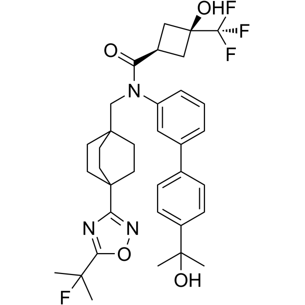
BMS-986339 is an orally active, potent FXR agonist. BMS-986339 forms H-bond with His298 and ASN287 residues. BMS-986339 can be used in the research of primary biliary cirrhosis (PBC), primary sclerosing cholangitis (PSC), and nonalcoholic steatohepatitis (NASH), anti-fibrosis.
For research use only. We do not sell to patients.
- Purity : 99.74%
- CAS No.: 2477873-64-4
- Formula: C35H41F4N3O4
- Molecular Weight:643.71
-
Storage:Powder -20°C, 3 years , 4°C, 2 years ; In solvent -80°C, 6 months , -20°C, 1 month

To place orders, for customer services and technical support, please contact: MedChemExpress USA
Tel: 609-228-6898 E-mail: [email protected] [email protected]


-
 Biological Activity
-
Chemical Information
-
Solvent & Solubility
- Protocols
- Purity & Documentation
- References
-
 Help & FAQs




Biological Activity
Description
IC50 & Target
[1]|
CYP2C8 8 μM (IC50) |
CYP2C9 13.5 μM (IC50) |
In Vitro
BMS-986339 (compound 32, 0.1 nM-10 μM, 24 h) reduces activation of genes expressing BSEP (bile salt export pump) in Huh-7 cells, and FGF19 in hepatocytes[1].
BMS-986339 inhibits cytochrome P450 activity (IC50: 8 μM (CYP2C8), 13.5 μM (CYP2C9)), and inhibits hERG channel in a patch clamp assay
(IC50: 4.5 μM)[1].
BMS-986339 inhibits transporters OATP1B3 and BSEP with IC50 values of 1.44 and 1.5 μM, and hUGT1A1 (IC50: 4.85 μM)[1].
MedChemExpress (MCE) has not independently confirmed the accuracy of these methods. They are for reference only.
In Vivo
BMS-986339 (p.o. or i.v., 5 mg/kg or 1 mg/kg) exhibits low clearance and a long elimination half-life in mice and rats[1].
MedChemExpress (MCE) has not independently confirmed the accuracy of these methods. They are for reference only.
-
Animal Model:Mouse bile duct ligation (BDL) model[1]
-
Dosage:0.3, 1, 3, and 10 mg/kg, once daily for 9 days.
-
Administration:Oral administration
-
Result:Induced Fgf15 and SHP (small heterodimer partner) gene expression to a similar extent in the ileum.
Decreased the ratio of hydroxyproline to the total protein content, and decreased the collagen levels.
-
Animal Model:Male C57BL6 mice, male Sprague-Dawley rat (pharmacokinetic assay)[1]
-
Dosage:5 mg/kg or 1 mg/kg
-
Administration:Oral administration, intravenous injection
-
Result:Pharmacokinetic profile of BMS-986339 (compound 32).
parameter male C57BL6 mice male Sprague-Dawley rat dose (mg/kg) i.v. 1 1 dose (mg/kg) p.o. 5 2 Vss (L/kg) i.v. 2.2 5.2 AUCtotal (μM•h) i.v. 16.4 6.6 AUCtotal (μM•h) p.o. 56.6 5.8 t1/2 h (i.v.) 16 18 Fp.o. 69 40


Chemical Information
-
CAS No. 2477873-64-4
-
Appearance Solid
-
Molecular Weight 643.71
-
Formula C35H41F4N3O4
-
Color White to off-white
-
SMILES
CC(F)(C1=NC(C23CCC(CC2)(CC3)CN(C4=CC(C5=CC=C(C=C5)C(C)(O)C)=CC=C4)C([C@H]6C[C@@](O)(C6)C(F)(F)F)=O)=NO1)C
-
Shipping
Room temperature in continental US; may vary elsewhere.
-
Storage
Powder -20°C 3 years 4°C 2 years In solvent -80°C 6 months -20°C 1 month


Solvent & Solubility
In Vitro:
DMSO : 100 mg/mL (155.35 mM; Need ultrasonic; Hygroscopic DMSO has a significant impact on the solubility of product, please use newly opened DMSO)
Please refer to the solubility information to select the appropriate solvent. Once prepared, please aliquot and store the solution to prevent product inactivation from repeated freeze-thaw cycles.
Storage method and period of stock solution: -80°C, 6 months; -20°C, 1 month. When stored at -80°C, please use it within 6 months. When stored at -20°C, please use it within 1 month.
Please refer to the solubility information to select the appropriate solvent. Once prepared, please aliquot and store the solution to prevent product inactivation from repeated freeze-thaw cycles.
Storage method and period of stock solution: -80°C, 6 months; -20°C, 1 month. When stored at -80°C, please use it within 6 months. When stored at -20°C, please use it within 1 month.
Concentration (start) × Volume (start) = Concentration (final) × Volume (final)


Purity & Documentation
-
Data Sheet (281 KB)
-
SDS (254 KB)
- English - EN (254 KB)
- Français - FR (254 KB)
- Deutsch - DE (254 KB)
- Norwegian - NO (254 KB)
- Español - ES (254 KB)
- Swedish - SV (254 KB)
- Italian - IT (254 KB)
- Korean - KR (254 KB)
- Portuguese - PT (254 KB)
-
Handling Instructions (2659 KB)
References


Complete Stock Solution Preparation Table
Please refer to the solubility information to select the appropriate solvent. Once prepared, please aliquot and store the solution to prevent product inactivation from repeated freeze-thaw cycles.
Storage method and period of stock solution: -80°C, 6 months; -20°C, 1 month. When stored at -80°C, please use it within 6 months. When stored at -20°C, please use it within 1 month.
| Optional Solvent | Concentration Solvent Mass | 1 mg | 5 mg | 10 mg | 25 mg |
|---|---|---|---|---|---|
| DMSO | 1 mM | 1.5535 mL | 7.7675 mL | 15.5349 mL | 38.8374 mL |
| 5 mM | 0.3107 mL | 1.5535 mL | 3.1070 mL | 7.7675 mL | |
| 10 mM | 0.1553 mL | 0.7767 mL | 1.5535 mL | 3.8837 mL | |
| 15 mM | 0.1036 mL | 0.5178 mL | 1.0357 mL | 2.5892 mL | |
| 20 mM | 0.0777 mL | 0.3884 mL | 0.7767 mL | 1.9419 mL | |
| 25 mM | 0.0621 mL | 0.3107 mL | 0.6214 mL | 1.5535 mL | |
| 30 mM | 0.0518 mL | 0.2589 mL | 0.5178 mL | 1.2946 mL | |
| 40 mM | 0.0388 mL | 0.1942 mL | 0.3884 mL | 0.9709 mL | |
| 50 mM | 0.0311 mL | 0.1553 mL | 0.3107 mL | 0.7767 mL | |
| 60 mM | 0.0259 mL | 0.1295 mL | 0.2589 mL | 0.6473 mL | |
| 80 mM | 0.0194 mL | 0.0971 mL | 0.1942 mL | 0.4855 mL | |
| 100 mM | 0.0155 mL | 0.0777 mL | 0.1553 mL | 0.3884 mL |


BMS-986339 Related Classifications
HY-150787 Related Classifications
 Powered by Bioz