



MI-1061
Based on 3 publication(s) in Google Scholar
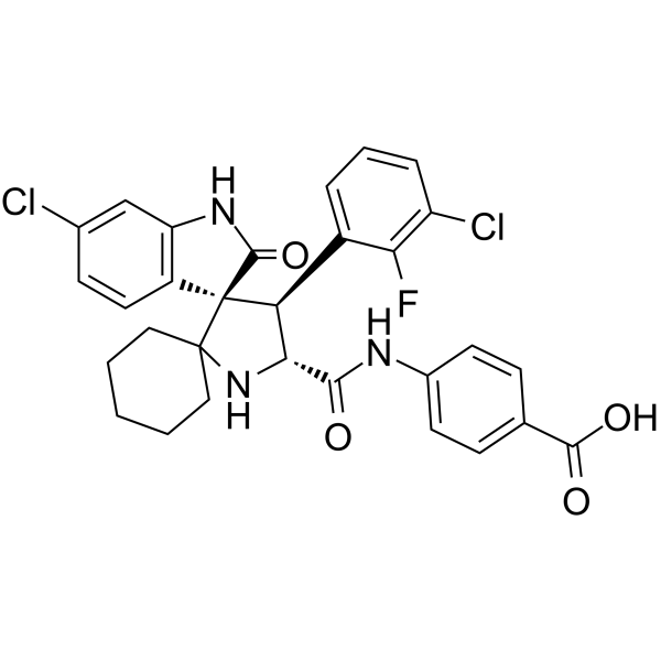
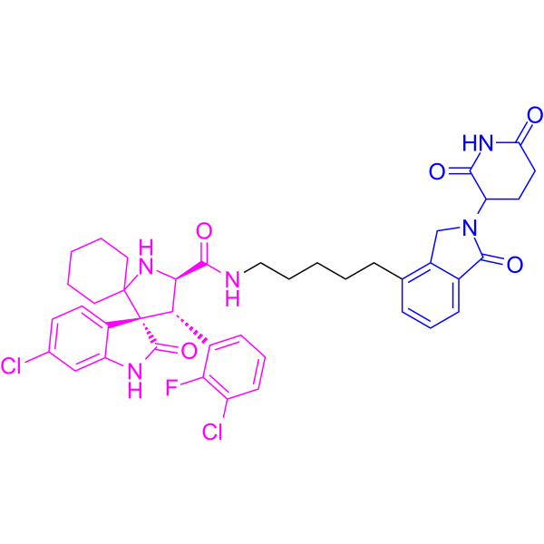
MI-1061 is a potent, orally bioavailable, and chemically stable MDM2 (MDM2-p53 interaction) inhibitor (IC50=4.4 nM; Ki=0.16 nM). MI-1061 potently activates p53 and induces apoptosis in the SJSA-1 xenograft tumor tissue in mice. Anti-tumor activity.
For research use only. We do not sell to patients.
- Purity : 99.08%
- CAS No.: 1410737-34-6
- Formula: C30H26Cl2FN3O4
- Molecular Weight:582.45
-
Storage:Powder -20°C, 3 years , 4°C, 2 years ; In solvent -80°C, 2 years , -20°C, 1 year

To place orders, for customer services and technical support, please contact: MedChemExpress USA
Tel: 609-228-6898 E-mail: [email protected] [email protected]


-
 Biological Activity
-
Chemical Information
-
Solvent & Solubility
- Protocols
- Purity & Documentation
- References
-
 Help & FAQs


-
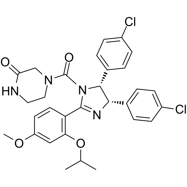
HY-10029
Nutlin-3aMDM2 Antagonist -
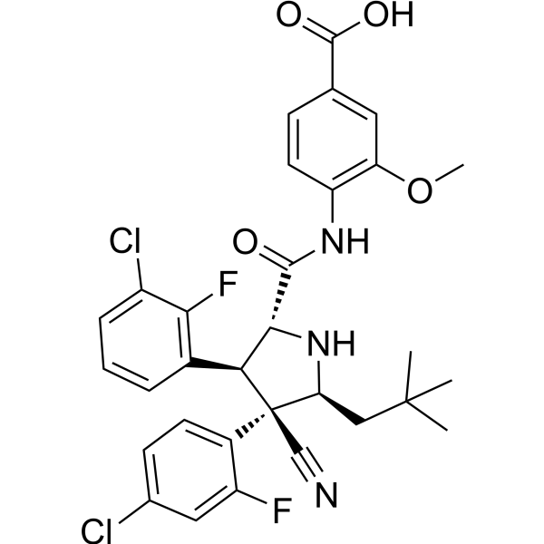
HY-15676
IdasanutlinMDM2 Inhibitor -
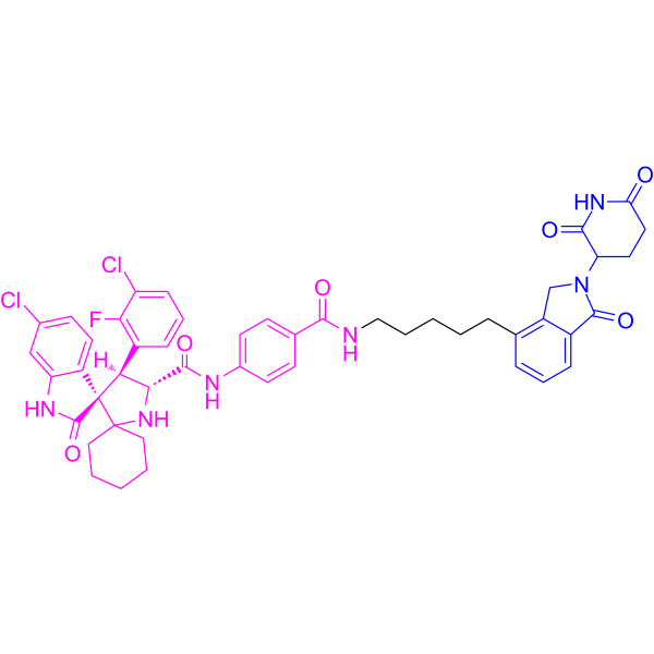
HY-134823
MD-222MDM2 PROTAC Degrader -
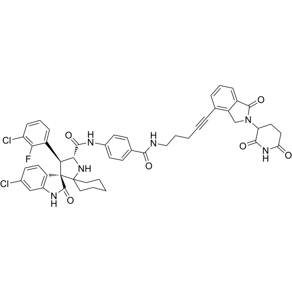
HY-114312
MD-224PROTAC MDM2 Degrader -
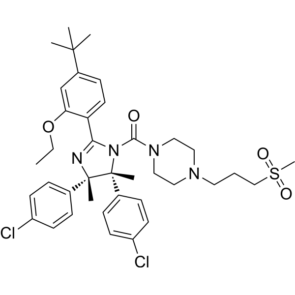
HY-10959
RG7112MDM2 Antagonist -
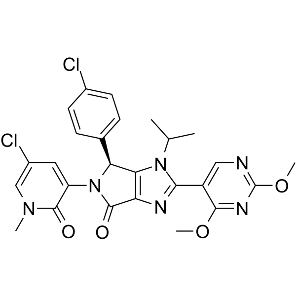
HY-18658
Siremadlinp53-MDM2 Inhibitor -
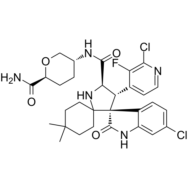
HY-101266
MilademetanMDM2 Inhibitor -
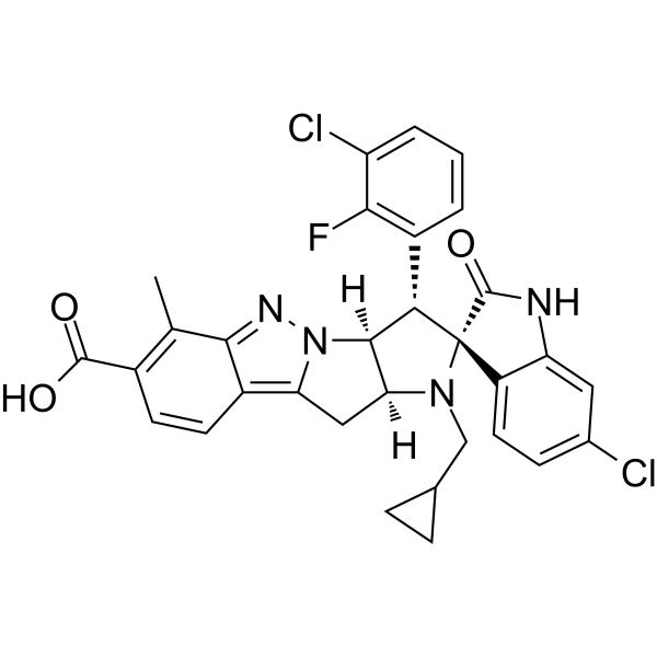
HY-152859
BrigimadlinMDM-2 Inhibitor -
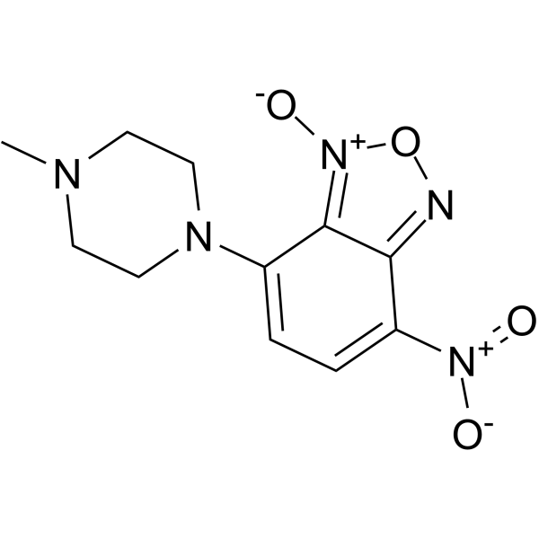
HY-14714
NSC-207895p53 Activator -
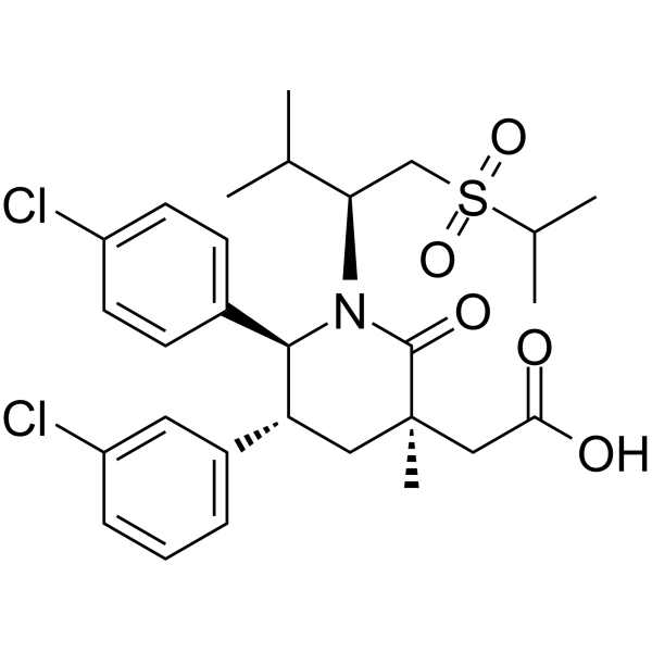
HY-12296
NavtemadlinMDM2-p53 Interaction Inhibitor -
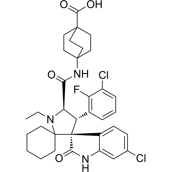
HY-101518
AlrizomadlinMDM2 Inhibitor -
HY-130122
MG-277PROTAC GSPT1 Degrader
Publications Citing Use of MedChemExpress (MCE) MI-1061
More

Biological Activity
Description
IC50 & Target
|
MDM2 |
Cellular Effect
|
Cell Line

|
Type | Value | Description | References |
|---|---|---|---|---|
| HCT-116 | IC50 |
>10 μM
Compound: 31, MI-1061
|
Growth inhibition of p53 -/- human HCT116 cells by WST8 assay
Growth inhibition of p53 -/- human HCT116 cells by WST8 assay
|
[PMID: 25496041] |
| HCT-116 | IC50 |
0.25 μM
Compound: 31, MI-1061
|
Growth inhibition of p53 +/+ human HCT116 cells by WST8 assay
Growth inhibition of p53 +/+ human HCT116 cells by WST8 assay
|
[PMID: 25496041] |
| KG-1 | IC50 |
>10 μM
Compound: MI-1061
|
Inhibition of cell growth in human KG-1 cells harbouring mutant p53 incubated for 4 days by lactate dehydrogenase-based WST-8 assay
Inhibition of cell growth in human KG-1 cells harbouring mutant p53 incubated for 4 days by lactate dehydrogenase-based WST-8 assay
|
[PMID: 30525597] |
| MDA-MB-231 | IC50 |
>3 μM
Compound: MI-1061
|
Antiproliferative activity against human MDA-MB-231 cells assessed as cell growth inhibition after 4 days in presence of control siRNA for 3 days followed by compound treatment by WST-8 assay
Antiproliferative activity against human MDA-MB-231 cells assessed as cell growth inhibition after 4 days in presence of control siRNA for 3 days followed by compound treatment by WST-8 assay
|
[PMID: 31560543] |
| MDA-MB-231 | IC50 |
>3 μM
Compound: MI-1061
|
Antiproliferative activity against human MDA-MB-231 cells transfected with CRBN siRNA LQ-021086-10-0002 assessed as cell growth inhibition after 4 days by WST-8 assay
Antiproliferative activity against human MDA-MB-231 cells transfected with CRBN siRNA LQ-021086-10-0002 assessed as cell growth inhibition after 4 days by WST-8 assay
|
[PMID: 31560543] |
| MDA-MB-231 | IC50 |
>3 μM
Compound: MI-1061
|
Antiproliferative activity against human MDA-MB-231 cells transfected with CRBN siRNA LQ-021086-11-0002 assessed as cell growth inhibition after 4 days by WST-8 assay
Antiproliferative activity against human MDA-MB-231 cells transfected with CRBN siRNA LQ-021086-11-0002 assessed as cell growth inhibition after 4 days by WST-8 assay
|
[PMID: 31560543] |
| MDA-MB-468 | IC50 |
>3 μM
Compound: MI-1061
|
Antiproliferative activity against human MDA-MB-468 cells assessed as cell growth inhibition after 4 days in presence of control siRNA for 3 days followed by compound treatment by WST-8 assay
Antiproliferative activity against human MDA-MB-468 cells assessed as cell growth inhibition after 4 days in presence of control siRNA for 3 days followed by compound treatment by WST-8 assay
|
[PMID: 31560543] |
| MDA-MB-468 | IC50 |
>3 μM
Compound: MI-1061
|
Antiproliferative activity against human MDA-MB-468 cells transfected with CRBN siRNA LQ-021086-10-0002 assessed as cell growth inhibition after 4 days by WST-8 assay
Antiproliferative activity against human MDA-MB-468 cells transfected with CRBN siRNA LQ-021086-10-0002 assessed as cell growth inhibition after 4 days by WST-8 assay
|
[PMID: 31560543] |
| MDA-MB-468 | IC50 |
>3 μM
Compound: MI-1061
|
Antiproliferative activity against human MDA-MB-468 cells transfected with CRBN siRNA LQ-021086-11-0002 assessed as cell growth inhibition after 4 days by WST-8 assay
Antiproliferative activity against human MDA-MB-468 cells transfected with CRBN siRNA LQ-021086-11-0002 assessed as cell growth inhibition after 4 days by WST-8 assay
|
[PMID: 31560543] |
| ML-2 | IC50 |
150.7 μM
Compound: MI-1061
|
Inhibition of cell growth in human ML-2 cells harbouring wild-type p53 incubated for 4 days by lactate dehydrogenase-based WST-8 assay
Inhibition of cell growth in human ML-2 cells harbouring wild-type p53 incubated for 4 days by lactate dehydrogenase-based WST-8 assay
|
[PMID: 30525597] |
| MOLM-13 | IC50 |
76.8 μM
Compound: MI-1061
|
Inhibition of cell growth in human MOLM-13 cells harbouring wild-type p53 incubated for 4 days by lactate dehydrogenase-based WST-8 assay
Inhibition of cell growth in human MOLM-13 cells harbouring wild-type p53 incubated for 4 days by lactate dehydrogenase-based WST-8 assay
|
[PMID: 30525597] |
| MOLM-14 | IC50 |
143.3 μM
Compound: MI-1061
|
Inhibition of cell growth in human MOLM-14 cells harbouring wild-type p53 incubated for 4 days by lactate dehydrogenase-based WST-8 assay
Inhibition of cell growth in human MOLM-14 cells harbouring wild-type p53 incubated for 4 days by lactate dehydrogenase-based WST-8 assay
|
[PMID: 30525597] |
| MONO-MAC-6 | IC50 |
>10 μM
Compound: MI-1061
|
Inhibition of cell growth in human MONO-MAC-6 cells harbouring mutant p53 incubated for 4 days by lactate dehydrogenase-based WST-8 assay
Inhibition of cell growth in human MONO-MAC-6 cells harbouring mutant p53 incubated for 4 days by lactate dehydrogenase-based WST-8 assay
|
[PMID: 30525597] |
| OCI-AML-5 | IC50 |
374.5 μM
Compound: MI-1061
|
Inhibition of cell growth in human OCI-AML5 cells harbouring wild-type p53 incubated for 4 days by lactate dehydrogenase-based WST-8 assay
Inhibition of cell growth in human OCI-AML5 cells harbouring wild-type p53 incubated for 4 days by lactate dehydrogenase-based WST-8 assay
|
[PMID: 30525597] |
| RS4-11 | IC50 |
140 nM
Compound: MI-1061
|
Inhibition of cell growth in human RS4-11 cells incubated for 4 days by Lactate dehydrogenase based WST-8 assay
Inhibition of cell growth in human RS4-11 cells incubated for 4 days by Lactate dehydrogenase based WST-8 assay
|
[PMID: 30525597] |
| SIG-M5 | IC50 |
105.6 μM
Compound: MI-1061
|
Inhibition of cell growth in human SIG-M5 cells harbouring wild-type p53 incubated for 4 days by lactate dehydrogenase-based WST-8 assay
Inhibition of cell growth in human SIG-M5 cells harbouring wild-type p53 incubated for 4 days by lactate dehydrogenase-based WST-8 assay
|
[PMID: 30525597] |
| SJSA-1 | IC50 |
0.1 μM
Compound: 31, MI-1061
|
Growth inhibition of human SJSA1 cells by WST8 assay
Growth inhibition of human SJSA1 cells by WST8 assay
|
[PMID: 25496041] |
In Vitro
MI-1061 achieves IC50=100 and 250 nM in the SJSA-1 and HCT-116 p53+/+ cell lines, respectively, and has IC50>10000 nM in the p53 knockout cell line HCT-116 p53–/–cell line[1].
MedChemExpress (MCE) has not independently confirmed the accuracy of these methods. They are for reference only.
In Vivo
MedChemExpress (MCE) has not independently confirmed the accuracy of these methods. They are for reference only.
-
Animal Model:SCID mice bearing SJSA-1 osteosarcoma xenografts[1]
-
Dosage:100 mg/kg
-
Administration:P.o.; daily for 14 days
-
Result:Demonstrated strong antitumor activity and achieved significant tumor regression.
- Detection of Apoptosis
- TUNEL staining for apoptotic DNA fragmentation
- Annexin V plus membrane-impermeant dye apoptosis staining
- Patient-Derived Xenograft (PDX)
- Apoptosis Solutions
- Splenic/Portal-Vein Liver Metastasis Xenograft
- Subcutaneous Cell-Line-Derived Xenograft
- Orthotopic Cell-Line Xenograft
- Intraperitoneal/Peritoneal Dissemination Xenograft


Chemical Information
-
CAS No. 1410737-34-6
-
Appearance Solid
-
Molecular Weight 582.45
-
Formula C30H26Cl2FN3O4
-
Color White to off-white
-
SMILES
O=C(O)C1=CC=C(NC([C@H](N2)[C@H](C3=CC=CC(Cl)=C3F)[C@]4(C(NC5=C4C=CC(Cl)=C5)=O)C62CCCCC6)=O)C=C1
-
Shipping
Room temperature in continental US; may vary elsewhere.
-
Storage
Powder -20°C 3 years 4°C 2 years In solvent -80°C 2 years -20°C 1 year


Publications (3)
-
Journal Impact Factor
-
Most Recent
-
Nat Commun
PROTAC repurposing uncovers a noncanonical binding surface that mediates chemical degradation of nuclear receptors. [Abstract]2025 Nov 6;16(1):9805. PMID: 41198675 -
Cell Rep
2022 May 31;39(9):110879. PMID: 35649362 -
Cells
PROTAC-Mediated Targeted Degradation of MDM2 Induces Tumor-Suppressive Signaling in Osteosarcoma Cells. [Abstract]2026 Mar 5;15(5):473. PMID: 41827906


Solvent & Solubility
In Vitro:
DMSO : 160 mg/mL (274.70 mM; Need ultrasonic; Hygroscopic DMSO has a significant impact on the solubility of product, please use newly opened DMSO)
Please refer to the solubility information to select the appropriate solvent. Once prepared, please aliquot and store the solution to prevent product inactivation from repeated freeze-thaw cycles.
Storage method and period of stock solution: -80°C, 2 years; -20°C, 1 year. When stored at -80°C, please use it within 2 years. When stored at -20°C, please use it within 1 year.
Please refer to the solubility information to select the appropriate solvent. Once prepared, please aliquot and store the solution to prevent product inactivation from repeated freeze-thaw cycles.
Storage method and period of stock solution: -80°C, 2 years; -20°C, 1 year. When stored at -80°C, please use it within 2 years. When stored at -20°C, please use it within 1 year.
Concentration (start) × Volume (start) = Concentration (final) × Volume (final)
In Vivo:
Select the appropriate dissolution method based on your experimental animal and administration route.
- For the following dissolution methods, please ensure to first prepare a clear stock solution using an In Vitro approach and then sequentially add co-solvents:
- To ensure reliable experimental results, the clarified stock solution can be appropriately stored based on storage conditions. As for the working solution for In Vivo experiments, it is recommended to prepare freshly and use it on the same day.
- The percentages shown for the solvents indicate their volumetric ratio in the final prepared solution. If precipitation or phase separation occurs during preparation, heat and/or sonication can be used to aid dissolution.
Add each solvent one by one: 10% DMSO 40% PEG300 5% Tween-80 45% Saline
Solubility: ≥ 4 mg/mL (6.87 mM); Clear solution
This protocol yields a clear solution of ≥ 4 mg/mL (saturation unknown).
Taking 1 mL working solution as an example, add 100 μL DMSO stock solution (40.0 mg/mL) to 400 μL PEG300, and mix evenly; then add 50 μL Tween-80 and mix evenly; then add 450 μL Saline to adjust the volume to 1 mL.
Preparation of Saline: Dissolve 0.9 g sodium chloride in ddH₂O and dilute to 100 mL to obtain a clear Saline solution.
Add each solvent one by one: 10% DMSO 90% (20% SBE-β-CD in Saline)
Solubility: 4 mg/mL (6.87 mM); Suspended solution; Need ultrasonic
This protocol yields a suspended solution of 4 mg/mL. Suspended solution can be used for oral and intraperitoneal injection.
Taking 1 mL working solution as an example, add 100 μL DMSO stock solution (40.0 mg/mL) to 900 μL 20% SBE-β-CD in Saline, and mix evenly.
Preparation of 20% SBE-β-CD in Saline (4°C, storage for one week): 2 g SBE-β-CD powder is dissolved in 10 mL Saline, completely dissolve until clear.
In Vivo Dissolution Calculator
Please enter the basic information of animal experiments:
-
-
-
-
Recommended: Prepare an additional quantity of animals to account for potential losses during experiments.
Please enter your animal formula composition:
-
%DMSO +
Recommended: Keep the proportion of DMSO in working solution below 2% if your animal is weak.
-
%+
-
+%Tween-80 + +
-
%Saline +
The co-solvents required include: DMSO, . All of co-solvents are available by MedChemExpress (MCE). , Tween 80. All of co-solvents are available by MedChemExpress (MCE).
Working solution concentration: 0.22 mg/mL
Method for preparing stock solution: mg drug dissolved in μL DMSO. Stock solution concentration: mg/mL.
1. Take μL DMSO stock solution;
2. Add μL .
μL , mix evenly;
3. Then add μL Tween 80, mix evenly;
4. Then add μL
Please ensure that the stock solution in the first step is dissolved to a clear state, and add co-solvents in sequence. You can use ultrasonic heating (ultrasonic cleaner, recommended frequency 20-40 kHz), vortexing, etc. to assist dissolution.


Purity & Documentation
-
Data Sheet (293 KB)
-
SDS (254 KB)
- English - EN (254 KB)
- Français - FR (254 KB)
- Deutsch - DE (254 KB)
- Norwegian - NO (254 KB)
- Español - ES (254 KB)
- Swedish - SV (254 KB)
- Italian - IT (254 KB)
- Korean - KR (254 KB)
- Portuguese - PT (254 KB)
-
Handling Instructions (2659 KB)
References


Complete Stock Solution Preparation Table
Please refer to the solubility information to select the appropriate solvent. Once prepared, please aliquot and store the solution to prevent product inactivation from repeated freeze-thaw cycles.
Storage method and period of stock solution: -80°C, 2 years; -20°C, 1 year. When stored at -80°C, please use it within 2 years. When stored at -20°C, please use it within 1 year.
| Optional Solvent | Concentration Solvent Mass | 1 mg | 5 mg | 10 mg | 25 mg |
|---|---|---|---|---|---|
| DMSO | 1 mM | 1.7169 mL | 8.5844 mL | 17.1689 mL | 42.9221 mL |
| 5 mM | 0.3434 mL | 1.7169 mL | 3.4338 mL | 8.5844 mL | |
| 10 mM | 0.1717 mL | 0.8584 mL | 1.7169 mL | 4.2922 mL | |
| 15 mM | 0.1145 mL | 0.5723 mL | 1.1446 mL | 2.8615 mL | |
| 20 mM | 0.0858 mL | 0.4292 mL | 0.8584 mL | 2.1461 mL | |
| 25 mM | 0.0687 mL | 0.3434 mL | 0.6868 mL | 1.7169 mL | |
| 30 mM | 0.0572 mL | 0.2861 mL | 0.5723 mL | 1.4307 mL | |
| 40 mM | 0.0429 mL | 0.2146 mL | 0.4292 mL | 1.0731 mL | |
| 50 mM | 0.0343 mL | 0.1717 mL | 0.3434 mL | 0.8584 mL | |
| 60 mM | 0.0286 mL | 0.1431 mL | 0.2861 mL | 0.7154 mL | |
| 80 mM | 0.0215 mL | 0.1073 mL | 0.2146 mL | 0.5365 mL | |
| 100 mM | 0.0172 mL | 0.0858 mL | 0.1717 mL | 0.4292 mL |


MI-1061 Related Classifications
HY-125858 Related Classifications
 Powered by Bioz