1038 Results for "

Acid Red

" in MedChemExpress (MCE) Product Catalog:
Products (1038)

1038 Results for "Acid Red" in MCE Product Catalog:

Cat. No.: HY-179112
CAS No.: 2826215-20-5
PROTAC IKKβ/NR4A1 degrader-1 is a highly efficient and effective dual-PROTAC degrader targeting IKKβ and NR4A1. PROTAC IKKβ/NR4A1 degrader-1 can increase the levels of caspase 3 and cleaved caspase 3 proteins, while the necroptosis marker RIP kinase remained unchanged, indicating that it can induce apoptosis. PROTAC IKKβ/NR4A1 degrader-1 can be used for the study of Acute myeloid leukemia (AML). Red: IKKβ/NR4A1 ligand (HY-13067); Blue: E3 ligase CRBN ligand (HY-14658); Black: Linker (HY-79577) .
loading...
    loading...
Cat. No.: HY-B1247R
CAS No.: 553-12-8
Synonyms: PPIX (Standard)
Protoporphyrin IX (Standard) is the analytical standard of Protoporphyrin IX. This product is intended for research and analytical applications. Protoporphyrin IX is a final intermediate in the heme biosynthetic pathway, which acts as a radiation sensitizer enhancing ROS generation even in a hypoxic state and inducing DNA damage. Protoporphyrin IX also acts as a photo sensitizer undergoing photobleaching that occurs through direct degradation by light irradiation. Protoporphyrin IX is formed and accumulated following 5-aminolevulinic acid (5-ALA) (HY-W000450) administration in the tumor cells of rats. Protoporphyrin IX causes selective improvement of basal cell carcinoma when activated red fluorescence of a peak wavelength at 405 nm. Protoporphyrin IX is promising for research of sonodynamic and photodynamic agents for a wide range of cancers, such as bladder cancer and nodular basal cell carcinoma .
loading...
    loading...
Cat. No.: HY-D3073
CAS No.: 2477607-64-8
Target:  

Fluorescent Dye

Research Areas:  

Others

DXB-NIR is a push-pull dioxaboron fluorescent probe with solvatochromic properties, featuring high fluorescence brightness, photostability, and a large two-photon absorption cross-section. DXB-NIR exhibits Ex/Em of 550/620–780 nm (with two-photon excitation at 930 nm). DXB-NIR quantifies local polarity via the intensity ratio I (>640)/I (<640) between the far-red channel (<640 nm) and the near-infrared channel (>640 nm), and it can simultaneously label the plasma membrane, endoplasmic reticulum, and lipid droplets in live cells. DXB-NIR monitors the increase in local polarity of various cell compartments under conditions of cholesterol depletion, starvation, and oxidative stress. DXB-NIR can be used for polarity imaging of biological membranes and lipid droplets, as well as studies related to cellular stress .
loading...
    loading...
Cat. No.: HY-D3086
CAS No.: 1668565-72-7
Target:  

Fluorescent Dye

Research Areas:  

Cancer

Mito-polarity is a mitochondria-targeted polarity probe. Mito-polarity can be used to detect mitochondrial polarity. The detection mechanism of Mito-polarity is based on an intramolecular charge transfer (ICT) system designed with a donor-π-bridge-acceptor (D-π-A) structure. Among them, the 467 nm green emission generated by the Coumarin (HY-N0709) structure is extremely sensitive to polarity changes, while the 642 nm red emission produced by the extended π-conjugation and ICT effect between the Coumarin and benzothiazine structures shows only a weak response, thus forming a ratiometric fluorescence response that is linearly correlated with solvent polarity. When Mito-polarity is excited at 405 nm in methanol, its excitation/emission wavelengths are Ex/Em = 405/467 nm and Ex/Em = 405/642 nm, with maximum absorption wavelengths of 426 nm and 561 nm .
loading...
    loading...
Cat. No.: HY-D3104
CAS No.: 2767443-23-0
Target:  

Fluorescent Dye

Research Areas:  

Others

ERNT is a fluorescent probe for monitoring endoplasmic reticulum (ER) polarity and detecting endoplasmic reticulum stress (ERS) during the progression of dynamic liver injury. ERNT functions based on a donor-π-acceptor (D-π-A) structure with an intramolecular charge transfer (ICT) effect. ERNT has multiple excitation/emission pairs: for in vitro cell imaging, the green channel has Ex/Em = 488/500-550 nm, and the red channel has Ex/Em = 488/570-620 nm; for in vivo liver imaging, Ex/Em = 520/620 nm; for solvent optical tests, Ex = 470 nm, with the maximum emission peak at 548 nm in low-polarity toluene and 668 nm in high-polarity DMSO. ERNT can evaluate the efficacy of hepatoprotective interventions by detecting changes in ER polarity, and also exhibits excellent photostability and low cytotoxicity .
loading...
    loading...
Cat. No.: HY-D3112
Target:  

Fluorescent Dye

Research Areas:  

Neurological Disease

P2-Aggregate is a polarity-sensitive fluorescent probe for protein aggregation, which is based on the Nile Red scaffold and modified with a morpholine ring to enhance water solubility. P2-Aggregate improves anti-precipitation ability in aqueous phase via morpholine ring modification, and enables site-specific labeling of target proteins by combining with HaloTag technology. Utilizing the property that its emission wavelength blue-shifts with decreasing local polarity (excited at 543 nm), P2-Aggregate quantitatively detects changes in local polarity during protein aggregation and polarity heterogeneity inside aggregates in living cells. P2-Aggregate can be used in the research of diseases associated with the aggregation of Htt-110Q and SOD1 proteins, such as Huntington's disease and amyotrophic lateral sclerosis .
loading...
    loading...
Cat. No.: HY-D3418
Target:  

Fluorescent Dye

Research Areas:  

Inflammation/Immunology

BOD-Tz-TMR is a fluorescent probe used for nitric oxide (NO) detection and atherosclerotic plaque imaging.BOD-Tz-TMR has a detection limit of 31 nM for NO. BOD-Tz-TMR functions via Förster resonance energy transfer (FRET) and bioorthogonal reactions, in which the tetrazine linker quenches fluorescence through the energy transfer to dark state (ETDS) mechanism. The interaction of BOD-Tz-TMR with NO triggers a ratiometric shift in fluorescence from BODIPY (Ex/Em: 488 nm/512 nm, green channel) to TMR (Em: 592 nm, red channel). BOD-Tz-TMR labels macrophages through a bioorthogonal reaction with BCN, enabling its targeted accumulation at atherosclerotic plaque sites to detect endogenous NO associated with lesion progression. BOD-Tz-TMR can be used in studies related to atherosclerosis .
loading...
    loading...
Cat. No.: HY-N12124
CAS No.: 1011244-19-1
Synonyms: Monascinol
Monascuspiloin (Monascinol) is an orally active compound extracted from red mold-fermented rice, with multiple biological activities including anti-androgenic, anti-inflammatory, hypolipidemic, and anti-cancer properties. Monascuspiloin attenuates the PI3K/Akt/mTOR signaling pathway, enhances AMPK phosphorylation, downregulates FASN/SREBP2, induces apoptosis, G2/M phase arrest and autophagy in prostate cancer cells, interferes with dihydrotestosterone-receptor binding, enhances radiation-induced DNA damage, stimulates endoplasmic reticulum stress, and alleviates hepatic oxidative stress. Monascuspiloin regulates hepatic metabolic pathways and modulates the expression of genes and proteins related to hepatic lipid metabolism. Monascuspiloin can be used in studies related to prostate cancer and alcoholic liver injury .
loading...
    loading...
Cat. No.: HY-P5640
CAS No.: 179264-81-4
Target:  

Bacterial Parasite

Research Areas:  

Infection

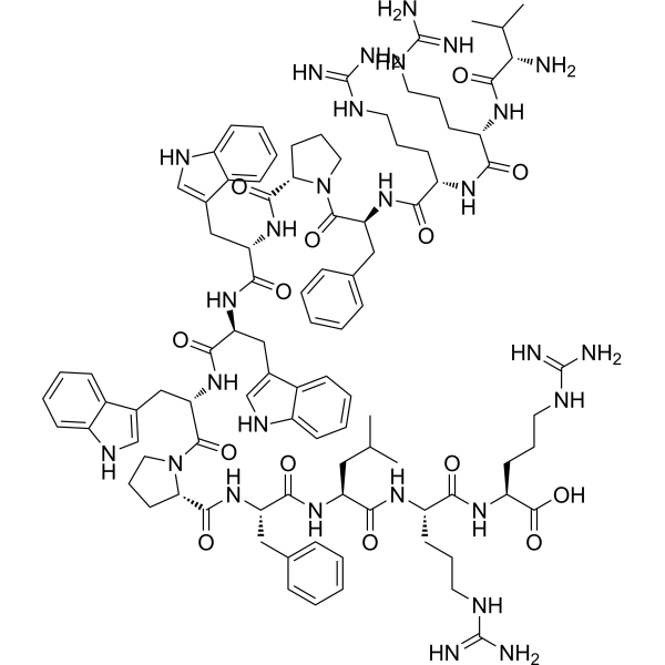
Tritrpticin is a porcine-derived antimicrobial peptide with properties such as membrane disruption and hemolysis. Tritrpticin disrupts the cell membranes of bacteria, fungi and Jurkat T cell leukemia cells and induces their death. Tritrpticin also enhances the efficacy of Metronidazole (HY-B0318) against *Trichomonas vaginalis*, reduces plasma endotoxin and inflammatory cytokine levels, restricts bacterial growth in blood and visceral tissues, decreases the mortality rate of septic shock in rats and enhances the therapeutic effect of ertapenem. Tritrpticin exhibits selective cytotoxicity against Jurkat T cell leukemia cells, while showing low toxicity to normal peripheral blood mononuclear cells and red blood cells, and can serve as a template for antimicrobial peptide design. Tritrpticin can be applied to research related to bacterial infections, fungal infections, trichomoniasis, septic shock and leukemia .
loading...
    loading...
Cat. No.: HY-W014394R
CAS No.: 82654-98-6
Protoporphyrin IX (Standard) is the analytical standard of Protoporphyrin IX. This product is intended for research and analytical applications. Protoporphyrin IX is a final intermediate in the heme biosynthetic pathway, which acts as a radiation sensitizer enhancing ROS generation even in a hypoxic state and inducing DNA damage. Protoporphyrin IX also acts as a photo sensitizer undergoing photobleaching that occurs through direct degradation by light irradiation. Protoporphyrin IX is formed and accumulated following 5-aminolevulinic acid (5-ALA) (HY-W000450) administration in the tumor cells of rats. Protoporphyrin IX causes selective improvement of basal cell carcinoma when activated red fluorescence of a peak wavelength at 405 nm. Protoporphyrin IX is promising for research of sonodynamic and photodynamic agents for a wide range of cancers, such as bladder cancer and nodular basal cell carcinoma .
loading...
    loading...
Cat. No.: HY-W581820
CAS No.: 10030-90-7
Synonyms: Ferrous succinate
Research Areas:  

Cardiovascular Disease

Iron(II) succinate (Ferrous succinate) is an orally active and organic acid iron supplement (ferrous salt). Iron(II) succinate improves hematological parameters in iron-deficiency anemia rats, including hemoglobin, red blood cells, hematocrit, mean corpuscular volume, mean corpuscular hemoglobin, and mean corpuscular hemoglobin concentration. Iron(II) succinate increases serum ferritin, serum iron levels, and transferrin saturation, while decreasing total iron-binding capacity in iron-deficiency anemia rats. Iron(II) succinate reduces the elevated heart, spleen, and kidney coefficients, and increases the decreased liver coefficient in iron-deficiency anemia rats. Iron(II) succinate replenishes the depleted iron content in the liver and spleen of iron-deficiency anemia rats. Iron(II) succinate can be used for the research of iron-deficiency anemia .
loading...
    loading...
Cat. No.: HY-153321A
CAS No.: 2649400-33-7
Synonyms: (R,R)-NX-5948; (R,R)-BTK-IN-24
Target:  

Drug Isomer PROTACs Btk

Research Areas:  

Inflammation/Immunology Cancer

(R,R)-Bexobrutideg is the (R,R)-enantiomer of Bexobrutideg (HY-153321). Bexobrutideg (NX-5948) is an orally active PROTAC that induces specific BTK protein degradation via a cereblon E3 ligase (CRBN) complex without degrading other cereblon neo substrates. Bexobrutideg mediates potent anti-inflammatory activity through BTK degradation, thereby inhibiting B cell activation. Bexobrutideg exhibits potent tumor growth inhibition in TMD8 xenograft models containing wild-type BTK or BTKi resistance mutations. Bexobrutideg is effective in a mouse model of collagen-induced arthritis (CIA). Bexobrutideg can cross the blood-brain barrier. NX-5948 consists of a target protein ligand, a linker, and a VHL E3 ubiquitin ligase (Red: BTK ligand (HY-170324); Blue: CRBN ligand (HY-171893); Black: linker) .
loading...
    loading...
Cat. No.: HY-169433
Naph-Se-TMZ is a PROTAC-like HDAC1 degrader. Naph-Se-TMZ induces ROS-dependent reduction in HDAC1 protein expression and total HDAC activity, and decreases cell viability in a dose-dependent manner. Naph-Se-TMZ exhibits activity in both TMZ-sensitive and TMZ-resistant glioma cells, and its cytotoxicity is reversed by the ROS scavenger N-acetylcysteine (HY-B0215). Naph-Se-TMZ can be used in studies related to glioblastoma .
Naph-Se-TMZ consists of a target protein ligand (red segment): Temozolomide (HY-17364), a DNA intercalator (blue segment): Nitro-Naphthalimide-C2-acylamide (HY-169437), and a molecular linker (black segment). Meanwhile, the activity control for the target protein ligand is Temozolomide-amino hydrochloride (HY-169439), and the DNA intercalator+linker is NNISC-2 (HY-169438).
loading...
    loading...
Cat. No.: HY-D0996
CAS No.: 181885-68-7
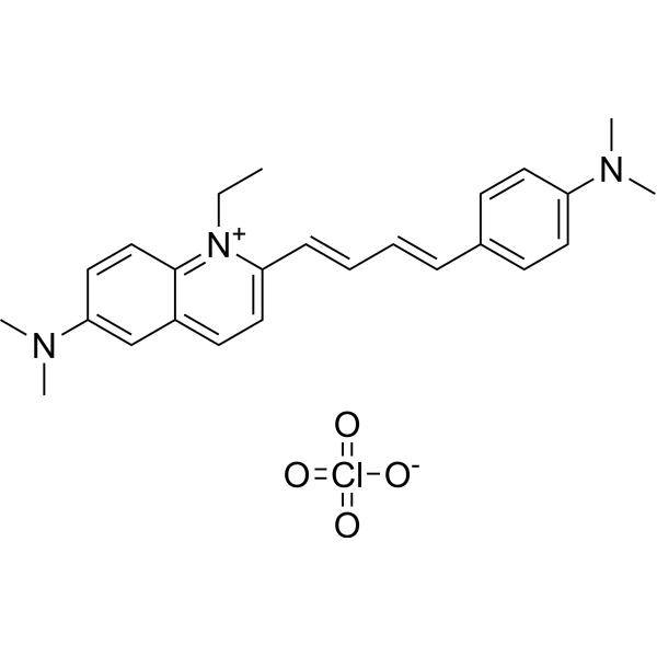
Lds-751 is a nucleic acid stain that mainly detects DNA. Lds-751 is a nucleic acid stain that mainly detects DNA. Lds-751 has a high affinity for DNA and fluorescence is enhanced after binding, but the maximum emission wavelength is 670nm. Lds-751 and Thiazole orange can be used for the differentiation of red blood cells, platelets, reticulocytes, and nucleated cells and can be stimulated at 488nm. Studies have shown that LDS-751 binds almost exclusively to mitochondria when incubated with nucleated living cells. After nucleated Acridine Orange (HY-101879) staining and LDS-751 treatment of cells, confocal microscopy revealed almost no co-location of the cells. Staining with Rhodamine 123 (HY-D0816), a dye known to bind polarized mitochondria, was almost identical to the pattern observed with LDS-751 .
loading...
    loading...
Cat. No.: HY-D1737
RADA is a fluorescent D-amino acid (FDAA) with high photostability and thermostability, which emits yellow-to-orange fluorescence. RADA shows low outer membrane permeability in wild-type Gram-negative Escherichia coli, but it targets penicillin-binding proteins and L,D-transpeptidases, mimics the interaction between acyl acceptors and enzyme intermediates, and integrates into peptidoglycan during biosynthesis. As a peptidoglycan labeling reagent, RADA metabolically integrates into the nascent peptidoglycan of live bacterial cells, labels the peptidoglycan at the poles and lateral walls of mycobacteria, and enables visualization of peptidoglycan synthesis and remodeling processes. RADA serves as a non-specific stain for fixed cells, is non-toxic to bacterial cells, and its red-shifted excitation/emission spectra reduce phototoxicity. RADA also supports virtual pulse-chase labeling experiments and stochastic optical reconstruction microscopy for sub-diffraction-limited imaging of bacterial cell walls .
loading...
    loading...
Cat. No.: HY-D2745
CAS No.: 1638544-48-5
BP Fluor 594 NHS ester is the most popular tool for modifying proteins or antibodies through the primary amines (Lys), amine-modified oligonucleotides, and other amine-containing biomolecules with BP Fluor 594 label. The labeling occurs most efficiently at pH 7-9 and forms a stable, covalent amide bond. BP Fluor 594 dye can be used for proteins labeling at high molar ratios without significant self-quenching, enabling brighter conjugates and more sensitive detection. BP Fluor 594 is bright, water-soluble, and pH-insensitive from pH 4 to pH 10 red-fluorescent dye with absorption and emission maxima at 590 and 617 nm, respectively. It can be used with the 561 nm and 594 nm laser lines. BP Fluor 594 dye conjugated to a variety of antibodies, peptides, proteins, tracers, and amplification substrates often used for generation of stable signal in imaging and flow cytometry.
loading...
    loading...
Cat. No.: HY-D3102
CAS No.: 1015251-87-2
Target:  

Fluorescent Dye

Research Areas:  

Others

CGTDP is a fluorescent probe used for selective labeling of active-site arginine residues, detection of local polarity, and monitoring of conformational changes in enzyme active sites. CGTDP contains a neutral red moiety serving as a polarity-sensitive fluorophore, and a phenylglyoxal unit acting as an arginine-specific labeling group; it undergoes a selective reaction via its α-dicarbonyl moiety with the guanidyl group of arginine residues in enzyme active sites to form a covalent linkage, thereby enabling the reporting of the local microenvironment at the labeled site. The excitation/emission wavelengths are Ex/Em = 398/607 nm and Ex/Em = 507/607 nm; these wavelengths blue-shift in organic solvents, such as Ex/Em = 375/569 nm and Ex/Em = 469/569 nm in THF, and Ex/Em = 375/587 nm and Ex/Em = 470/587 nm in acetonitrile. When labeled onto creatine kinase, its excitation/emission wavelengths are Ex/Em = 493/590 nm .
loading...
    loading...
Cat. No.: HY-131027
Purity:  99.87%
Synonyms: JF646, Azide
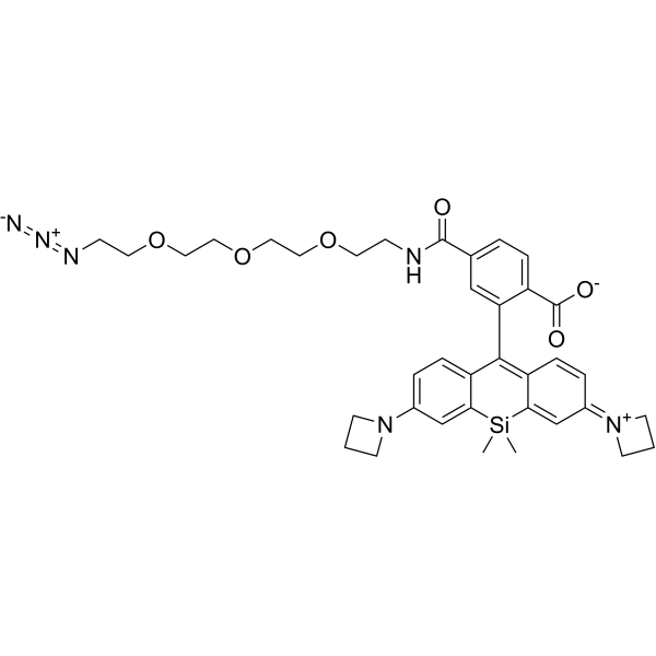
Janelia Fluor 646, Azide (JF646, Azide) is a red fluorogenic fluorescent dye containing a click chemistry group Azide. Janelia Fluor 646, Azide can be used for live-cell imaging experiments . Janelia Fluor fluorescent dyes can be chemically conjugated to the specific HaloTag substrate (a chloroalkane ligand) to form a fluorescent ligand, rather than binding directly to the HaloTag protein itself. Janelia Fluor products are licensed under U.S. Pat. Nos. 9,933,417, 10,018,624 and 10,161,932 and other patents from Howard Hughes Medical Institute. Janelia Fluor 646, Azide is a click chemistry reagent, it contains an Azide group and can undergo copper-catalyzed azide-alkyne cycloaddition reaction (CuAAc) with molecules containing Alkyne groups. It can also undergo strain-promoted alkyne-azide cycloaddition (SPAAC) reactions with molecules containing DBCO or BCN groups (Ex/Em = 646/664 nm).
loading...
    loading...
Cat. No.: HY-184603
Bovine serum albumin-modified gold nanoclusters (BSA-Au NCs) are nanomaterials combining gold nanoclusters and bovine serum albumin (BSA). Composed of several to dozens of gold atoms forming the core of the gold nanoclusters, BSA acts as a protective ligand, coating the surface of the gold nanoclusters. BSA-Au NCs are widely used due to their high luminescence properties and stability over a wide pH range. When Au(III) ions are introduced into a BSA solution, BSA acts as a scaffold protein, isolating and capturing Au ions, similar to the biomineralization behavior of inorganic ions in organisms in nature. The captured Au ions are then reduced in situ by BSA to form Au NCs. The synthesized Au NCs consist of 25-Au atoms and are stable in BSA as BSA-Au NCs, exhibiting strong red fluorescence. BSA-Au NCs possess good biocompatibility and their surface is easily modified or functionalized, making them attractive for many biomedical applications.
loading...
    loading...
Cat. No.: HY-D1602
CAS No.: 217075-07-5
Target:  

Fluorescent Dye

Research Areas:  

Others

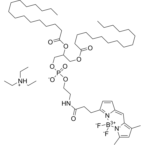
BODIPY FL-DHPE is a fluorescent lipid probe composed of phosphatidylethanolamine (DHPE) labeled with a BODIPY FL fluorophore, which is used for fluorescent labeling of cell membranes, differentiation between liposome membrane fusion and endocytic pathways, and studies of membrane dynamics. The two C16 alkyl chains of BODIPY FL-DHPE insert into the lipid bilayer of membranes, and its fluorescence exhibits concentration dependence: it exists as monomers and emits green fluorescence at low mole fractions; it forms dimers with a red-shifted emission peak at high mole fractions; this spectral change is independent of enzymes, pH, and membrane potential. When liposomes enter the plasma membrane via membrane fusion, the dye is diluted, leading to attenuation of the dimer peak and enhancement of the monomer peak; when entry occurs via endocytosis, the dye remains in endosomes at high concentration with no change in the dimer peak, allowing differentiation between the two uptake pathways. Ex = 488 nm; monomer Em = 515 nm, dimer Em ≈ 620 nm .
loading...
    loading...

